Digestive system Small Intestine Small intestine Is the


")


- Slides: 19
Digestive system Small Intestine
Small intestine : Is the site of food digestion , nutrition , absorption , and endocrine secretion. This small intestine is large about 5 meters and consist of 3 segments : Duodenum , Jejunum and Ileum.
Mucous membrane : The lining of small intestine shows permanent folds , plicae circularis , consisting of mucosa and submucosa and having circular or spiral form , it is the characteristic feature of jejunum. Intestinal villi are long outgrowth of the mucosa projecting into the lumen. In the duodenum they are leaf shaped , gradually assuming fingerlike as they reach the ileum.
Between the villi are small openings of simple tubular gland called intestinal gland (crypt) or glands of Lieberkuhn. The intestinal glands contain stem cells , absorptive cells , goblet cells and paneth and enteroendocrine cells.
Absorptive cells : Are tall columnar cells with an oval nucleus in the basal half of the cell , at the apex there is what's called the striated (Brush border ) which is due to densely packed microvilli. The microvilli increase the surface area of contact of the small intestine with the nutrients. It has been calculated that plicae circularis increase the surface area about 3 folds , the villi increase it 10 folds and the microvilli increase it 20 folds. Together these processes increase the surface area about 600 folds.
The functions of the columnar intestinal cells is to absorb the nutrient. Disaccharidases and peptidases which are bound to microvilli in the brush border hydrolyze disaccharide and dipeptides. Lipid digestion occur by the action of pancreatic lipase and bile.
*Goblet cells : located among absorptive cells , they are less in number in the duodenum and increase in number as they approach the ileum. These cells produce acid glycoprotein of the mucin whose main function is to protect and lubricate the lining of the intestine.
*Paneth cells : In the basal part of I. glands , have secretary granules in the apical cytoplasm , these granules contain lysozyme which have antibacterial activity. *M (microfold )cells : Are specialized epithelial. cells overlying lymphoid follicles. these cells have numerous basal membrane invagination that form pits containing intraepithelial lymphocyte and Ag-presenting cells. M cells endocytose Ag and transport it to the underlying macrophage and lymphocyte , the basement membrane under M cells is discontinuous , facilitating transit between the L. P and M cells.
The LP penetrate the core of I villi In the Duodenum the submucosa contain mucous secreting glands called duodenal or Brunner gland which secret alkaline mucous to protect the duodenal mucosa from acidic gastric fluid and to bring the intestinal contents to the optimum PH for Pancreatic enzymes. Also it secretes urogastrone
The LP of the ileum contain aggregation of lymphatic nodules called Payer's patches , an important components of GALT , they are 30 in number each one contain 10 -200 nodules. The muscularis is well developed composed of inner circular and outer longitudinal.
Vessels and Nerves : The blood vessels penetrate the muscularis and form large plexus in the submucosa , from the submucosa , branches extend through the muscularis mucosa and LP into the villi. At the tips of villi , venules arise from the capillaries and run in the opposite direction reaching the veins in the submucosal plexus.
Lymphatic vessels begin as closed tubes in the core of villi , they are important in lipid absorption , because the blood circulation can not easily accept the lipoproteins , Another important function of the I is the rhythmic movement of the villi , this movement occur by the contraction of smooth muscle in the villi , these contraction can propel the lymph to the mesenteric lymphatics.
Innervation of I is formed by an intrinsic and extrinsic components , the intrinsic one is the Auerback and Meissner's plexus which also receive information about the degree of expansion of the I (mechanoreceptors) and regarding the composition of the I content (chemoreceptors). While the extrinsic component are the parasympathetic which increase the motility of GIT and sympathetic nervous system which decrease the motility of GIT.
Large intestine The mucous membrane of large I has no folds except in the rectal part. No villi are present , the I glands are long coiled with high number of goblet cells and absorptive cells. The absorptive cells are columnar with irregular microvilli. The function of Large I is for water absorption , formation of fecal mass and production of mucous.
The mucous is hydrated gel that lubricates the surface and cover the bacteria. The LP is rich in lymphoid cells as part of GALT due to the abundant of bacteria. The muscularis comprises longitudinal and circular fibers , the fibers of the outer longitudinal layer aggregate in three thick longitudinal bands called teniae coli.
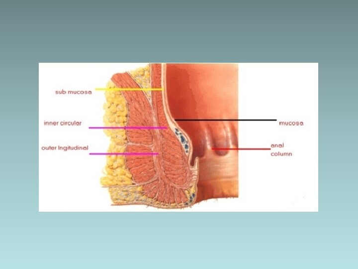
In the anal region , the mucosa form a series of longitudinal folds, rectal columns of Morgagni about 2 cm above the anal opening , the mucosa is replaced by stratified squamous epithelial. In this region , the LP contain a plexus of large veins that when dilated produces hemorrhoids.
Appendix : Is evagination of the cecum , characterized by small , irregular lumen due to the presence of abundant lymphoid follicles in its wall. Although its general structure is similar to that of large I. It contains fewer and shorter I gland has no teniae coli.