Small Intestine Large Intestine and anal cannel 32409


. Small intestine, donkey. Mesothelium of serosa tunica muscularis")
. Small intestine, donkey. Intestinal absorptive cells Simple columnar")
. Small intestine, donkey Absorptive cells Enteroendocrine")
 To see the distribution of EC")


. Small intestine, donkey. Enteroendocrine cells")
, dog. Goblet cell Intestinal crypts (glands")
, dog. Enteroendocrine cell Intestinal crypts (glands")
. Small intestine (jejunum), dog. Absorptive cells epithelial")
. Small intestine (jejunum), dog. lamina subglandularis stratum granulosum")
. Small intestine (jejunum), dog. Crypts of Lieberkuhn submucosa tunica")
. Small intestine (jejunum), pig. . submucosa Absorptive cells")
. Small intestine (jejunum), pig. central lacteal Brush border")
 – Small intestine, puppy (dog). Epithelium Lamina propria Muscularis")
 – Small intestine, puppy (dog). Lymph nodules Intestinal crypts")
 – Small intestine, puppy (dog). Dividing cells Epithelium Lamina")

‐‐‐Large intestine (cecum), dog. villi are absent Epithelium")
‐‐‐Large intestine (cecum), dog. villi are absent Intestinal")
, cat. Epithelium Lamina propria Muscularis mucosa (developing)")
, cat. Intestinal crypts (glands Or Crypts of")
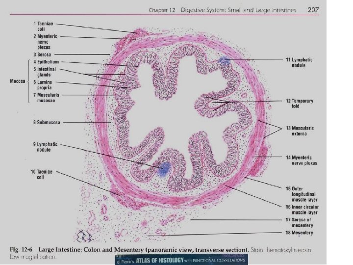
. Large intestine (colon), horse. the taenia coli;")
‐‐‐Large intestine (colon), dog. Intestinal crypts (glands Or")
‐‐‐Large intestine (colon), dog. Enteroendocrine cells Intestinal crypts")
. Anal region, dog. the recto‐‐‐anal junction and regional skin")
. Anal region, dog. keratinized stratified squamous epithelium")
. Anal region, dog the circumanal glands (perianal glands) in")
 glands skin large")

 glands skeletal muscle")


 Submucosa (with")
- Slides: 39
Small Intestine, Large Intestine and anal cannel 32409 Small intestine Large intestine Small intestine
General Structure of the Digestive Tract central lacteal rat 32409 Epithelium with goblet cells and absorptive cells Lamina propria Muscularis mucosa Submucosa Muscularis externa (tunica muscularis) Serosa
Slide #37 (Ed 904‐ 64 b). Small intestine, donkey. Mesothelium of serosa tunica muscularis the myenteric (“Auerbach’s) plexus located between the inner circular and outer longitudinal smooth muscle layers of the tunica muscularis neuronal cell bodies of the submucosal (“Meissner’s) plexus submucosal (Brunner’s) glands intestinal crypts (glands of Lieberkuhn) muscularis mucosa lamina propria central lacteal muscularis mucosa
Slide #37 (Ed 904‐ 64 b). Small intestine, donkey. Intestinal absorptive cells Simple columnar epithelium Paneth cells Goblet cell Paneth cell brush” or “striated” border = High density of non-branching microvilli of uniform length. Microfilaments are the cytoskeleton component in microvilli
Paneth cells Slide #37 (Ed 904‐ 64 b). Small intestine, donkey Absorptive cells Enteroendocrine Cells (EC cells) Mitotic figures
Enteroendocrine cells in fundic stomach, rabbit (toluidine blue) To see the distribution of EC cells among glands 244 Chief cells Parietal cell Enteroendocrine cells
monkey 146 Duodenum, Enteroendocrine cell Crypts of Lieberkühn Goblet and absorptive cells, Muscularis mucosa Lamina propria. Submucosal Brunner's glands.
152 Duodenum Brunner’s glands Absorptive cells Goblet cells Paneth cell Enteroendocrine cell Intestinal villus
Slide #37 (Ed 904‐ 64 b). Small intestine, donkey. Enteroendocrine cells
DEMO SLIDE BOX 57 ‐‐‐ Small intestine (ileum), dog. Goblet cell Intestinal crypts (glands Or Crypts of Lieberkuhn Smooth Muscle in Intestinal villus Peyer’s patches lamina propria of the villi central lacteals
DEMO SLIDE BOX 57 ‐‐‐ Small intestine (ileum), dog. Enteroendocrine cell Intestinal crypts (glands Or Crypts of Lieberkuhn
DEMO SLIDE BOX 225 (C‐‐‐H‐‐‐ 73 ). Small intestine (jejunum), dog. Absorptive cells epithelial Modifications = Brush border and goblet cells central lacteal Strips of Muscularis mucosa lamina subglandularis muscularis mucosa submucosa
DEMOSLIDE BOX 225 (C‐‐‐H‐‐‐ 73 ). Small intestine (jejunum), dog. lamina subglandularis stratum granulosum stratum compactum muscularis mucosa submucosa Enteroendocrine cells Paneth cells lamina subglandularis Absorptive cells
DEMOSLIDE BOX 225 (C‐‐‐H‐‐‐ 73). Small intestine (jejunum), dog. Crypts of Lieberkuhn submucosa tunica muscularis lamina subglandularis muscularis mucosa Submucosal (“Meissner’s”) Smooth muscle cells serosa. Mesothelium of serosa myenteric (“Auerbach’s”) plexuses
Slide #192 (Pf 5‐‐‐ 84 d). Small intestine (jejunum), pig. . submucosa Absorptive cells lamina propria muscularis mucosa tunica muscularis
Slide #192 (Pf 5‐‐‐ 84 d). Small intestine (jejunum), pig. central lacteal Brush border of absorptive cells Intestinal absorptive cells Simple columnar epithelium Smooth muscle cells of the muscularis mucosa lamina propria Goblet cell Paneth cells Enteroendocrine cells
Slide #89 (83 DF 1) – Small intestine, puppy (dog). Epithelium Lamina propria Muscularis mucosa Submucosa tunica muscularis Serosa
Slide #89 (83 DF 1) – Small intestine, puppy (dog). Lymph nodules Intestinal crypts (glands Or Crypts of Lieberkuhn Small intestinal villi central lacteal
Slide #89 (83 DF 1) – Small intestine, puppy (dog). Dividing cells Epithelium Lamina propria myenteric (“Auerbach’s”) plexuses Muscularis mucosa (developing) Submucosa with lymphoid cell infiltration tunica muscularis Serosa mesothelium
Compare luminal surfaces of the small and large intestines small intestines Villi large intestines NO Villi
DEMO SLIDE BOX 167 (C 003‐‐‐H‐‐‐ 29)‐‐‐Large intestine (cecum), dog. villi are absent Epithelium Lamina propria Muscularis mucosa Submucosa tunica muscularis Smooth muscle Serosa Lymph nodules Peyer’s patches Intestinal crypts (glands Or Crypts of Lieberkuhn
DEMO SLIDE BOX 167 (C 003‐‐‐H‐‐‐ 29)‐‐‐Large intestine (cecum), dog. villi are absent Intestinal crypts (glands Or Crypts of Lieberkuhn Epithelium Lamina propria Muscularis mucosa absorptive cells and goblet cells Enteroendocrine cells
DEMO SLIDE BOX 61. Large intestine (colon), cat. Epithelium Lamina propria Muscularis mucosa (developing) Submucosa with lymphoid cell infiltration tunica muscularis Serosa
DEMO SLIDE BOX 61. Large intestine (colon), cat. Intestinal crypts (glands Or Crypts of Lieberkuhn absorptive cells goblet cells Epithelium Lamina propria Muscularis mucosa Submucosa
DEMO SLIDE BOX 171 (E 3‐‐‐H‐‐‐ 38). Large intestine (colon), horse. the taenia coli; a gross thickening of the longitudinal layer of tunica muscularis in the horse. taenia coli Lymph nodules Intestinal crypts (glands Or Crypts of Lieberkuhn
DEMO SLIDE BOX 168 (C 003‐‐‐H‐‐‐ 38)‐‐‐Large intestine (colon), dog. Intestinal crypts (glands Or Crypts of Lieberkuhn Epithelium with goblet cells and absorptive cells Lamina propria Muscularis mucosa Submucosa Muscularis externa (tunica muscularis) Serosa
DEMO SLIDE BOX 168 (C 003‐‐‐H‐‐‐ 38)‐‐‐Large intestine (colon), dog. Enteroendocrine cells Intestinal crypts (glands Or Crypts of Lieberkuhn
Slide #43 (K 9‐‐‐ 1). Anal region, dog. the recto‐‐‐anal junction and regional skin rectum goblet cells located in the crypts rectum nonkeratinized stratified squamous epithelium regional skin circumanal glands (perianal glands) which can become neoplastic
Slide #43 (K 9‐‐‐ 1). Anal region, dog. keratinized stratified squamous epithelium
Slide #43 (K 9‐‐‐ 1). Anal region, dog the circumanal glands (perianal glands) in this region CCT Walled off with CCT parasite external anal sphincter = bundles of skeletal muscle
DEMO SLIDE BOX 62‐‐‐Rectum and anal canal, dog. the circumanal (perianal) glands skin large invagination of the anal sac. rectum simple columnar epithelium
Again the simple columnar epithelium is missing. Note the approximate recto‐‐‐anal junction. At this junction, there is an abrupt change to nonkeratinized stratified squamous epithelium. Near the junction of nonkeratinized and keratinized stratified squamous epithelium, observe the large invagination of the anal sac. The inner circular layer of the tunica muscularis is somewhat thickened to form the internal anal sphincter. The skeletal muscle fibers are part of the external anal sphincter. The anal canal is continuous with the skin of the perineal region (note the change in epithelium to keratinized stratified squamous); see the circumanal (perianal) glands
DEMO SLIDE BOX 62‐‐‐Rectum and anal canal, dog. the circumanal (perianal) glands skeletal muscle fibers are part of the external anal sphincter skin keratinized stratified squamous skin nonkeratinized stratified squamous epithelium rectum tunica muscularis (smooth muscle) is somewhat thickened to form the internal anal sphincter. large invagination of the anal sac. rectum
Large intestine or Colon, monkey
MUCOCUTANEOUS JUNCTIONS
Slide 66: Recto‐anal junction External anal sphincter Internal anal sphincter Anus Rectum
Slide 66 : Recto‐anal junction Keratinized stratified squamous epithelium Hair follicles Sebaceous glands Gland ducts Apocrine glands Anus
Digestive tract distinguishing features human summary Mucosa (epithelium, lamina propria, muscularis mucosa) Submucosa (with submucosal plexuses) Muscularis (Inner circular and outer longitudinal layers, with myenteric plexuses between them) Adventitia/ Serosa Esophagus (upper, middle, lower) Nonkeratinized stratified squamous epithelium; cardiac glands at lower end Small esophageal glands (mainly mucous) Both layers striated muscle in upper region; both layers smooth muscle in lower region; smooth and striated muscle fascicles mingled in middle region Adventitia, except at lower end with serosa Stomach (cardia, fundus, body, pylorus) Surface mucous cells, and gastric pits leading to gastric glands with parietal and chief cells, (in the fundus and body) or to mucous cardiac glands and pyloric glands No distinguishing features Three indistinct layers of smooth muscle (inner oblique, middle circular, and outer longitudinal) Serosa Small intestine (duodenum, jejunum, ileum) Plicae circulares; villi, with enterocytes and goblet cells, and crypts/glands with Paneth cells and stem cells; Peyer patches in ileum Duodenal (Brunner) glands (entirely mucous); possible extensions of Peyer patches in ileum No distinguishing features Mainly serosa Large intestine (cecum, colon, rectum) Intestinal glands with goblet cells and absorptive cells No distinguishing features Outer longitudinal layer separated into three bands, the teniae colo Mainly serosa, with adventitia at rectum Anal canal Stratified squamous epithelium; longitudinal anal columns Venous sinuses Inner circular layer thickened as internal sphincter Adventitia