Lymphoid or lymphatic tissues which mainly consist of
 tissues, which mainly consist of dense accumulations of lymphocytes,")


 nodule. Lymph node, rabbit - H&E")
 form a delicate network between the capsule and trabeculae.")


 labeled \"sc,")


 corpuscle, showing")


- Slides: 62
• Lymphoid (or lymphatic) tissues, which mainly consist of dense accumulations of lymphocytes, are widely distributed in the body. • The lymphocytes may be concentrated into 1 - nodules e. g. mucosa-associated lymphoid tissue (MALT), examples: the tonsils and Peyer's patches; in the ileum and the vermiform appendix. or bronchus-associated lymphoid tissue (BALT), or may be 2 - scattered as in diffuse lymphoid tissue. • Nodules are spherical aggregates of small lymphocytes lacking a C. T. capsule. They may reach a diameter of about 1 mm. They may be homogenous or may be organized into an outer dark cortex containing small lymphocytes and a light central area, the germinal center, which is sensitive to antigens and contains lymphoblasts and lymphocytes. • Lymphoid tissues represent the sites of proliferation and differentiation of lymphocytes. • Lymphoid organs may be defined as anatomical "entities" which consists chiefly of lymphoid tissues.
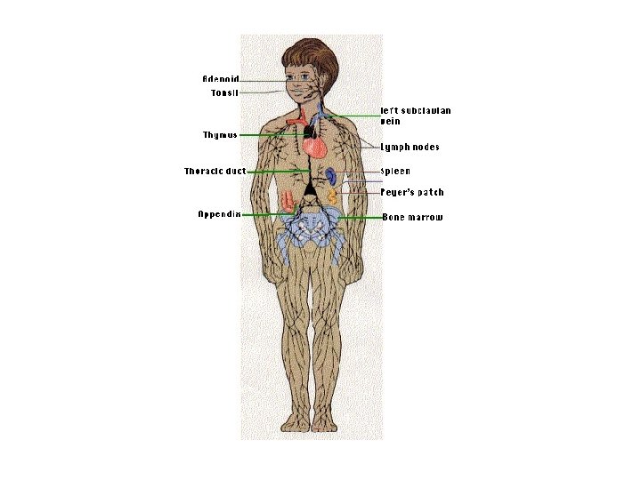
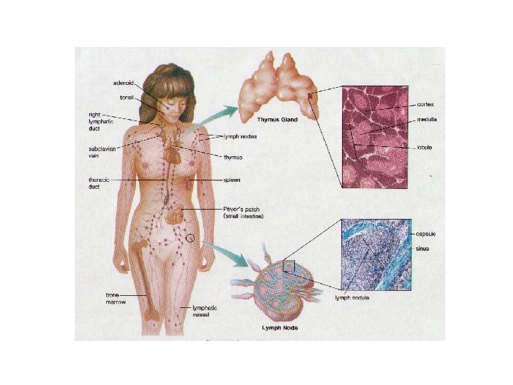
Lymphoid organs: Thymus: is a primary lymphoid organ in that it supplies other lymphoid organs and tissues with T-lymphocytes. Inserted into the blood and lymph vascular system, monitor the internal environment of the body. Lymph Nodes Spleen : the spleen and lymph nodes (secondary lymphoid organs) Tonsils Lymphoid organs and tissues with the exception of the thymus, have in common the presence of large numbers of cells, primarily lymphocytes, and a thin supporting stroma composed of a reticular fiber meshwork.
Mucosa-Associated Lymphoid Tissue: Tonsils, Gut-Associated Lymphoid Tissue The mucosal lining of the alimentary canal and airways is in many ways specialized to facilitate the exchange of substances between the external environment and the body. mucosa-associated lymphoid tissue (MALT), Lymphoid tissue located beneath the mucosal epithelia, protects the body against pathogens that may enter the body via the mucosa. The task that the immune cells of the MALT have to accomplish is different from that of other parts of the immune system. We do need a defense against pathogens, but it would not be a good idea to mount an immune response against components of the food. Immune cell activation therefore differs between the MALT and other lymphoid tissues.
Mucosa-Associated Lymphoid Tissue Often MALT consists of small accumulations of lymphoid cells or one to a few lymph follicles beneath the epithelium and possibly extending into the submucosa. The tonsils and Peyer's patches are large accumulations of lymphoid tissue with associated specialisations of the epithelium.
The tonsils: are accumulations of lymphoid tissue surrounding the openings of the digestive and respiratory tracts. The tonsils and smaller accumulations of lymphoid tissue. • The tonsils share some histological features with lymph nodes: cells in the tonsils are supported by a fine network of reticular fibres and high-endothelial (postcapillary ~) venules function in the "homing" of circulating lymphocytes - this is actually a shared feature of all lymphoid tissues and organs.
Gut-Associated Lymphoid Tissue - GALT Small accumulations of lymphocytes or solitary lymph follicles are found scattered in beneath the epithelium throughout the gastrointestinal tract. However, the most prominent accumulations occur in the ileum and appendix in the form of Peyer's patches. • In the ileum, they form domeshaped protrusions into the lumen. Beneath the epithelial lining of the domes, Peyer's patches extend from the lamina propria to the submucosa. Within Peyer's patches, lymph follicles with germinal centers are typically located deep in the submucosa
Aggregate Lymph Nodule Vermiform appendix. H&E stain. Magnification 40 x. • Aggregate nodules appear when two or more lymph nodules blend together as seen here in the vermiform appendix. Note the germinal centers.
Lymph node and its composition
Lymph Nodes are small, flattened, oval or bean shaped organs, which are situated in the course of the collecting lymph vessels. Their size is variable (from a few mm to more than 2 cm). Each lymph node is formed of: C. T. stroma or framework and A parenchyma of lymphocytes: The lymphocytes are arranged into a peripheral cortex and a central medulla. The stroma (formed of capsule, trabeculae and reticular network. ): The capsule and trabeculae of lymph nodes are formed by connective tissue. some smooth muscles are present at its hilum. The trabeculae extend from the deep surface of the capsule. They divide the cortex of the lymph node into several compartments. The reticular network is present in the background of the lymph node. It is formed of reticular cells and of reticular fibres. It can be demonstrated by silver stain. The cortex: is formed of cortical lymphatic follicles and cortical lymphatic sinues. The medulla is formed of medullary cord and medullary sinuses.
Afferent lymph vessels penetrate the capsule and empty into the subcapsular space. The lymph continues thereafter through cortical and medullary sinuses towards the efferent lymph vessels, which emerge from the hilus of the lymph node. The walls of the sinuses can be traversed freely by all components of the lymph, which allows lymphocytes to enter/leave the lymphoid tissue (as part of their constant circulation) or to get in contact with antigens/antigenpresenting cells that may arrive with the lymph.
Lymphocytes which are located in the outer cortex of the lymph node are likely to represent B-lymphocytes. They are organised into spherical masses - lymphoid nodules or follicles. • • • Germinal centres; sites within the cortex at which B-lymphocytes have been stimulated to proliferate (by contact with an antigen) appear lighter than the surrounding tissue and allow you to identify the centres of lymphoid nodules. The medullary cords mature B-lymphocytes (plasma cells) are located in cord-like extensions of the lymphoid tissue into the medulla, which are continuous with the cortocal follicles. T-lymphocytes are located in the more diffuse tissue between the nodules and in the paracortex, i. e. the deep part of the cortex. Lymph node, rabbit - H&E
Magnified lymphoid (follicle) nodule. Lymph node, rabbit - H&E
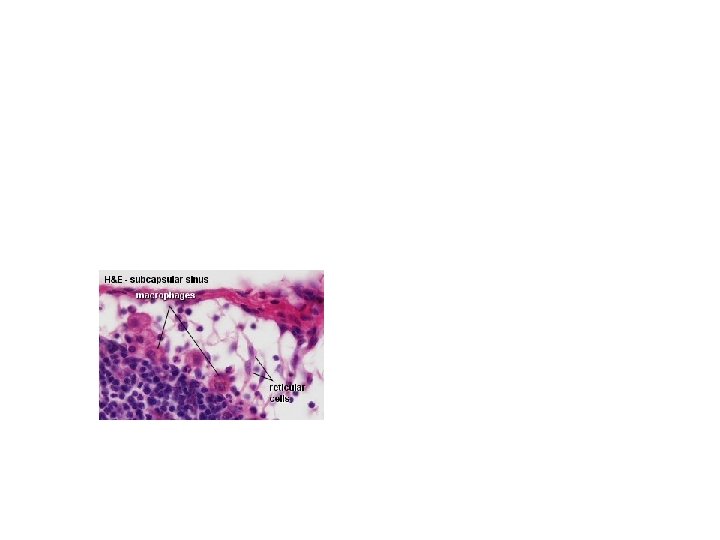
Reticular cells (and reticular fibres) form a delicate network between the capsule and trabeculae. Only their large and light nuclei are easily visible in the microscope. The cytoplasm of reticular cells is only weakly eosinophilic. Lymphocytes and macrophages are **housed in the network of reticular cells and the reticular fibres formed by them. **The processes of reticular cells and reticular fibres extend into and criss-cross within the sinuses.
Macrophages are found scattered within the lymphoid tissue. In many preparations they are difficult to distinguish from the reticular cells, But if an H&E stain turns out nice, macrophages can be distinguished from the reticular cells in the sinus system of the lymph node. Lymph node - H&E, carbon injected This slide illustrates the distribution of macrophages in lymph nodes - take a quick look at them. Note that most of them are located in the paracortex and medulla.
The medulla is formed of medullary cord and medullary sinuses.
Spleen
The spleen is, like the lymph nodes, a discriminatory filter. Unlike the lymph nodes, the spleen is inserted into the blood stream. The spleen clears the blood of aged blood cells and foreign particles and is the site of immune reactions to blood-borne antigens. The spleen is not essential to life in adult individuals. Other organs can take over its functions if the spleen is removed. Stroma: • The spleen is surrounded by a capsule of dense connective tissue from which branched trabecula extend into the parenchyma of the spleen (sounds familiar). reticular connective tissue The parenchyma of the spleen: is termed the pulp of the spleen. Most of the pulp of a fresh, unfixed spleen is a soft, dark red mass, the red pulp. It consists of large, irregular, thin-walled blood vessels, the splenic sinusoids, interposed between sheets and strands of reticular connective tissue, the splenic cords (of Billroth). Within the red pulp small, oval or rounded greyish white areas, the white pulp, is formed by lymphoid tissue (possibly a nodule with a peripherally placed central artery), . The white pulp surrounds the central arteries as a periarterial lymphoid sheath (PALS). Lymphocytes of the PALS are likely to be T-lymphocytes. In addition, we see macrophages and plasma cells in the PALS. Lymph nodules, formed by Blymphocytes, are present along the course of the central arteries. The central arteries are typically located in the periphery of the nodule.
spleen, human H&E lymphoid tissue, • overview, red pulp, white pulp. X 04
spleen, human H&E lymphoid tissue, • X 10
The normal histologic appearance of the spleen. Note the small lymphocytes centered around the splenic arteriole at the center, forming the white pulp. Around this is the red pulp comprised of many splenic sinusoids. The spleen acts as a filter, removing old red blood cells and RBC inclusions. The spleen also acts as a storage area for platelets
Spleen H&E stain. Magnification 40 x. At very low magnification a panoramic view of the spleen. The number "1" identifies a portion of the white pulp composed of splenic corpuscles or splenic nodules, while the number "2" indicated the red pulp. The pointer on the left designates the capsule, and the remaining two pointers each indicate a structure called a central artery.
Spleen H&E stain. Magnification 40 x. Note the capsule indicated by the pointer on the upper left, and the connective tissue trabeculus indicated by the pointer in the right central portion of the image
Spleen H&E stain. Magnification 100 x. Note the connective tissue of the capsule and a trabeculus.
Spleen H&E stain. Magnification 400 x. Basically, the splenic capsule is a dense interlacing connective tissue, but contains in addition to the usual connective tissue components, a few smooth muscle cells. • The pointer indicates the nucleus of one of the smooth muscle cells.
Spleen H&E stain. Magnification 100 x. Without reference to the previous images, what are the structures designated by the three pointers? From left to the right, they are: • Capsule • Trabeculus • Central artery
Spleen H&E stain. Magnification 100 x. Note the splenic nodule (splenic corpuscle) labeled "sc, " a trabeculus shown by the pointer on the left side and a central artery indicated by the left central pointer.
Spleen H&E stain. Magnification 100 x. A splenic nodule's three components are indicated in this image. Find the pale germinal centers the central artery and a structural region known as the marginal zone. The pointer shows the latter in the lower right quadrant.
Spleen H&E stain. Magnification 400 x. Note the detail of a central artery indicated by the pointer. Also examine the parenchymal cells in the vicinity of the central artery.
thymus, human, adult. H&Elymphoid tissue, overview. X 04
The thymus • is situated in the upper parts of the thorax, behind the sternum and the upper four costal cartilages, in the anterior and superior mediastina. The size of the thymus changes in the course of life. It weighs about 10 -15 g at birth and reaches its top weight (about 30 -40 g) at puberty. After puberty a progressive involution (see below) occurs, which leaves a middle-aged person with a thymus weighing about 10 g. The thymus consists of a right and left lobe which are joined by connective tissue.
Thymus H&E stain. Magnification 40 x. At very low magnification some of the structural components of the thymus are shown. • Note the capsule indicated by the pointer in the upper right quadrant, the cortex designated by the letter "c" and the medulla indicated by the letter "m. " This lobe of the thymus is divided into small lobules by thin connective tissue septa.
Thymus, foetal human - H&E This lobe of the thymus is divided into small lobules by thin connective tissue septa Identify the connective tissue capsule and septa, a lobule at low magnification. Note the lobular morphology of the organ, the dense darkly staining cortex and the pale staining region of the medulla. Identify lymphocytes and thymic (Hassall's) corpuscles. They look pretty much like a sliced (very, very small) onion.
thymus, young human, foetal H&E lymphoid tissue, overview 20 th week of gestation. X 04
Thymus, young - cortex
Thymus, young - medulla • Note the detail of the thymic (Hassall's) corpuscle, showing the concentrically arranged acidophilic layers and the relationship of these structures to the medullary region
Thymus H&E stain. Magnification 400 x. • In the medulla the pointer designates a stellate epithelial reticular cell. Note also, the presence of a thymic corpuscle.
Diffuse Lymphoid Tissue Esophagus. H&E stain. Magnification 40 x. As indicated by the pointer note the scattered infiltration of the lamina propria by many lymphocytes
Diffuse Lymphoid Tissue Large intestine. H&E stain. Magnification 40 x. Note the homogenous infiltration by lymphocytes into the mucosa of the large intestine.
Diffuse Lymphoid Tissue Large intestine. H&E stain. Magnification 100 x. As indicated by the pointer note the lymphocytes of the diffuse lymphoid tissue.
Thymus H&E stain. Magnification 40 x. Also at very low magnification a panoramic view of a portion of a lobe of the thymus. Note the lobular morphology of the organ, the dense darkly staining cortex and the pale staining region of the medulla.
Thymus H&E stain. Magnification 40 x. Note the detail of the thymic corpuscle, showing the concentrically arranged acidophilic layers and the relationship of these structures to the medullary region
H&E stain. Magnification 100 x respectively. Note the detail of the thymic corpuscle, showing the concentrically arranged acidophilic layers and the relationship of these structures to the medullary region
Thymus H&E stain. Magnification 400 x. At high magnification examine the structure of the thymic corpuscles and the lymphocytes of the medullary region.
Thymus H&E stain. Magnification 100 x. At increased magnification, note the darkly staining cortex, the pale medulla with its thymic or Hassall's corpuscles.
Thymus H&E stain. Magnification 40 x. Within the medulla, one of several thymic corpuscles (Hassall's corpuscles) is indicated by the pointer. Note that even at very low magnification these characteristic acidophilic structures are readily seen.
Thymus H&E stain. Magnification 100 x respectively. Note the detail of the thymic corpuscle, showing the concentrically arranged acidophilic layers and the relationship of these structures to the medullary region
thymus, young human, foetal. H&Elymphoid tissue, cortex 20 th week of gestation. X 40
thymus, young human, foetal. H&Elymphoid tissue, medulla. 20 th week of gestation. X 40
tonsil, human. H&Elymphoid tissue, overview, lymph follicles, tonsilar crypt. X 02
tonsil, human. H&Elymphoid tissue, reticulated stratified. squamous epithelium. X 40
Diffuse Lymphoid Tissue Vermiform appendix. H&E stain. Magnification 40 x. Note the particularly heavy infiltration of lymphocytes with in the mucosa and submucosa of the organ, and the large lymph nodule in the center of the image
Palatine Tonsil H& E stain. Magnification 40 x. At very low magnification note the nonkeratinized stratified squamous epithelium covering the free surf ace of the palatine tonsil. The pointer in the center of the image designates a structure known as the crypt
Palatine Tonsil H&E stain. Magnification 40 x. The pointer in the center of the image indicates a lymph nodule. Note that it contains a pale staining area known as the germinal center. Also note the epithelium covering the free surface.
Palatine Tonsil H&E stain. Magnification 40 x. The basal region of a palatine tonsil. The pointer in the lower left quadrant indicates a structure known as the basal capsule, while the pointer on the right shows one of the connective tissue septa.
Palatine Tonsil H&E stain. Magnification 400 x. At high magnification examine the detail of the supporting reticular fibrillae, the reticular cells and the lymphoid parenchymal cells. The latter are a combination of lymphocytes, plasma cells and mast cells although they are not easily identified in this image.
Palatine Tonsil Mallory's stain. Magnification 40 x. A panoramic view of a palatine tonsil. Note the crypt, and as indicated by the pointer, a lymph nodule. Well shown is the blue staining connective tissue that supports the covering epithelium.
Palatine Tonsil Mallory's stain. Magnification 40 x. The basal area of the palatine tonsils showing the basal capsule and one of the septa. Compare with image 4
Palatine Tonsil Mallory's stain. Magnification 400 x. As indicated by the pointer examine thin blue stained meshwork of reticular fibrillae that supports the parenchymal cells. Compare with image 5.