The Small Intestine THE SMALL INTESTINE The longest


- Slides: 32

The Small Intestine

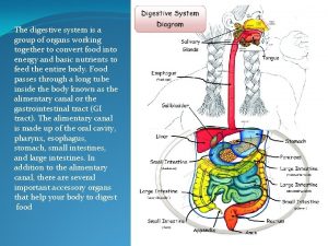
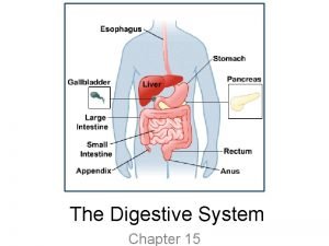
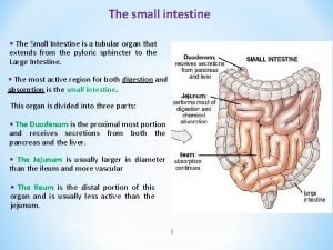
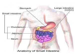
THE SMALL INTESTINE • The longest segment of the digestive tube (4 -7 m) • Function: complete digestion & permit absorption.

• Divided into three parts: • The duodenum. • The jejunum (proximal twofifths). • The ileum (distal threefifths).

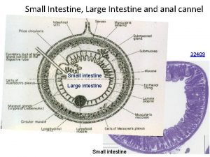
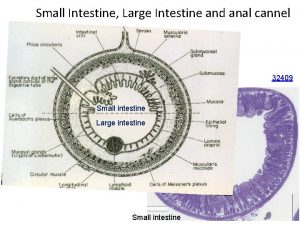
How to increase surface area? Microvilli Plicae circularis Intestinal villi

Modification of the luminal surface

• Longest, thinner and fingerlike in the jejunum. • Finger-like in the ileum. • Maximal food absorption is in jejunum, then duodenum and least in ileum.

THE MUCOSA 1 -Intestinal epithelium: • Simple columnar with microvilli • covers the villi and lines the crypts of lieberkuhn. villus crypts

• Each villus is formed of: Central core of loose CT rich in lymphoid cells, capillary loops, blindly ended lymphatic (lacteal) and few sm ms.

2 - Lamina propria: • Intestinal glands; Simple branched tubular (crypts of Lieberkuhn). • Loose CT forming the villi core& filling the spaces between crypts. • Contains large number of macrophages, mast cells and lymphocytes (WHY? ).

• 3 - Muscularis mucosa: inner cir& outer long sm m layer underlying the bottom of the crypt.

Cells covering the intestinal surface& villi 1 -Absorptive columnar cells. 2 - Goblet cells. 3 - Enteroendocrine cells. 4 MCells (Microfold Epithelial cells).

1 - Absorptive columnar cells(enterocytes) LM: • Tall columnar cells • Prominent striated border luminal surface (closely packed microvilli).

EM: • Prominent brush border • Prominent cell coat (WHY? ); digestive enzymes are attached to it (Disacharidases & peptidase) • Tight junctions along the lateral border (WHY? ); to separate luminal content from lamina propria to allow contact with digesive enzymes BB

ØCell organelles: • Mitochondria • Lysosomes • Small Golgi complex • Few r. ER • Well developed s. ER (fat absorption). • Inclusion: lipid droplets

2 -Goblet cell: LM: • Pale, expanded apical cytoplasm • Narrow basophilic part

EM: • Few apical microvilli at the peripheral zone of the apex. • Apical secretory granules. Function: • Secret a viscid glycoprotein (WHY)? SG

3 -Enteroendocrine cells: Ø Few in the villi but more abundant in the deep intestinal crypts. Ø S-cells; secretin Ø Mo cells; motilin

4 - M- Cells (Microfold Epithelial cells) • Site: specialized cells in epith of the ileum overlying Peyer’s patches.

• EM: broad cells, few loosely packed short microvilli. • Basal cell membrane: deep invaginations forming pockets occupied by lymphocytes and pseudopodia of macrophage between itself and the underlying basal lamina.

Function: Ø They are antigen processing cells. Ø Endocytose (take in) antigens in the intestinal lumen transporting them to cells of the mucosal immune system (Peyer’s patches) Ø Induce an appropriate immune response.

B- The Intestinal glands • Lined by the 4 cells : 1. Undifferentiated Columnar Cells (Stem Cells): 2. Paneth Cells. 3. Absorptive columnar cells. 4. Goblet cells

Undifferentiated Columnar Cells (Stem Cells) • Columnar cells with oval basal nuclei. • Located in the lower half of the intestinal crypts.

Paneth Cells • Confined to the bottom of the intestinal crypts. • LM: Ø Pyramidal cells with ovoid basal nuclei Ø Basophilic cytoplasm near the cell base Ø Eosinophilic zymogen granules in the apex.

• Function: secrete lysozymes (digest the wall of certain bacteria & control the microbial flora in the intestines).

What are the protein secreting cells in the GIT? 1. Serous cells of serous acini in salivary glands (parotid & submandibular). 2. Pancreatic acinar cells. 3. Chief cells of the fundic glands. 4. Paneth cells in the small intestinal glands. LM; apical acidophilia(EM; zymogen gr), basal basophilia (EM; r. ER)

What are the histological adaptations for protection of different parts against gastric acidic secretion? 1. Lower esophageal sphincter ( formed of musculosa of lower third) to prevent reflux. 2. Mucosal (lamina propria) glands in esophagus. 3. Cardiac glands of stomach secrete neutral mucous for protection. 4. Surface mucous cells of stomach; insoluble thick mucous. 5. Brunners glands in submucosa of doudenum; alkaline mucous secretion to neutralize acidic chyme.

Paneth Cell

THE SUBMUCOSA • Formed of relatively dense CT containing blood capillaries, lymphatics and nerves. • The jejunum does not show any particular additional structures in its submucosa.

• In the duodenum: the first part is occupied by Brunner’s glands: • Cpd tubulo-alveolar glands. • Ducts open in the bottom of the crypts.

Brunner’s Glands • Secrete alkaline mucous to protect the intestinal mucosa from acidic gastric chyme and create a suitable p. H for the activity of the digestive intestinal enzymes.

ØEnumerate immunological cells of the small intestine 1 - Goblet cells; adherence of pathogens. 2 - Paneth cells; secrete lysozymes that breakdown bacterial wall. 3 - M-(microfold cells); antigen processing cells that activate immune reaction. 4 - Macrophages & lymphocytes in lymphoid follicles & loose lymphoid tissue.

• In the ileum: • The corium contains aggregations of lymphoid nodules called the Payer’s patches. • Penetrate through the m m and appear in the submucosa. Ileum Villi P P cryp ts mm
 Esophagus stomach small intestine large intestine
 Histology of duodenum
 Arterial supply of small intestine
 5 steps in digestion
 Teeth formula
 Intestine main function
 Intestinal villus
 Frog body parts and functions neisd
 Git layers
 Small intestine
 Small intestine peristalsis
 Small intestine
 Cardiac sphincter and pyloric sphincter
 Small intestine mechanical digestion
 Ligament of treitz
 Galt
Galt The microscopic structure of the small intestine
 Function of digestive system
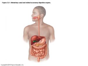
 Alimentary canal figure
 Plicae circulares
 The small intestine extends from the
 Lumen of small intestine
 Yellowish structures that serve as an energy reserve frog
 Parts of the large intestine
 Plicae circulars
 Parts of small intestine
 Digestive enzymes and their functions table
 Is the small intestine a physical or chemical change
 Kidneys are part of the excretory system in a human body
 Special epithelium
 N
N Digestion combining form
 Alimentary cannel
