ANATOMY OF INTESTINE The small intestine measures about

ANATOMY OF INTESTINE The small intestine measures about 20 times the length of the animal. It is composed of three sections: the duodenum, jejunum, and ileum. The small intestine receives the secretions of the pancreas and the gallbladder, which aid digestion. Most of the digestive process is completed here, and many nutrients are absorbed through the villi (small finger-like projections) into the blood and lymphatic systems.

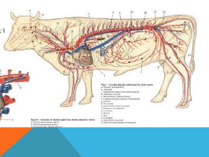
ANATOMY OF RUMINANT INTESTINE

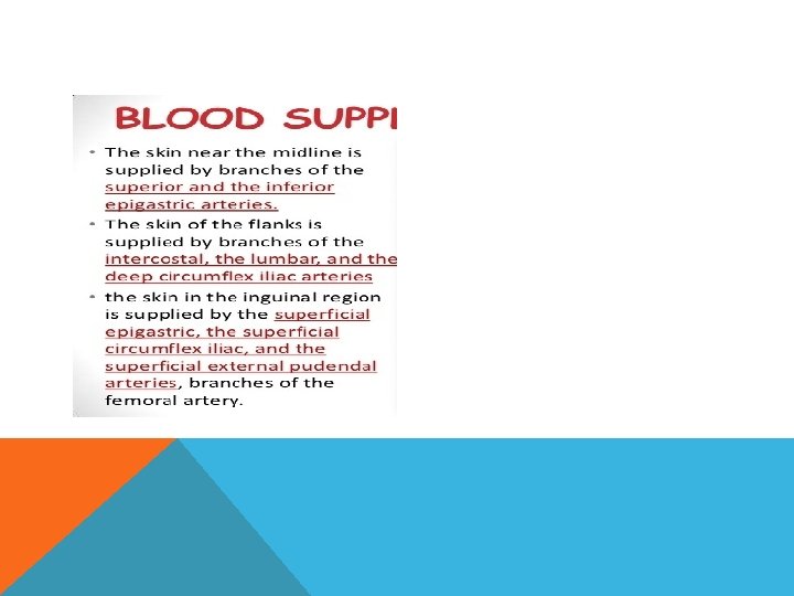
BLOOD SUPPLY v. The duodenum is supplied proximally by a branch of the common hepatic artery called the gastroduodenal artery vthe jejunum and the ileum is from a single artery known as the superior mesenteric artery v. The superior mesenteric vein collects blood from the venous arcades of the small intestine and merges with the splenic vein posterior to the head of the panc reas to form the portal vein.
")
INNERVATION The duodenum receives both sympathetic and parasympathetic innervation. The vagus nerve (CN X)

LIGAMENTS vduodenum is short- short mesoduodenum vjejunum and ileum are less closely fixedmesntric

SURGICAL APPROACH
. 1. paracostal")
SURGICAL APPROACH Position of various left flank incisions (see also Figure below). 1. paracostal (18– 25 cm), cranial in sublumbar fossa: rumenotomy (essential to be as far cranial as possible in large-framed cow and short surgeon); 2. left flank abomasopexy (Utrecht technique) or exploratory laparotomy (25 cm); 3. low flank incision in recumbent cow or heifer for caesarean section, where it is anticipated that it will be difficult to bring uterine wall to flank (35 cm); 4. standard caudal left flank (35– 40 cm) and 5. oblique flank incision (35– 40 cm) for caesarean section in standing animal.

SURGICAL APPROACH
- Slides: 10