12 02 2015 Histology of Glands Dr Archana


:")


 ØCheeks")


 Palatine b) Parotid c) Submandibular")
 Mammary gland b)")
 Simple tubular gland b) Simple")
 Smaller in size b)")
 Serous cell")
- Slides: 44
12. 02. 2015 Histology of Glands Dr. Archana Rani Associate Professor Department of Anatomy KGMU UP, Lucknow
Introduction A gland is an organ that consists of specialized secretory cells. Secreted material is usually a liquid. Epithelial in origin. Present in singly or in groups. Unicellular or Multicellular 2 types: Ø Exocrine Ø Endocrine
Unicellular glands Single cell distributed among non-secretory cells.
Multicellular glands Formed by invagination of epithelial layer in deeper tissue to form a diverticulum: Proximal part forms duct. Distal part forms secretory element.
Exocrine: pour their secretions onto an epithelial surface, directly or through ducts. Endocrine (ductless): pour their secretions directly into the blood.
Classification of exocrine glands Based on shape & branching pattern of duct Based on mode of release of their product Based on the nature of their secretion
Classification based on shape & branching pattern of duct Based on shape of secretory units (tubular or alveolar) & branching pattern of ducts. 2 types: Simple Compound
Contd… Simple glands: Ø Simple tubular Ø Simple coiled tubular Ø Simple branched alveolar Compound glands: Ø Compound tubular Ø Compound alveolar Ø Compound tubuloalveolar
Simple glands
Compound glands
Crypt of Lieberkuhn Brunner glands Sweat glands Submandibular gland Fundic glands of stomach Meibomian glands Mammary gland
Classification based on mode of secretion • Merocrine: secretion is released by exocytosis of secretory granules (pancreas, parotid gland, protein component of milk from mammary gland). • Apocrine: apical portion of cell along with secretory product is pinched off (lipid component of milk from the mammary gland). • Holocrine: entire cell is shed along with secretory product (sebaceous glands).
• Modes of secretion
• Modes of secretion Merocrine
• Modes of secretion Merocrine: secretion by exocytosis
• Modes of secretion Merocrine: secretion by exocytosis Apocrine
• Modes of secretion Merocrine: secretion by exocytosis Apocrine: secretion by loss of cytoplasm
• Modes of secretion Merocrine: secretion by exocytosis Apocrine: secretion by loss of cytoplasm Holocrine
• Modes of secretion Merocrine: secretion by exocytosis Apocrine: secretion by loss of cytoplasm Holocrine: secretion through loss of entire cell
Classification based on the nature of secretion • Serous glands • Mucous glands • Mixed glands
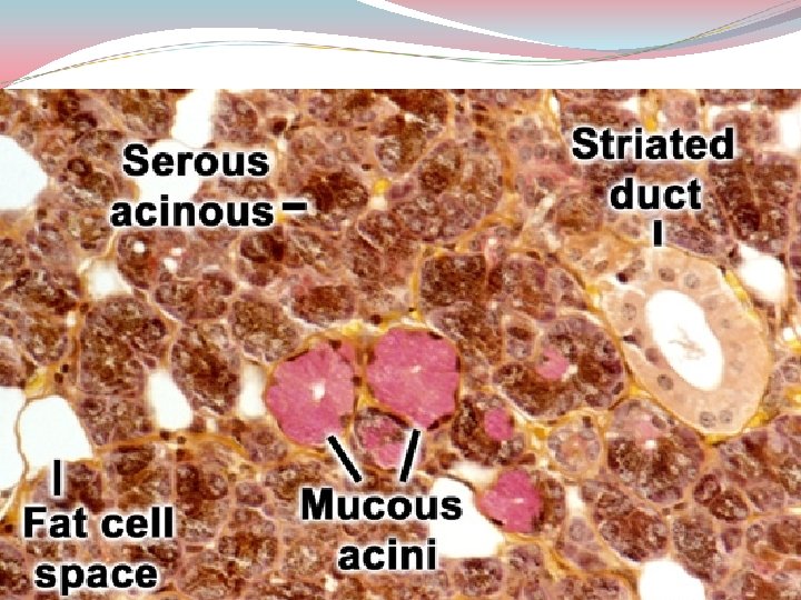
Serous glands Secretion: thin, watery, rich in enzymes, protein in nature. Cytoplasm: granular (stain darkly from pink to dark purple with H/E stain) & rich in r. ER. Nuclei: rounded & basally placed.
Mucous glands Secretion: contains mucopolysaccharides which collects in the apical part of cell. Cytoplasm: stain very lightly with H/E stain & therefore gives empty look. Nuclei: basally placed & flattened (mucoid pushes the nuclei).
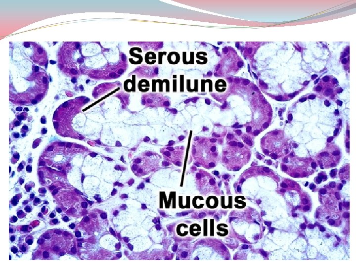
Mixed glands Contain both serous & mucous secretory units. Sometimes serous cells form crescentic caps on mucous acini called as serous demilunes.
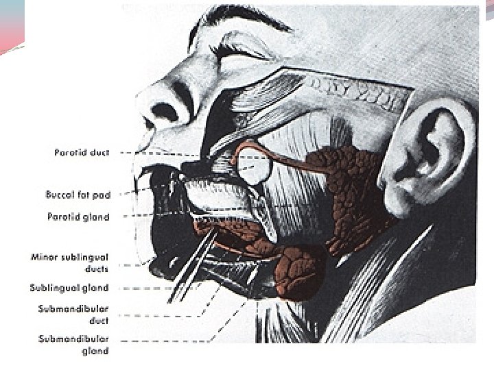
Salivary glands
Major glands Lie completely outside the alimentary tract & connected to it by an excretory duct. Compound alveolar or tubuloalveolar in type. Parotid, submandibular, sublingual.
Minor glands Small salivary glands situated in the mucous membrane of: ØLip (labial) ØCheeks (buccal) ØSoft palate (palatine) ØTongue (lingual)
Functions of Salivary glands Secrete saliva composed of water, mucus, proteins, salts, salivary amylase (ptyalin), immunoglobulins (Ig. A) & lactoperoxidase. Serves to moisten food. Lubricates & moistens oral mucosa and lip. Initiates the digestion of carbohydrates.
Basic organization
Different types of acini Mucous Serous Mixed
Features of serous & mucous cells Serous cell EM LM Mucous cell
Differences between serous & mucous acini
Differences between serous & mucous cells
Parotid gland
Submandibular gland
Sublingual gland
References 1. di. Fiore’s Atlas of Histology with functional Correlations, 12 th Edition. 2. Textbook of Human Histology. Inderbir Singh, 1 st Edition. 3. Textbook of Histology. GP Pal, 3 rd Edition.
MCQ 1. All are major salivary glands except: a) Palatine b) Parotid c) Submandibular d) Sublingual
MCQ 2. Holocrine type of secretion is a feature of: a) Mammary gland b) Parotid gland c) Sebaceous gland d) Pancreas
MCQ 3. Sweat gland is an example of: a) Simple tubular gland b) Simple coiled tubular gland c) Simple branched tubular gland d) Simple alveolar gland
MCQ 4. All are true about serous acini except: a) Smaller in size b) Stain dark c) May present as demilune d) Wide lumen
MCQ 5. Expelling of secretion through contraction is a feature of: a) Serous cell b) Mucous cell c) Serous demilune d) Myoepithelial cell