Classification of Exocrine Glands Type of Secretions Produced


- Slides: 11
Classification of Exocrine Glands: Type of Secretions Produced Type of secretion produced �Serous secretions �Watery �Contain a high concentration of enzymes �Pancreatic secretion �Mucous secretions �Thick, viscous �Composed of glycoproteins � Mucus membranes (GI and respiratory tracts) �Mixed exocrine glands contain both mucous and serous components �Salivary glands
Connective Tissue Forms metabolic and structural connections between tissues Found ______ in the body and represents most abundant tissue by weight. Examples: Blood, tendons, fat, cartilage, bone Some systems are almost exclusively composed of connective tissue Skeletal, integumentary Derived from _______ Is vascularized. Loose & Adipose = good supply, Dense = poorly supplied Form and function may vary can be elastic and flexible, rigid, semisolid, liquid Reserve for energy, protective sheath, provides framework of structural
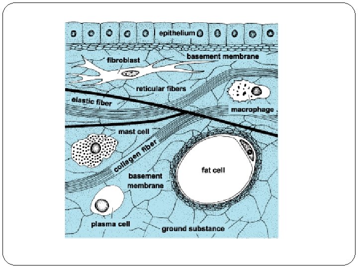
Connective Tissue Components 3 major components of connective tissue: Extracellular fibers, Ground substance, Cells Extracellular Matrix = Extracellular fibers + Ground substance Matrix surrounds and separates the cells, providing important structural and nutritional support to them, allowing them to be farther apart than epithelial cells. 1. Extracellular fibers Collagenous, Reticular, Elastic 2. Ground substance Liquid, solid, or gel 3. Cells Fixed cells Fibroblasts, Adipocytes (fat cells), Reticular cells Wandering cells Mast cells, Leukocytes (white blood cells), Macrophages (fixed and wandering)
Substance An amorphous, homogenous material that ranges from ______ to _______. Composed of glycoproteins called _________ (GAGs) hyaluronic acid Help to orient fiber formation in connective tissue. Is medium through which cells exchange nutrients and waste with the bloodstream. Acts as ______ absorbing cushion and helps to protect the delicate cells that it surrounds.
Fibers of Connective Tissue Collagenous: Most common fiber found in the body Strong, thick bands composed of collagen (structural protein). Organized into bundles Resist pulling forces, so they are found in tendons and ligaments that are continuously being pulled and stretched. wavy appearance when not stretched Sometimes called white fibers Density and arrangement can vary depending on tissue function. Loose around organs to dense within tendons.
Fibers of Connective Tissue Reticular: Composed of collagen, but are not thick Thin, delicate, branched into complicated networks. Form support for around highly cellular organs endocrine glands, lymph nodes, spleen, bone marrow, liver Found around blood vessels, nerves, muscle fibers, capillaries Elastic: Composed primarily of protein elastin. Are branched and form networks Can stretch and contract. Found in tissues that stretch: vocal cords, lungs, skin, blood
Major Cell Types of Connective Tissue Fixed Cells: Remain in the connective tissue Produce and maintain the matrix Fibroblast: secrete fibers and ground substance of the matrix Can reproduce and are metabolically active. Name is based on location o Chondroblast (cartilage), osteoblast (bone), etc. As the cells mature and the matrix is formed, cells become less active and are called –cytes. o Chondrocyte, osteocyte, fibrocyte o Can revert back to blast if more matrix is needed.
Major Cell Types of Connective Tissue Fixed cells continued. Adipose cells/Adipocytes: Found throughout connective tissue Resemble fibroblasts early on, but as they age they become filled with lipid and swell. nucleus gets pushed to the side Adipocytes clustered together form adipose tissue. found all over, but is prominent under the skin, behind the eyes, around the kidneys, and in the abdomen Reticular Cells: Flat, star-shaped cells that form net-like connections with other cells Manufacture of reticular fibers. Found in tissues of the immune system:
Major Cell Types of Connective Tissue Wandering Cells: Move in and out of connective tissue as needed. Leukoctyes: (white blood cells) Found in blood and move into connective tissue during periods of infection. Squeeze through the simple squamous epithelium of blood vessels (diapedesis) Important in immune function- engulf and digest invaders or produce antibodies against them Mast cells Carry histamine and heparin granules which initiate inflammatory
Major Cell Types of Connective Tissue Wandering cells continued. . Macrophages: Phagocytotic scavengers that may be either fixed or transient in connective tissue. Engulf microbes, dead cells and debris that are digested by the macrophage’s lysosomes drawn to sites of infection where they engulf invaders Given different names depending on locations Kupffer cells in liver, microglial cells in brain