Volume 86 No 2 2017 GASTROINTESTINAL ENDOSCOPY 299


, and most lesions")

, At the")

. 16 patients had more")
 were minor : included severe pain")


- Slides: 46
Volume 86, No. 2 : 2017 GASTROINTESTINAL ENDOSCOPY 299
Background and Aims: § Despite recent advances in the diagnosis and treatment of esophageal cancer, more than half of malignancies involving the esophagus are inoperable at presentation because of locally advanced or metastatic disease. § The prognosis of esophageal cancer is dismal, with a 5 -year survival rate below 20%. § Esophageal stenosis and esophagorespiratory fistula are frequent adverse events (AEs), usually associated with weight loss, nutritional deficiency, and low quality of life.
§ A variety of treatment modalities has been used to palliate dysphagia in patients with advanced esophageal tumors such as endoscopic ablation, radiotherapy, brachytherapy, endoluminal stents, and surgery. § Self-expandable metallic stents (SEMSs) are considered the mainstay palliative treatment of dysphagia for patients with advanced esophageal cancer. § SEMSs provide rapid, efficient, and lifelong relief of dysphagia in most patients and are the most common palliative treatment modality performed worldwide.
§ However, migration, bleeding, fistula, and tumor or hyperplastic tissue overgrowth are common long-term AEs of definitive SEMSs and represent a major concern, especially in patients with better prognosis and longer survival. § The incidence of long-term esophageal stent-related AEs varies widely in the literature, with reported values ranging from 20% to 50%. § However, most data concerning late AE rates come from relatively small cohorts with reported median stenting times shorter than 3 months.
§ The aim of this study was to evaluate the rate of AEs among patients who survived longer than 6 months with esophageal stents in place and the endoscopic management of these AEs. § In addition, we wanted to study possible predictive factors associated with SEMS-related AEs in this clinical setting.
Methods; § We conducted a retrospective analysis of a prospectively collected database including all patients submitted to esophageal stent for palliation of malignant strictures or malignant fistulas between February 2009 and February 2014 at the Cancer Institute of the University of São Paulo. § For the purpose of this study, only patients who remained with the stent longer than 6 months were included in the analysis. § Patients with benign stenosis or fistula because of anastomotic leakage were excluded.
Stent implantation; § All procedures were done under the supervision of an anesthesiologist. Patients were placed in the supine position or left lateral decubitus. § Before stent insertion, an endoscopy was performed to evaluate the tumor length and location.
§ When it was possible to traverse the tumor with a conventional or slim endoscope, a stiff guidewire was used to guide the stent insertion § When the tumor was not traversable and thin endoscopes were not available, a hydrophilic guidewire preloaded into a biliary catheter was used to traverse the stricture under fluoroscopic monitoring, and iodine-based dye was used to estimate tumor length (dilation was avoided). External markers or lipiodol injection in the esophageal walls was used to mark the tumor’s limits.
§ Stent length was chosen to cover at least 2 cm beyond each tumor edge. § Stents were deployed under fluoroscopic control. § For tumors of the upper third close to the cricopharyngeus, endoscopic control was also used to ensure the best positioning of the proximal flare. § An esophagogram with iodine-based solution was obtained immediately after stent placement to confirm the adequate flow of contrast to the stomach and to confirm fistula sealing when it was present.
§ For lesions located in the proximal esophagus, we used a dedicated model of a partially covered stent with a lowprofile, proximal flare shape (Choostent; MI Tech, Pyeongtaek, Korea). § For lesions located in the mid-esophagus, we used a standard partially covered stent or, less often, a fully covered stent, with an 18 - to 23 -mm body diameter and 23 - to 28 -mm flare diameter (Wallflex and Ultraflex [Boston Scientific, Watertown, Mass], Evolution [Wilson Cook Medical, Letchworth, UK], Hanarostent [MI Tech], or Endoflex [Voerde, Germany]). § If it was predicted that the stent would traverse the esophagogastric junction, we used a partially covered stent with an antireflux valve (Hanarostent Esophagus Valve; MI Tech).
§ Patients received pre-emptive analgesia with morphine at a dose of 10 mg orally 3 times a day associated with a nonopioid analgesic for 14 days. Ondemand simple analgesics (eg, paracetamol) to control mild pain were allowed after this period.
Follow-up; § Clinical success was defined when patients were able to resume oral intake of liquid and solid foods. § For patients with malignant fistula, clinical success was defined when patients were able to tolerate liquid and solid intake without coughing or choking. § Sealing of the fistula was confirmed by esophagogram performed just after stent implantation.
§ Follow-up time was defined from the day of stent implantation until the last clinical visit or death. After stent placement routine visits were scheduled at 1, 3, 6, 9, and 12 months, and a standard questionnaire was applied for all patients. § For patients who missed the clinic appointment, contact by telephone was made for a follow-up interview. § The presence of any symptoms that suggested stent malfunctioning (recurrence of dysphagia, coughing related to liquid swallowing, pain, etc) was an indication for endoscopic re-evaluation.
Ø The expected AEs were; § § § § migration overgrowth/ingrowth food impaction bleeding esophageal perforation stent-induced fistulas severe pain severe gastroesophageal reflux
Ø AEs were classified into; § minor when the stent dysfunction could be managed clinically or endoscopically and no life-threatening situation was present. § major when it was a life-threatening situation or when a major intervention such as surgery was required. § AE-related death was defined as patient death caused by a stentrelated AE.
Management of stent dysfunction; § If there was a distal dislodgment but the stent remained at the esophagus, endoscopic traction was achieved using rat-tooth forceps. § Stents that migrated into the stomach were completely removed with a rat-tooth forceps and a biliary pusher, as previously described. § Tumor ingrowth or overgrowth was treated with argon plasma coagulation, dilation, or the combination of both. In refractory cases a second stent was placed. § Stent-induced fistula was managed by placement of another stent.
Statistical analysis; § Statistical significance was defined as P< 0. 05. § Statistical modeling and tests were performed with SPSS software, version 21. 0 (SPSS Statistics for Windows, Version 21. 0, IBM Corp. , Armonk, NY).
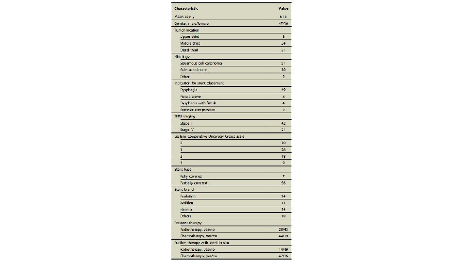
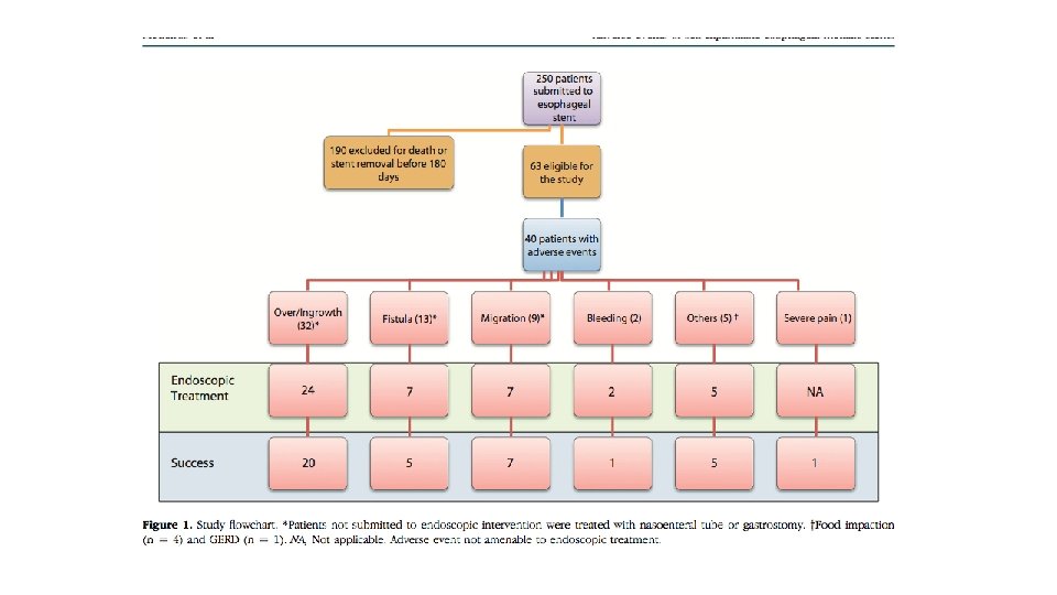
RESULTS; Ø Patient characteristics; § From February 2009 to February 2014, 250 patients were submitted to esophageal stent insertion and 63 patients fulfilled the inclusion criteria (Fig. 1). § There was a predominance of men (74. 6%), and patient mean age was 61. 4 years (range, 42 -79). Performance status according to the Eastern Cooperative Oncology Group (ECOG) was 0 (n = 10), 1 (n=26), 2 (n =18), and 3 (n =9).
§ The most common cancer was squamous cell carcinoma (80. 9%), and most lesions were located in the middle esophagus (53. 9%). § Regarding stents placed initially, 56 were partially covered (88. 8%) and 7 were fully covered. Demographic characteristics are shown in Table 1.
Follow-up; § The indication for stent placement was dysphagia because of esophageal malignancy in 49 patients (77. 7%), malignant fistula in 8 patients (12. 6%), dysphagia associated with fistula in 4 patients (6. 3%), and extrinsic compression in 2 patients (3. 1%). § Clinical success was achieved in all patients. The median stent patency (until death or stent dysfunction) was 7. 1 months (standard deviation, 3. 8). Only 4 patients had their stent removed during the follow-up.
§ Mean follow-up time was 10. 7 months (range, 6. 1 -25), At the end of follow-up; § 37 patients (58. 7%) had a functioning stent and were accepting oral intake. § 26 patients (41. 2%) were not accepting oral intake: 20 because of cachexia and 6 because of stent dysfunction refractory to endoscopic treatment. A nasoenteric tube or gastrostomy feeding was used for these patients.
§ There were 2 cases of extrinsic esophageal compression caused by metastatic breast cancer and lung cancer; § The first patient had 3 episodes of overgrowth and ingrowth. All episodes were endoscopically managed with success, and the patient survived 25 months with a patent stent. § The patient with lung cancer had 2 episodes of overgrowth, both successfully treated by endoscopy. Dysphagia recurred 12. 8 months after stent placement, when a nasoenteric tube was placed; the patient died 10 days later.
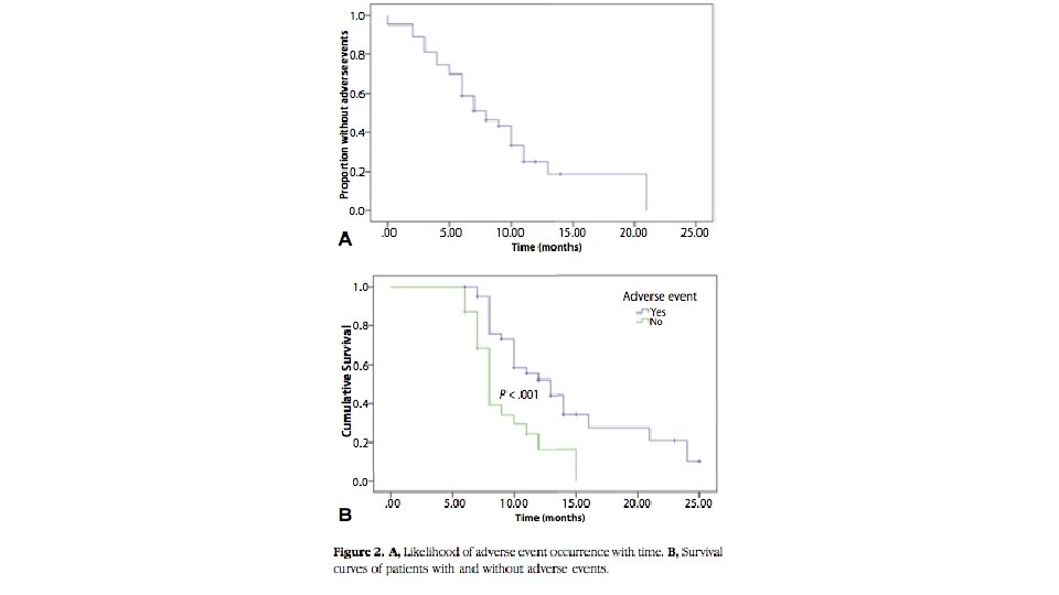
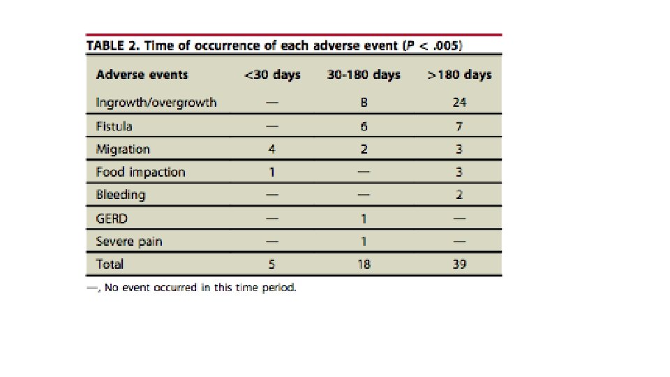
Adverse events; § AEs occurred in 40 patients (63. 5%). 16 patients had more than 1 AE, and 5 patients had recurrence of a previously treated AE (4 patients with recurrence of ingrowth/overgrowth and 1 patient with recurrence of migration). § There were a total of 62 AEs, with a mean of 1. 5 AEs per patient. 5 AEs occurred within 30 days of stenting, 18 occurred between 30 and 180 days, and 39 occurred after 180 days (P =0. 042). Table 2 shows the occurrence of each AE according to 3 different periods of time. Figure 2 A shows the likelihood of AE occurrence with time.
§ Most AEs (n = 47, 75. 8%) were minor : included severe pain (1), severe reflux (1), migration (9), ingrowth/ overgrowth (32), and food impaction (4). § There were 15 major AEs: 13 esophageal fistulas and 2 bleedings. Endoscopic treatment was attempted in 9 of 15 major AEs, with success in 6. Three major AEs were fatal: 2 patients had esophagorespiratory fistulas and died from pulmonary sepsis and 1 died because of tumor bleeding. Figure 1 summarizes AEs and endoscopic interventions.
Management of stent dysfunction; § Endoscopic management was not attempted in every stent-related AE. When patients were considered too ill (usually because of cachexia) to be submitted to an endoscopic intervention, they were palliated with a nasoenteral tube or a gastrostomy. 26 patients of our cohort advanced to this situation. § When endoscopic treatment was attempted, the success rate was 84. 5% and the mean number of reinterventions per patient was 1. 6 (range, 0 -10). One patient with severe pain required opioid analgesia (morphine) for 4 months after stent placement.
q The management of the 3 most common AEs are briefly described as follows; § 32 patients were managed for ingrowth or overgrowth: 11 were treated with argon plasma coagulation and/or dilation, 13 patients required a second stent, and 8 received palliative care (nasoenteral tube). § For the 13 patients with stent-induced fistula: 7 received a second SEMS (6 with clinical success) and 6 received palliative care (nasoenteral tube or gastrostomy). § 9 patients experienced migration: 5 had their stents successfully repositioned with endoscopy, 2 had their stents completely removed and received a second stent, and 2 received palliative care (nasoenteral tube).
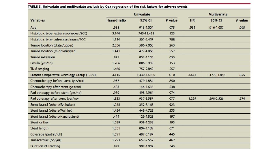
Risk factors for Aes; § The univariate analysis showed that ECOG status, age, and radiotherapy after stent placement presented a trend to higher risk of AEs. However, on multivariate analysis only ECOG status was associated with AEs. Patients with ECOG status other than 0 had an increased risk of Aes. § There was no statistically significant correlation between AE and chemotherapy, stent features, duration of stenting, or tumor characteristics (Table 3)
§ Patients submitted to chemotherapy before stenting did not present higher migration rate (13. 6% vs 10. 5%, P >. 999). § Patients submitted to radiotherapy before stenting did not present higher migration (10% vs 14%, P >. 999) or “de novo” fistula rates (25% vs 18. 6%, P =. 739). § Patients who presented AEs survived longer than patients without AEs (Fig. 2 B).
DISCUSSION; § Metallic esophageal stenting is the most common palliative intervention for malignant dysphagia and malignant esophageal fistula. In spite of being an efficient and safe method and providing rapid relief of dysphagia, AEs may occur in up to half of the patients in some series. This is a concern, especially in patients with longer survival. It is important to point out that with modern palliative treatment for squamous cell carcinoma, survival can reach up to 19 months. § In this study we present data related only to patients who remained with esophageal stents in place for longer than 6 months
§ The overall AE rate in our cohort was 63. 5%. Thirty-four of 63 patients (53. 9%) had to undergo endoscopic reintervention (mean, 1. 6 reinterventions per patient). § In spite of the high incidence of AEs, 75. 8% were minor and 84. 8% of all AEs could be managed clinically or endoscopically with success. § Most AEs occurred after 180 days. § the median survival of patients with AEs was longer than patients without AEs (13. 2 vs 7. 9 months, P <. 001).
§ AE rate in our cohort was higher than those reported in other studies. § Homann et al analyzed 133 patients with esophageal SEMSs and found an overall incidence of delayed AEs of 53. 4%. However, the cut-off definition for delayed AEs was only 4 weeks. In support of our results, the authors found that the median survival in patients submitted to endoscopic therapy was longer than in patients who did not require reintervention.
§. Schoppmann et al retrospectively evaluated the safety and efficacy of 70 patients with long-term esophageal stenting (>120 days) for various indications. The overall AE rate was 30%, and endoscopic reintervention was necessary in 17. 1% of patients. However, their cohort included patients with benign stenosis and postoperative leakage, and 60% of stents were placed in a temporary intention. § The higher AE rate in our study probably reflects our cohort that included only palliative patients with advanced disease who remained stented at least more than 180 days.
§ The impact of chemoradiation on the esophageal SEMS results is contradictory in the literature. Some studies report longer survival for patients palliated with esophageal SEMSs who also received chemotherapy and/or radiotherapy compared with those treated with stent alone. § However, some evidence supports adjuvant therapy increasing stent-related AEs, especially migration. In our study we found a trend to higher AE rates in patients submitted to radiotherapy, albeit not statistically significant (P =. 08).
§ Migration was the third most common AE in this series. The efficacy of antimigration measures is always a matter of debate. To minimize the risk of migration, we prefer a partially covered stent in the palliative setting. Curiously, all 9 patients who experienced stent migration had a partially covered stent, and none of the patients with fully covered stent presented this AE. § Suture and Ovesco clipping would be our preference, but they are not yet available in our country. § Standard clips seem too fragile to keep the stent in place, although it is a simple and low-cost technique that could be applied to patients at risk for migration. § In our practice, when we judge that there is a higher risk of migration (eg, stenting for malignant fistula without stenosis), we use a temporary external fixation method with a modified Shim’s technique as previously described by our group.
§ We are not sure about the use of antimigration measures in a routine fashion. § we had 2 migrations before 30 days in 62 patients, or a 3. 2% 30 -day migration rate. This finding could argue against the routine use of antimigration measures.
§ in a retrospective study of patients with malignant strictures submitted to SEMS placement, Fuccio et al demonstrated that longer stents protected against the occurrence of AEs. This finding was not confirmed in our study. § On the other hand, poor performance status (ECOG) was associated with increased AEs. We hypothesize that poor performance status could be associated with neoplasia that present a more aggressive biologic behavior that could theoretically explain a higher rate of tumor ingrowth, overgrowth, fistulization, and bleeding.
§ Alternative therapies like intraluminal brachytherapy have emerged in recent years. The relief of dysphagia is delayed when compared with SEMS insertion, but better improvement in dysphagia and lower AE rates were identified with brachytherapy treatment. § Stent insertion combined with single-dose brachytherapy has been reported and seems to be feasible and safe, besides providing a longer dysphagia-free period and longer survival.
§ The main drawback of long endoscopic stenting is dysphagia recurrence, through ingrowth/overgrowth, migration, fistula, or food impaction. Nevertheless, endoscopic reinterventions are efficient, safe, and acceptable.
§ Restenting was performed in 23 patients: § § 13 for ingrowth/overgrowth, 7 for fistulas, 2 for migrations, 1 for severe reflux( The patient with severe reflux received an antireflux valve stent). § A combination of methods (argon plasma coagulation, dilation, foreign body removal) was applied to treat other patients. § No surgery was required to treat stent-related AEs.
§ It is possible to infer that an early endoscopic approach for stent-related AEs can improve the quality of life in patients with advanced gastroesophageal tumors palliated with SEMSs.
Conclusion; § our data demonstrate that long-term esophageal stenting in patients with malignant disease involving the esophagus; ü ü ü is related to a relatively high prevalence of AEs. AE was not related to higher mortality rate. most cases were minor events that could be managed endoscopically. Fatal AEs were uncommon. Performance status was the only predictive factor for Aes. § Our data suggest that metallic stenting is a valid option for the treatment of malignant esophageal conditions, even when survival longer than 6 months is expected.