University of Calgary Undergraduate Medicine RESPIRATORY COURSE OCCUPATIONAL


 for key findings associated with")
 · arterial blood gases ·")
 of the")


 after exposure")


- Slides: 74
University of Calgary - Undergraduate Medicine RESPIRATORY COURSE OCCUPATIONAL LUNG DISEASE: CASE PRESENTATIONS Kenneth Corbet MD FRCPC Community Health Sciences
Inhalation of Air Contaminants: Diagnostic Approach 1. describe recent and remote inhalational exposures: smoke, chemicals, mineral dusts and organic/biological 2. assess symptoms, signs, chest radiograph, spirometry, and blood gases 3. identify the level(s) of the respiratory tract that are likely involved, and any systemic effects 4. consider occupational lung disease in your diagnosis (refer to Table in Core Document)
History In addition to a general medical history, ask about: • the patient’s respiratory symptoms • current and past exposures to air contaminants • the temporal relationship between exposure and symptoms • other persons with similar symptoms • the impact of symptoms on the patient’s activities
Physical Examination Examine the patient (inspection, percussion, auscultation, palpation) for key findings associated with respiratory diseases.
Investigations · chest radiograph · spirometry (FVC, FEV 1%) · arterial blood gases · · · • • lung volumes diffusion capacity peak flow monitoring methacholine challenge testing bronchoalveolar lavage (BAL)
Differential Diagnosis Based on the type of air contaminant and the level(s) of the respiratory tract involved, consider occupational causes in the diagnosis
Case Presentation #1 Acute Inhalational Exposure
Occupational Lung Disease: Case Presentation #1 Hank is a 36 year old man who presents to the Emergency department at 9 PM. He is usually quite healthy, but over the past few hours he has felt progressively ill, with occasional chills, myalgia, and cough. He has worked at local metal recycling smelter in the 'melting room' for the last two years. Ongoing problems with ventilation - smokes and fumes can get 'pretty thick' at times.
Occupational Lung Disease: Case Presentation #1 How would you describe the air contaminants? What level of the respiratory tract is involved? What are possible occupational diagnoses?
Occupational Lung Disease: Case Presentation #1 How would you describe the air contaminants? Smokes, fumes - possibly chemicals What level of the respiratory tract is involved? What are possible occupational diagnoses?
Occupational Lung Disease: Case Presentation #1 How would you describe the air contaminants? Smokes, fumes - possibly chemicals What level of the respiratory tract is involved? Cough can originate from all levels of the respiratory tract; note systemic symptoms What are possible occupational diagnoses?
Occupational Lung Disease: Case Presentation #1 Findings 1. 1 Physical Examination • • mild fever (38. 5 C) mild pharyngeal redness chest clear, no distress or tachypnea HR 90, no murmurs or bruits Investigations • mild increase in WCB • normal spirometry • normal blood gases
Chest Radiograph 1. 1 International Labour Organization
Inhalational Fevers Self-limited syndrome of mild fever, leukocytosis, myalgia; onset usually 4 -6 hours after exposure, resolves 24 -48 hours; no apparent sequelae in regards to lung pathology or function. Metal Fumes zinc, copper, manganese Organic Dusts grain dust, moldy silage Plastics Teflon (fluorinated) Endotoxins contaminated humidifiers
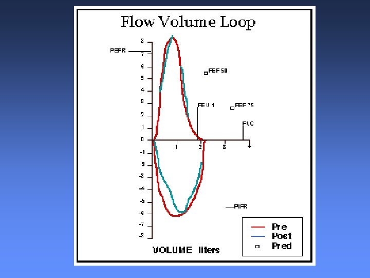
Occupational Lung Disease: Case Presentation #1 Findings 1. 2 Physical Examination • occasional wheezes, afebrile • scant phlegm, black specks, no blood Investigations • chest radiograph normal, normal WBC • blood gases - mild respiratory alkalosis • FVC 104% predicted; FEV 1 81% predicted; FEV 1/FVC = 62%
Occupational Lung Disease: Case Presentation #1 How would you describe the air contaminants? What level of the respiratory tract is involved? What are possible occupational diagnoses?
Occupational Lung Disease: Case Presentation #1 How would you describe the air contaminants? Smokes, fumes - possibly chemicals What level of the respiratory tract is involved? Wheezing and obstructive pattern on spirometry suggests small airway involvement What are possible occupational diagnoses?
Occupational Lung Disease: Case Presentation #1 - Findings 1. 2 Airways Injury - Reactive Airways Disease Symptoms occur with 24 hours after single, high intensity exposure to irritant gas, smoke, fume, or vapour Cough, wheeze, and dyspnea Spirometry may show small airway obstruction methacholine challenge + If airways reactivity and symptoms persist > 6 months = Reactive Airways Dysfunction Syndrome (RADS)
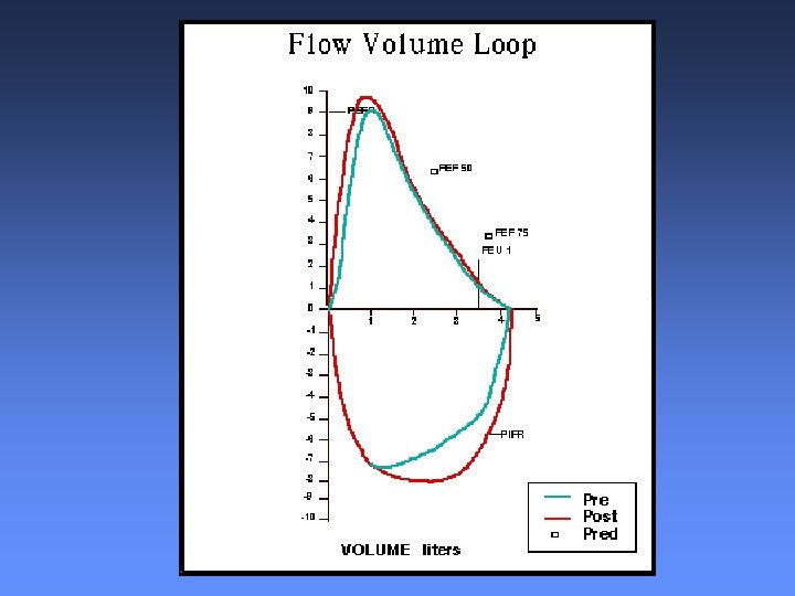
Occupational Lung Disease: Case Presentation #1 Findings 1. 3 Physical Examination • mild distress, tachypneic, tachycardic • scattered crackles, occasional wheezes Investigations • mild hypoxemia on ABG • mixed obstructive and restrictive pattern on spirometry
Chest Radiograph 1. 3 International Labour Organization
Occupational Lung Disease: Case Presentation #1 How would you describe the air contaminants? What level of the respiratory tract is involved? What are possible occupational diagnoses?
Occupational Lung Disease: Case Presentation #1 How would you describe the air contaminants? Smokes, fumes - possibly chemicals What level of the respiratory tract is involved? Chest x-ray changes suggest parenchymal involvement, can’t rule out small airways. What are possible occupational diagnoses?
Chemical Pneumonitis - ARDS • onset within hours (up to 36 hours) after exposure • progressive respiratory distress, hypoxemia, diffuse interstitial/air space changes on CXR • interstitial fibrosis, bronchiolitis obliterans or reactive airways disease may persist after initial recovery • high index of suspicion required based on intensity of exposure and nature of industrial process
Some agents that produce chemical pneumonitis: acrolein hydrogen sulfide cadmium nitrogen dioxide chlorine ozone fire smoke phosgene hydrogen chloride sulphur dioxide
Case Presentation # 2 Abnormal Chest Radiograph
International Labour Organization
Occupational Lung Disease: Case Presentation #3 Bill is a 65 -year-old retired accountant who presents for a periodic medical exam. He reports only slight dyspnea on exertion, no cough or sputum; he has never smoked. For each of a series of possible chest radiographs, what is a possible occupational cause, and what would you ask Bill on a more detailed history?
Chest Radiograph 2. 1 International Labour Organization
Asbestos Fiber Courtesy of Dr. Francis Green
Chest Radiograph 2. 2 International Labour Organization
Distribution of Irregular Opacities In Asbestosis International Labour Organization
INTERSTITIAL FIBROSIS - ASBESTOSIS Courtesy of Dr. Francis Green
ASBESTOSIS Courtesy of Dr. Francis Green
Chest Radiograph 2. 3 International Labour Organization
Courtesy of Dr. Francis Green
Particle Deposition Courtesy of Dr. Francis Green
Dust Nodule Courtesy of Dr. Francis Green
Distribution of Rounded Opacities In Silicosis International Labour Organization
Parenchymal Dust Deposition Courtesy of Dr. Francis Green
Progressive Massive Fibrosis Courtesy of Dr. Francis Green
Progressive Massive Fibrosis Courtesy of Dr. Francis Green
Courtesy of Dr. Francis Green
Courtesy of Dr. Francis Green
Courtesy of Dr. Francis Green
Courtesy of Dr. Francis Green
Courtesy of Dr. Francis Green
Courtesy of Dr. Francis Green
Courtesy of Dr. Francis Green
Courtesy of Dr. Francis Green
Courtesy of Dr. Francis Green
Courtesy of Dr. Francis Green
Courtesy of Dr. Francis Green
Courtesy of Dr. Francis Green
Courtesy of Dr. Francis Green
Courtesy of Dr. Francis Green
Courtesy of Dr. Francis Green
Courtesy of Dr. Francis Green
Courtesy of Dr. Francis Green
Courtesy of Dr. Francis Green
Courtesy of Dr. Francis Green
Courtesy of Dr. Francis Green
Courtesy of Dr. Francis Green
Courtesy of Dr. Francis Green
Courtesy of Dr. Francis Green
Courtesy of Dr. Francis Green
Courtesy of Dr. Francis Green
Courtesy of Dr. Francis Green