The Senses Chapter 10 What is a sensation


 a. Free nerve endings b. Merkel")


 and Chronic (Slow) • Acute –")


 Regeneration every 30 -60 days Adaptation")


")


 tube –")


, breaks down into OPSIN and")


")


- Slides: 65
The Senses Chapter 10
What is a sensation? A stimulus that has been received by the brain bringing awareness to the organism. (Usually in the Thalamus) What is perception? How the brain interprets the sensation. (Located in your Cerebral Cortex)
The Receptors • • Function: Pick up stimuli and send signal to CNS. Two categories: General & Special Neuron Types: Unipolar, few bipolar (eye/nose) Functional types within the categories: Chemoreceptors Thermoreceptors Mechanoreceptors Nociceptors Photoreceptors
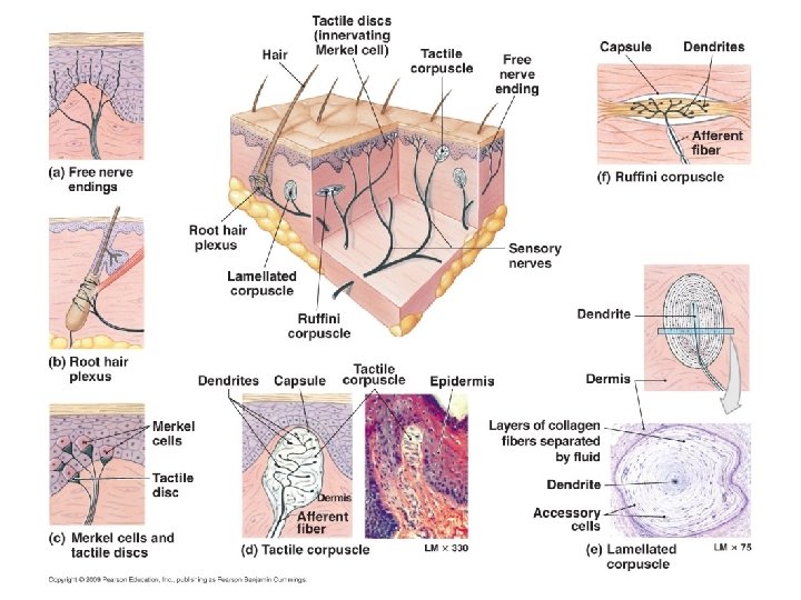
Cutaneous General Receptors 1. Touch and Pressure (mechanoreceptors) a. Free nerve endings b. Merkel discs • The receptive field increases c. Meissner corpuscles with depth in the skin. • The number of receptors varies d. Pacinian corpuscles with body part. e. Ruffini corpuscles f. Hair receptors 2. Temperature (thermoreceptors) - Warm and cold free nerve endings 3. Pain Receptors (nociceptors) - free nerve endings
What is “sensory adaptation”? Receptors have decreased signals to where eventually signals are stopped, where awareness is gone. Ex: wearing clothes, watch, jewelry, hot tubs etc. In order to depolarize again, a stronger stimulus is needed. Some receptors quickly adapt, slowly adapt or adapt very little.
Nociceptors • • Widely distributed except in brain. Very little adaptation if at all Function: Protection Triggered by: Chemicals, ischemia, and mechanical stimulation from tissue damage. • Highly sensitive in somatic vs. visceral areas • Referred pain – derived from common nerve pathways. Ex: Neurons from viscera converge with neurons from somatic. Pain in left arm when heart attack is occurring.
Referred Pain
Pain Nerve Fibers • Two types: Acute (Fast) and Chronic (Slow) • Acute – Sharp pains, impulses travel on myelinated fibers for quick response; precise location can be determined. • Chronic – dull, throbbing, aching pains, impulses carried on unmyelinated fibers; can be deep or superficial. Precise location of pain can be hard to determine.
Pain Regulation • Pain awareness = Thalamus • Pain Intensity, tolerance, location and response = Cerebral Cortex • Pain signaling neurotransmitters: Substance P and Glutamate • Natural anesthetics = enkephalins, serotonin, and endorphins • Analgesics – Medication for pain relief
Natural Pain Killer Physiology
And now for the main attraction: The Special Senses
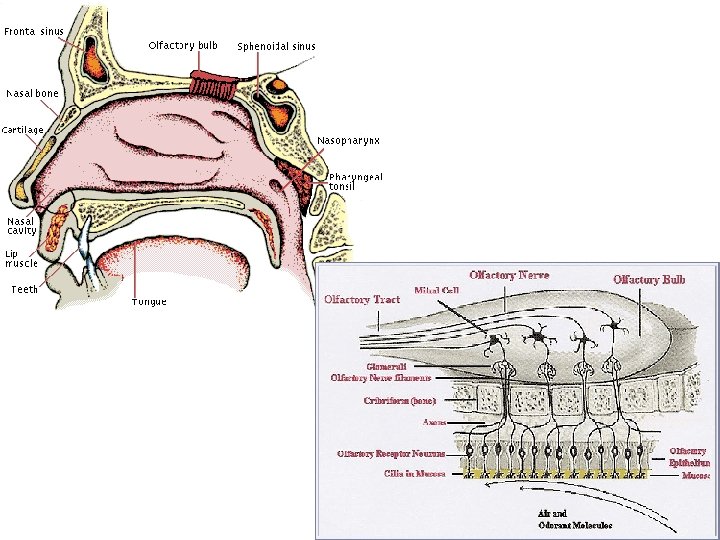
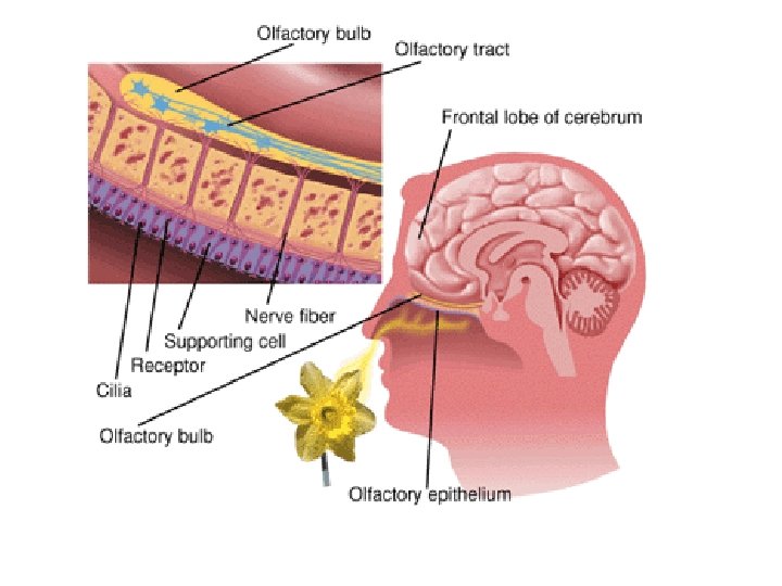
Smelling • • • Chemoreceptor cells (bipolar neurons) Regeneration every 30 -60 days Adaptation occurs quickly Low threshold (only a few molecules needed to detect) Watery mucus lines cells to act as solvent. Olfactory cells – ciliated, axons extend through Cribriform plate into brain. Olfactory bulb sends impulses to olfactory cortex region of brain. Anosmia – loss of sense of smell Helps with taste perception.
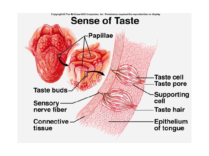
Tasting • • Chemoreceptor cells Regeneration every 3 -6 days Taste buds contain the “Gustatory” cells. Several gustatory cells per taste bud. Found on tongue and around mouth cavity. Gustatory Cortex in the parietal lobes. Papillae are the structures on the tongue where the taste buds are. • Salivary glands provide the solvent so chemical can be tasted. • FIVE primary taste sensations: S, S, S, B, U! • Adaptation occurs quickly, but threshold varies by taste sensation. Ex: Sour & bitter has the lowest threshold…why?
Tastes can be found all over the tongue not just the areas as mapped out below:
The human tongue contains four kinds of papillae, three of which (highlighted in blue) contain taste buds. Foliate papillae - a series of folds on the sides of the tongue in the back Circumvallate papillae - are organized in an inverted V at the back of the tongue Fungiform papillae - are found scattered across the top front and middle of the tongue Filiform papillae - are found across the top of the tongue
The Ear Section 7 and 8
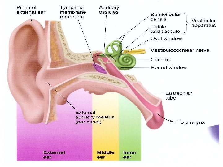
Major Ear Structures External: • Auricle or Pinna • External auditory meatus Middle: • Tympanic membrane • Auditory ossicles **Know the • Eustachain Tube function of Inner: each • Labyrinth (bony and membranous) structure!! • Cochlea • Organ of Corti • Semicircular canals • Perilymph and Endolymph • Oval and Round window • Utricle and Saccule
3 bones: malleus, incus and stapes
The Ear • Mechanoreceptors • Function: Hearing and Balance • Eustachian (Auditory) tube – pathway from ear to throat a. Function: Equalizes pressure between middle and external ear b. Otitis media – infection of the middle ear - Ear tubes can help alleviate pain by draining pus or fluid build-up
Ear Tubes
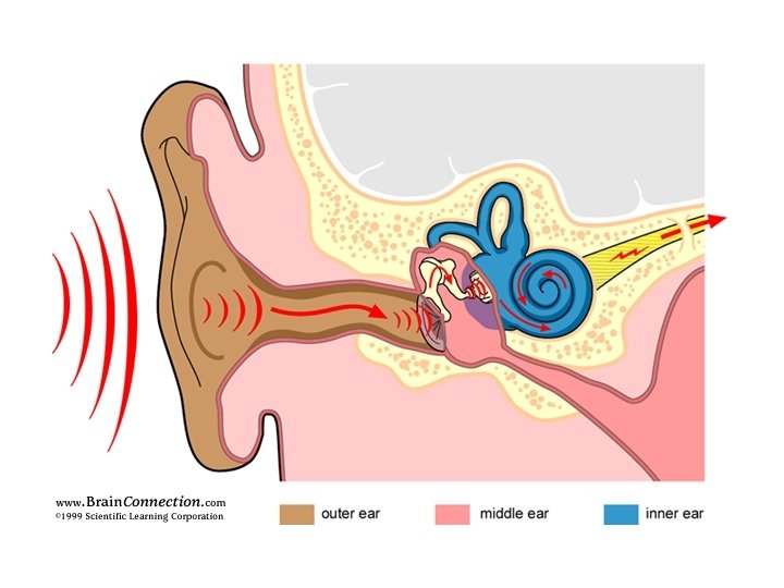
How do vibrations in the air stimulate receptor cells that will send messages to the brain?
See it animated!! Be able to describe the steps to hear a sound!
Great Animation!
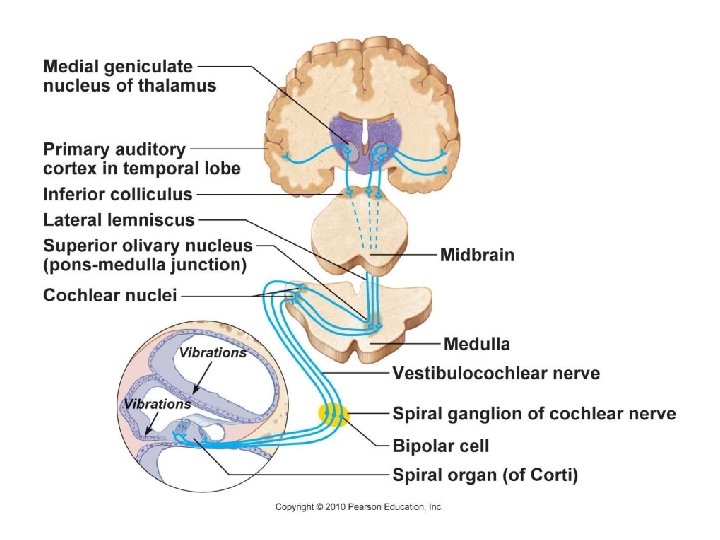
Deafness • Any hearing loss • Two types: 1. Conduction – something hampers sound conduction (ex: excess wax, ear drum damage, otitis media or otosclerosis) 2. Sensorineural – damage to neural structures at any point from cochlea, auditory nerve, or even auditory cortex.
Decibels – unit to measure the amount of sound intensity. The amount of sound exposure determines damage to hearing cells. Apps to measure decibels!
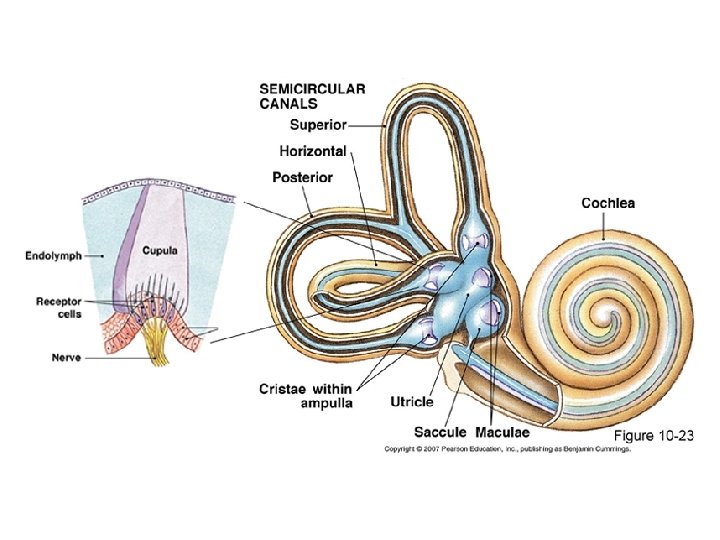
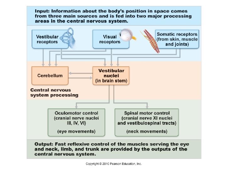
Equilibrium and Balance 1. Static – body/head position when body is still. Sensitive to gravitational forces. Ex: moving head up, down, or tilting to side. 2. Dynamic – body/head position awareness when body is in motion. Ex: rotating head side to side, spinning, running, twirling of body
Static Equilibrium • Location: Vestibule of Semicircular Canals. • Inside vestibule: Utricle and Saccule • Inside the U and S: Macula which has tiny receptor hairs. • Otoliths (Ca. CO 3 grains) move on the gelatinous material on top of cells • Grains bending hairs sends signal to auditory cortex in temporal lobe.
See it animated!
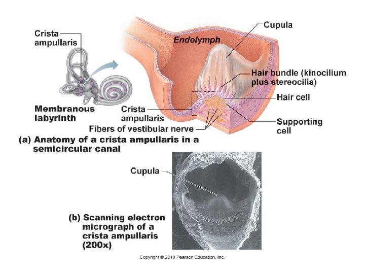
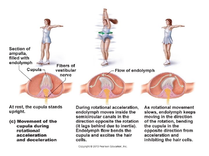
Dynamic Equilibrium • Labyrinth region of Semicircular Canals • Ampulla region contains hair cells that will bend Cupula when fluid flows over it depolarizing hair cells. - Crista ampullaris (hair cell region) - Cupula (gelatinous material)
Meniere’s Syndrome: a labyrinth disorder; causing vertigo, nausea and vomiting. Cause may be from distortion of membranous labyrinth from excessive endolymph. Motion Sickness: Due to sensory input mismatch. OTC medications depress vestibular inputs
Equilibrium Video
The Eye Section 10. 9
Accessory Eye Anatomy • Eyelid – protection • Conjunctiva – clear membrane that lines eye surface except for cornea • Lacrimal Gland – tear secretion, protection, Lysozyme, lubrication • Extrinsic muscles – 6 outer eyeball muscles • Meibomian and Ciliary glands: line eyelids and eyelashes lubricated
Styes, Chalazions, Conjunctivitis
Optic disc
Outer Tunic – Fibrous layer • Cornea – outermost transparent cover, no blood vessels, transplants work very well. • Sclera – white of the eye, tough tendon-like tissue, protects and gives shape • Optic Nerve – sends sensory input to occipital lobe
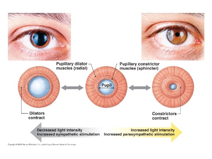
Middle Tunic – Vascular Layer • Choroid Coat – underneath sclera, blood vessels, brown pigment absorbs excess light • Ciliary Body – smooth muscle fibers that change lens shape; attached with suspensory ligaments to hold lens • Lens – focuses image onto retina • Iris – 2 smooth muscles (radial & circular), pigment gives color, controls pupil size • Aqueous Humor – watery fluid • Pupil – the hole
Lens works much like a camera does…
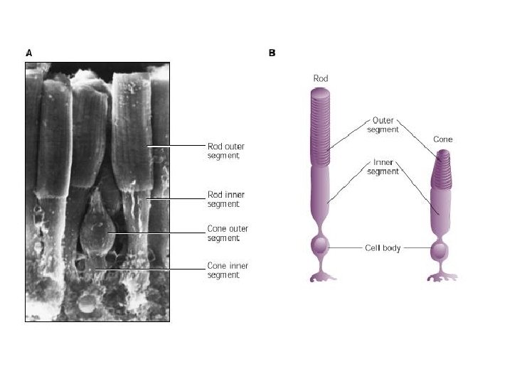
Inner Tunic- Nervous Layer • Retina – back wall of eyeball, contains photoreceptor cells. 1. Rods – long, thin, dim-light and peripheral vision 2. Cones – short, blunt, high acuity color vision • Macula lutea – lateral to optic disc, in center is fovea centralis • Fovea Centralis – sharpest vision, no rods • Optic Disc – no vision (blind spot), optic nerve entrance, no photoreceptors • Vitreous Humor – jelly-like fluid, maintains shape
Neural pathway link!
Visual Pigments • In the Rods: Rhodopsin (visual purple), breaks down into OPSIN and RETINAL. You need this pigment to see in dim light! • Retinal is made from Vitamin A • Breakdown of Rhodopsin occurs in bright light almost immediately, rods less sensitive. • In dim light, Rhodopsin is regenerated from opsin and retinal with the help of ATP. • In the Cones: 3 Color pigments found: erythrolabe, cyanolabe, chlorolabe
Eye Disorders • Myopia – Nearsightedness, poor distance • Hyperopia – Farsightedness, poor close-up • Astigmatism – Irregularly shaped cornea or tilted lens • Colorblindness – lacking one or more cone types. Sex-linked condition • Nightblindness – rod function impaired, usually Vit. A deficiency • Cataract – Cloudy lens due to changes in lens proteins. • Glaucoma – Abnormally high intraocular pressure due to buildup of aqueous humor.
Myopia Nearsightedness
Hyperopia Farsightedness
Astigmatism
Cataracts
Glaucoma
Macular Degeneration Macula breaks down. Most common cause of blindness for people over 50.
LASIK (Laser-Assisted In Situ Keratomileusis)
Retinal Implants & “Bionic Eye”
Optic disc