The Integumentary System Layers of the Skin The

")


 is the layer of the")
 there is another layer called")


 • The stratum corneum is a keratinized stratified")
 • The stratum corneum is Latin for 'horny")

 • The stratum granulosum is Latin for granular layer. •")
 • The stratum spinosum is Latin for \"spiny\".")
 • The stratum basale is the deepest of the")


")


- Slides: 48
The Integumentary System
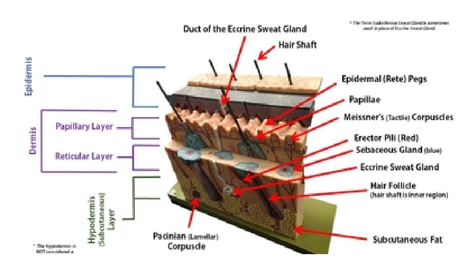
Layers of the Skin The skin is composed of 2 primary layers: 1) the epidermis and 2) the dermis The apical (outer most) layer is called the epidermis. The prefix "epi-" means "above". Lying underneath the epidermis is the dermis. Each of these primary layers is broken down into subsequent layers.
Layers of the Skin • The outermost surface of your skin (the superficial region) is made of a thick epithelial tissue called epidermis. • The word "derm" means "skin". • The prefix "epi-" means "above".
• Epidermal tissues that lie toward the outer part of the body or that line tracts, have a layer of dead skin cells for added protection. • This type of epithelial tissue is referred to as keratinized stratified squamous epithelial tissue
The epidermis consists of 4 cell types • Keratinocytes - The most abundant cell type in the epidermis. • They are created in the stratum basale and pushed upward toward the skin's surface. • These cells make keratin and act to protect deeper layers of soft tissue. • These cells die as they approach the surface of the skin. • Once they are dead and shriveled up, they are called corneocytes.
The epidermis consists of 4 cell types • Melanocytes - Pigmented cells of the stratum basale region that produce melanin which protects from UV radiation.
The epidermis consists of 4 cell types • Merkel Cells - function as touch receptors in association with sensory nerve endings (Merkel disc); located at epidermal-dermal junction
The epidermis consists of 4 cell types • Langerhans’ Cells - epidermal macrophage-like cells that help activate the immune system via receptor-mediated endocytosis
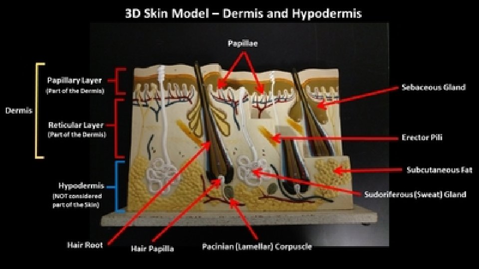
DERMIS • Beneath the epidermis, (deep to the epidermis) is the layer of the dermis. • The dermis is composed of connective tissue.
hypodermis • Below the skin (deep to the skin) there is another layer called the hypodermis. • "Hypo-" means "below". • This layer is composed of areolar connective tissue and adipose tissue. • The hypodermis is NOT considered to be part of the skin itself.
Thick Skin and Thin Skin • thick skin… • is located in regions of your body that has a lot of physical contact with the outside world. • fingertips, palms and soles o feet. • These extra layers help to protect the soft tissues from abrasions.
Thick Skin and Thin Skin • While the epidermis is thicker in thick skin than in thin skin. • But the layer of the dermis is THINNER in thick skin than in thin skin. • Thick skin does not contain hairs, sebaceous glands, or sweat glands.
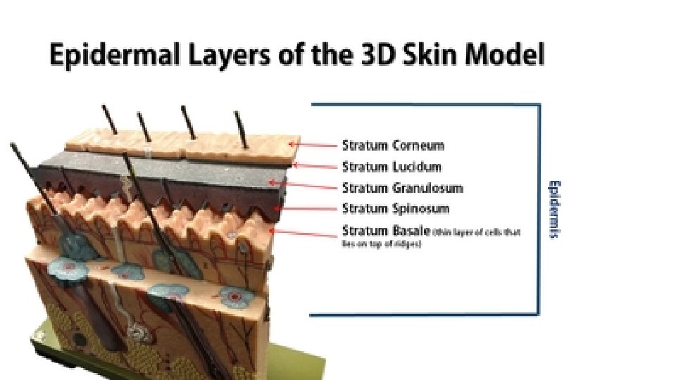
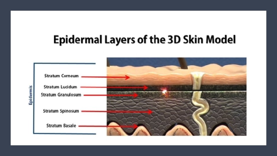
epidermal layers The epithelium of thick skin contains 5 epidermal layers, whereas thin skin contains only 4 layers. The epidermal layers of thick skin starting from the most superficial layer are the 1) stratum corneum 2) the stratum lucidum 3) the stratum granulosum 4) the stratum spinosum 5) the stratum basale. You can easily remember the layers of the epidermis using the acronym: Come, Let's Get Sun-Burned! Thin skin contains the same layers, except for the stratum lucidum, which is missing.
1. The Stratum Corneum (Horny Layer) • The stratum corneum is a keratinized stratified squamous epithelium that contains four types of cells. • The four cells types cells that make up the stratum corneum are 1) keratinocytes 2) melanocytes 3) tactile epithelial cells 4) dendritic cells
1. The Stratum Corneum (Horny Layer) • The stratum corneum is Latin for 'horny layer’. • Its odd name comes from the dead squamous cells called corneocytes that make up the apical surface of the tissue. • The corneocytes form several layers of flattened (squamous) cells with no nuclei and no organelles. • This is the layer that includes the final keratin product, which is a combination of cytokeratin and keratohyaline.
2. *The Stratum Lucidum • The stratum lucidum is Latin for "clear layer". • This epidermal layer gets its name from translucent appearance of the dead skin cells that make it up. *This layer only exists in the thick skin of the soles of your feet, your palms and your fingertips.
Stratum Granulosum (Granular Layer) • The stratum granulosum is Latin for granular layer. • In this layer, the keratinocytes have become squamous cells that contain granules of keratohyaline, a precursor to the extracellular keratin that protects the skin tissue from abrasion.
Stratum Spinosum (Prickle Cell Layer) • The stratum spinosum is Latin for "spiny". After forming in the basal cell layer, keratinocytes migrate upwards to form the stratum spinosum. • In this layer, they develop short projections that attach via desmosomes to adjacent cells. • The stratum spinosum is also known as the "prickly layer" because of these characteristic spines. • The cells in this layer produce cytokeratin, an intermediate filament precursor to keratin. • The cells in this layer experiences shrinking of its microfilaments during the slide staining process.
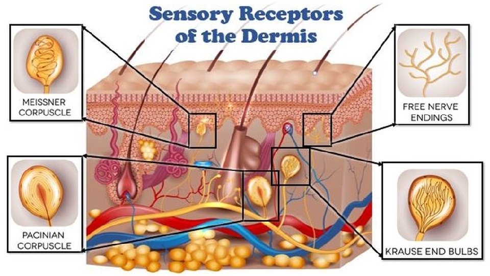
Stratum Basale (Basal Layer) • The stratum basale is the deepest of the 5 epidermal layers. • The primary cell of the epidermis is the keratinocyte stem cells which give a continuous supply of new cells to replace old dead ones. • The stratum basale also contain sensory nerves called Meissner's (Tactile) Corpuscles that are sensitive to light tactile sensations (light touch). • This layer also contains melaocytes which produce the pigment melanin which acts to filter out harmful UV radiation.
Functions of the Integumentary System 1. Protection 2. Body Temperature Regulation 3. Excretion 4. Production of vitamin D 5. Sensory Reception
The primary function of your skin is to provide a physical protective barrier against harmful environmental elements, dehydration and physical forces. Functions of the Integumenta ry System : Protection The epithelium allows your internal organs and soft tissues to be somewhat shielded from the outside world. It provides a barrier that is able to withstand some wear and tear. It can get bumped, bruised or scraped and it is able to heal relatively quickly.
Functions of the Integumentary System : Protection • Epidermis – • composed of keratinized stratified squamous epithelial tissue which provides layers of small, squamous cells which are rapidly and easily replaced.
Functions of the Integumentar y System : Protection • protects you from harmful elements • Bacteria • Viruses • Chemicals • Disease-causing Microbes • Reactive Substances • Irritants • Ultraviolet Radiation (UV Radiation) From The Sun • The UV protection of the epidermis comes from the presence of melanin produced in specialized cells called melanocytes.
YOU SHALL NOT PASS! • Epithelial tissue is selectively permeable, allowing only certain substances to enter the body at any time. • The skin is our first line of defense against harmful elements in our environment, such as. Your skin functions to prevent excess water loss from the body.
Body Temperature Regulation MAINTAINING HOMEOSTASIS • The dermis of the skin contains a vast network of blood vessels that are under the control of the nervous system.
Body Temperature Regulation • When your body gets cold, your nervous system will constrict these blood vessels, thereby restricting the blood flow to your extremities, as well as to the outer surfaces of your body, in an effort to keep the blood warm.
• The sweat glands are also activated by the nervous system and secrete sweat in response to heat. • When the sweat reaches the surface of the skin, the skin is cooled as the sweat droplet evaporates. Body Temperature Regulation
Metabolic Functions • Skin cells also play a role in some chemical conversions. Vitamin D is a steroid that gets converted into calcitriol in the body, Calcitriol is a hormone that assists in the metabolism of calcium. • This is why you will sometimes see "with vitamin D" marked on products containing calcium. • Vitamin D is made using a substance called "a precursor" that is synthesized from cholesterol by epidermal cells.
Cutaneous Sensations • Our epithelium is our interface will the outside world. • Our physical sensation of touch, pressure, pain and temperature all come from specialized sensory receptor cells in our epithelium. • Our skin gathers sensory information about the outside world that gets relayed to the brain. • Your skin has sensory receptors for temperature, touch and pain.
Epidermal tissue is categorized as a keratinized stratified squamous epithelial tissue. The most abundant type of cell found in the epidermis are keratinocytes. Keratinocytes produce keratin which is a fibrous protein that gives strength to epidermis.
• Keratinocytes form in the deepest portion of the epithelium (the stratum basale) and then are continuously pushed outward until they die and eventually get sloughed off of the body. • We loose millions of these dead skin cells every day! your epidermis is completely replaced every 35 -45 days.
• Melanocytes are specialized cells that lie within the skin. They produce a brown pigment (melanin) that functions to provide protection from damaging UV radiation from the sun. • The pigment filters out the UV light. • When your skin is exposed to more sunlight, your melanocyte darken the skin by increasing the production of melanin. • Melanocytes - make melanin - makes skin dark. • Protects from UV Light. UV light is carcinogenic. This is why getting too much sun cause skin cancer.
keratin • The keratin protects the deeper tissue layers not only by providing a physical barrier to the outside world, but they provide antibiotics and enzymes that act to detoxify the harmful chemicals to which our skin is exposed.
• The keratinocytes undergo physical changes as they mature and move closer to the surface of your skin. • The keratinocytes are created from stem cells that exist in the stratum basale. • As these cells are pushed up by the production of new cells beneath them, they make their way to higher (more apical) epithelial layers. KERATINOCYTES
• When the keratinocytes reach the stratum spinosum (the layer superficial to the stratum basale) they begin to produce keratin. • Once they reach the stratum granulosum, these cells are busy making lots of keratin which fills their cytoplasm. KERATINOCYTES
KERATINOCYTES • The keratinocytes begin to die as they become part of the stratum lucidum, becoming smaller and clear. • They begin to loose their organelles as they die.
CORNEOCYTES • By the time these dead keratinized squamous epithelial cells reach the skin's surface, they are completely dead and are characterized as "corneocytes". • The corneocytes are basically dead, flat sacs completely filled with fibrous keratin.
Glandular Epithelium • Epithelial cells that produce secretions are called gland cells or glandular epithelial cells. • The gland cells will come together to form glandular epithelial tissues.
Glandular Epithelium • Epithelial cells that produce secretions are called gland cells or glandular epithelial cells. • The gland cells will come together to form glandular epithelial tissues.
Glandular Epithelium • Sweat glands function in thermoregulation to secrete sweat onto the surface of the skin to us cool.
• Sebaceous glands secrete an oily substance called sebum directly int o hair follicles. • It lubricates the skin and hair, prevents water loss, from the skin, and acts as a bactericidal agent. Glandular Epithelium