The Integumentary System The Skin The Integumentary System


")


")


 Second-degree (epidermis and dermis, with blistering) Third-degree (full thickness,")


- Slides: 52
The Integumentary System: The Skin
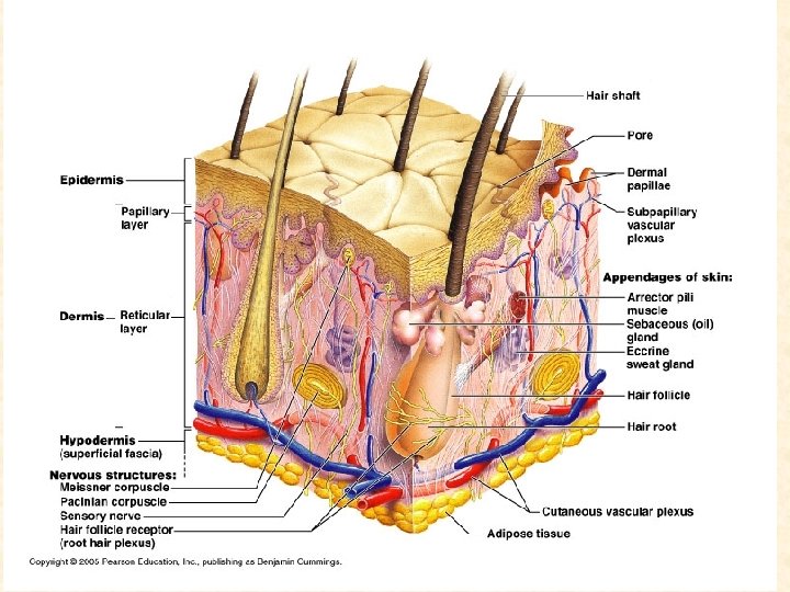
The Integumentary System Integument is skin n Skin and its appendages make up the integumentary system n A fatty layer (hypodermis) lies deep to it n Two distinct regions n ¨ Epidermis ¨ Dermis
Skin Your skin is your largest organ n It is your first line of defense against disease and damage n It is made of several layers n
Functions of skin n Protection ¨ Cushions and insulates and is waterproof ¨ Protects from chemicals, heat, cold, bacteria ¨ Screens UV n n Synthesizes vitamin D with UV Regulates body heat Prevents unnecessary water loss Sensory reception (nerve endings)
Layers of Skin n Epidermis ¨ Uppermost layer of skin Contains dead and living cells n New cells are replaced daily old ones slough off (they shed) n n Dermis¨ Inner thicker layer of skin ¨ Contains: n n Blood vessels Nerve receptors Hair follicles Sebaceous glands Sweat glands ¨ Oil glands ¨ Wax glands ¨
Remember… n Four basic types of tissue ¨Epithelium – epidermis just discussed ¨Connective tissue - dermis ¨Muscle tissue ¨Nervous tissue
Dermis n n Strong, flexible connective tissue: your “hide” Rich supply of nerves and vessels Critical role in temperature regulation (the vessels) Two layers (see next slides) ¨ Papillary – includes dermal papillae- makes fingerprints ¨ Reticular – “reticulum” (network) of collagen and reticular fibers
Skin Layers n Subcutaneous Layer- lies below the dermis layer, not really a skin layer ¨ Contains n n Fat cells Fibers used to attach skin to muscle
*Dermis layers *Dermal papillae * *
Skin Color n Skin pigments ¨ Melanin- dark skin pigment (causes human skin colors) ¨ Where there are darker colors on the skin there is a higher concentration of melanin in that area Freckles n Moles n birthmarks n
Skin color n Three skin pigments ¨ Melanin: the most important ¨ Carotene: from carrots and yellow vegies ¨ Hemoglobin: the pink of light skin n Melanin ¨ Variations in color ¨ Protection from UV light by producing vitamin D
How does our skin protect us? n Forming ¨ callus- a thickened area of epidermis caused by rubbing or pressure ¨ Blisters- area of skin when the layers of skin separate due to excessive friction or intense heat ¨ Melanin produces vitamin D- “natural sunblock”
Sweat glands n n Prevent overheating 500 cc to 12 Liters/day! (is mostly water) Humans most efficient (only mammals have) Produced in response to stress as well as heat
Fingerprints, palmprints, footprints n n Dermal papillae lie atop dermal ridges Elevate the overlying epidermis into epidermal ridges Are “sweat films” because of sweat pores Genetically determined Flexion creases n Deep dermis, from continual folding Fibers n n Collagen: strength and resilience Elastic fibers: stretch-recoil ¨ n Striae: stretch marks Tension lines (or lines of cleavage) ¨ The direction the bundles of fibers are directed The dermis is the receptive site for the pigment of tattoos
Disorders of the integumentary system n Burns ¨ Threat to life n Catastrophic loss of body fluids n Dehydration and fatal circulatory shock n Infection ¨ Types n First degree – epidermis: redness (e. g. sunburn) n Second degree – epidermis and upper dermis: blister n Third degree - full thickness Infections n Skin cancer n
Burns First-degree (epidermis only; redness) Second-degree (epidermis and dermis, with blistering) Third-degree (full thickness, destroying epidermis, often part of hypodermis)
SKELETAL SYSTEM BONES & JOINTS
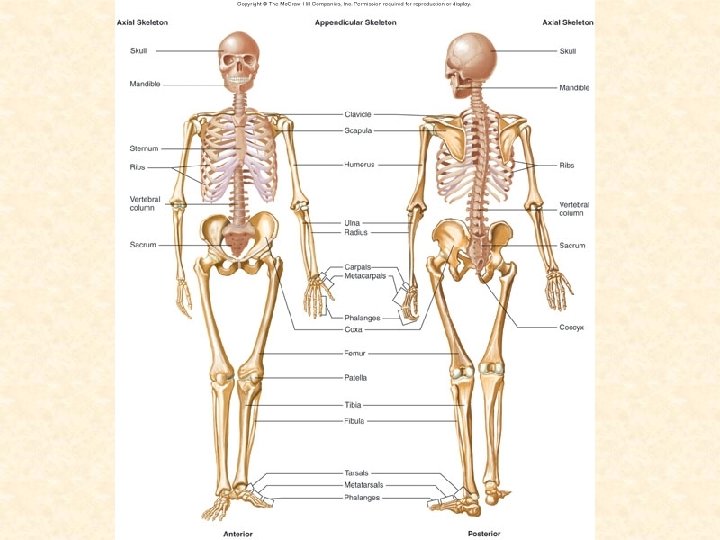
Functions of the Bones 1. Support 2. Protection 3. Movement 4. Storage (minerals like calcium and phosphorous) 5. Hematopoiesis (makes new blood cells in the bone marrow)
Fig. 6. 21
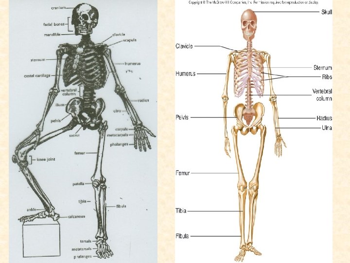
Bones n n n The adult human skeleton has 206 bones Bones store minerals (calcium) and make blood cells (in the center of the bone, bone marrow) Are made of mineral deposits of calcium and phosphorous There small spaces that contain blood vessels (veins, arteries, and capillaries) The blood vessels and surrounding bone materials makes up an osteon (bone cell)
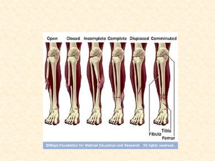
Broken Bones • A broken bone is a fractured bone: • Bones can break in many ways: • • • Incomplete fracture- when there is only a crack, but not a break Complete fracture- when the bone breaks completely in two Comminuted fracture- fractures that splinter the bone
JOINTS
JOINTS = a site where 2 or more bones come together with or without movement
JOINTS 1. 2. 3. Hinge joints = bend in only one direction, like a door hinge flexion & extension movements possible e. g. : elbow, knee Gliding Joints= allows bones to slide across one another, and allows some twisting movement Like the bones between the vertebrae Ball & socket joints = ball – shaped head of one bone fits into a socket – like concavity of another = free movements possible: e. g. shoulder and hip
Joints 4. Pivot Joint=allows for some circular movement e. g. The two bones of the forearm near the elbow 5. Fused Joints= do not allow any movement They become permanently fused together e. g. Bones of the skull
Muscles
My muscles are important because they… n n n n Hold my organs in place Hold my bones together so that I can move Help me chew my food Open and close my eyelids Pump my blood Allow me to run and play Help me to smile!
Muscles & the Skeleton Skeletal muscles cause the skeleton to move at joints n They are attached to skeleton by tendons. n Tendons transmit muscle force to the bone. n Tendons are made of collagen fibres & are very strong & stiff n
Functions of the Muscular System n The characteristics of muscle tissue enable it to perform some important functions, including: § Movement – both voluntary & involuntary § Maintaining posture § Supporting soft tissues within body cavities § Guarding entrances & exits of the body § Maintaining body temperature
Muscular System n This system is responsible for movement of the body: ¨ But it cannot do this alone. Help is required from the nerves, joints, and bones. n n –They work together as an orchestra, each playing their part in perfect harmony Muscles work by pulling never pushing
Muscular System Muscles are responsible for all types of body movement n 3 basic muscle types are found in the body n ¨ Skeletal muscle ¨ Cardiac muscle ¨ Smooth muscle
Types of Muscles n Voluntary: muscles you can control, they are subject to one’s will ¨ Skeletal muscles- move your skeleton/bones ¨ Have striations- they have a banded pattern, appears layered
Characteristics of the Skeletal Muscles n Most are attached by tendons to bones n Striated – have visible banding n Voluntary – subject to conscious control
Anatomy of skeletal muscles epimysium tendon perimysium Muscle Fascicle Surrounded by perimysium Skeletal muscle Surrounded by epimysium endomysium Skeletal muscle fiber (cell) Surrounded by endomysium Play IP Anatomy of Skeletal muscles (IP p. 4 -6)
Characteristics of Cardiac Muscle n n Similar to skeletal muscle cardiac muscle has striations These striations are not as pronounced in cardiac muscles as in skeletal muscle It is the only involuntary muscle that is striated Found only in the heart
Smooth Muscle Characteristics • Has no striations • Involuntary – no conscious control • Found mainly in the walls of hollow organs • Stomach, intestines, blood vessels, and other internal organs • The heart is involuntary, but not smooth muscle • Slow, sustained and tireless • They are constantly in motion and do not tire as do skeletal muscles
Smooth Muscle
Comparison of Muscle Tissue
How Muscles Work n Skeletal muscles work in pairs ¨ When your bicep contracts your triceps relax ¨ When the triceps contract your bicep relaxes n Many skeletal muscles extend across at least one joint to 2 different bones ¨ The Sartorius (longest muscle in body) attaches to the top of the hip bone and extends across the hip and knee connecting to the tibia
How Muscles Work n Some muscles do not extend across joints but rather movement in skin or other muscles ¨ Tongue and throat muscles do not extend over joints but they move to allow swallowing
Temporalis Frontal Obicularis oculi Obicularis oris Masseter Sternoclediomastoid
Functions of Muscles n Muscles keep you warm by contracting ¨ When you are cold your body shivers in an attempt to warm you up by increasing muscle contractions and thereby producing heat n They keep your body erect (standing up) ¨ Even when you are still, standing or sitting, your muscles are working and thereby can keep your body warm ¨ This is why yoga is so effective in working muscles, prolonged muscle contractions
More Muscle Functions n When muscles in your skin contract making the hair follicles stand upright your skin around the hair follicle also stands up and causes… ¨ Goose bumps
Goose Bumps
How Muscles Work n Muscles need energy to function, so where does that energy come from ¨ Energy is stored in the body as glucose (sugar) ¨ It is carried by the blood to the muscles where the muscles break down the glucose into usable energy n In order to break down glucose the body needs oxygen
How Muscles Work n When the muscles break down sugar, with the use of oxygen, it is aerobic respiration ¨ The oxygen comes to the muscles from the lungs via arteries so that they muscles can break the sugar down into usable energy for the body
How Muscles Work When you work or play hard, like when running, you begin to breathe faster in attempts to bring more oxygen into your blood so that your muscles can continue to work (break down sugar) n When you do not have sufficient oxygen your muscles undergo lactic acid fermentation producing lactic acid in the muscles n
How Muscles Work n A build up of lactic acid in your muscles causes them to become sore or cramp up ¨ …have you gotten tired or sore muscles the day following a hard workout or a day of hard play? ¨ This is also how you get a Charlie horse