Integumentary System Layers of skin Pigments Quiz Integumentary


 – Hypodermis (superficial fascia) • • Subcutaneous layer deep")


 cells • Star-shaped macrophages that patrol deep epidermis – Are key")
 cells • Sensory receptors that sense touch")


 1. – – – Deepest of")
 2. – – Several cell layers")
 3. – – Four to six")
 4. – – – Found only")
 5. – – – 20– 30")
")


 lines in reticular layer are caused by many collagen")


 •")

- Slides: 38
Integumentary System • Layers of skin • Pigments • Quiz
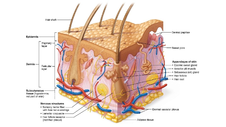
Integumentary System • Integumentary system consists of: – – – Skin Hair Nails Sweat glands Sebaceous (oil) glands
Structure of skin • Skin consists of two distinct regions: – Epidermis: superficial region • – Consists of epithelial tissue and is avascular Dermis: underlies epidermis • Mostly fibrous connective tissue, vascular
Structure of skin (sort of) – Hypodermis (superficial fascia) • • Subcutaneous layer deep to skin Not part of skin but shares some functions Mostly adipose tissue that absorbs shock and insulates Anchors skin to underlying structures: mostly muscles
Epidermis Cells of the Epidermis • Epidermis consists mostly of keratinized stratified squamous epithelium Keratinized=
Epidermis Four cell types found in epidermis: Keratinocytes 1. • • Produce fibrous keratin (protein that gives skin its protective properties) Major cells of epidermis Tightly connected by desmosomes Millions slough off every day
Melanocytes 2. • • Spider-shaped cells located in deepest epidermis Produce pigment melanin, which is packaged into melanosomes – Melanosomes are transferred to keratinocytes, where they protect nucleus from UV damage
3. Dendritic (Langerhans) cells • Star-shaped macrophages that patrol deep epidermis – Are key activators of immune system
4. Tactile (Merkel) cells • Sensory receptors that sense touch
Layers of the Epidermis • Epidermis is made up of four or five distinct layers – – Thick skin contains five layers (strata) and is found in high-abrasion areas (hands, feet) Thin skin contains only four strata
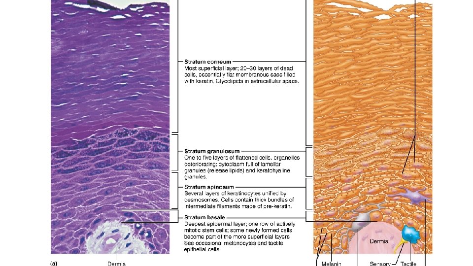
Layers of the Epidermis • Five layers of skin 1. 2. 3. 4. 5. Stratum basale Stratum spinosum Stratum granulosum Stratum lucidum (only in thick skin) Stratum corneum
Layers of the Epidermis Stratum basale (basal layer) 1. – – – Deepest of all epidermal layers (base layer) Layer that is firmly attached to dermis Consists of a single row of stem cells that actively divide (mitotic), producing two daughter cells each time Layer also known as stratum germinativum because of active mitosis 10– 25% of layer also composed of melanocytes
Layers of the Epidermis Stratum spinosum (prickly layer) 2. – – Several cell layers thick Cells contain weblike system of intermediate prekeratin filaments attached to desmosomes • – – Allows them to resist tension and pulling Keratinocytes in this layer appear spikey, so they are called prickle cells Scattered among keratinocytes are abundant melanosomes and dendritic cells
Layers of the Epidermis Stratum granulosum (granular layer) 3. – – Four to six cells thick, but cells are flattened, so layer is thin Cell appearance changes • • • – Cells flatten, nuclei and organelles disintegrate Keratinization begins Cells also accumulate lamellar granules, a waterresistant glycolipid that slows water loss Cells above this layer die • Too far from dermal capillaries to survive
Layers of the Epidermis Stratum lucidum (clear layer) 4. – – – Found only in thick skin Consists of thin, translucent band of two to three rows of clear, flat, dead keratinocytes Lies superficial to the stratum granulosum
Layers of the Epidermis Stratum corneum (horny layer) 5. – – – 20– 30 rows of flat, anucleated, keratinized dead cells Accounts for three-quarters of epidermal thickness Though dead, cells still function to: • • Protect deeper cells from the environment Prevent water loss Protect from abrasion and penetration Act as a barrier against biological, chemical, and physical assaults
Layers of the Epidermis • Cells change by going through apoptosis (controlled cell death) – – Dead cells slough off as dandruff and dander Humans can shed ~50, 000 cells every minute
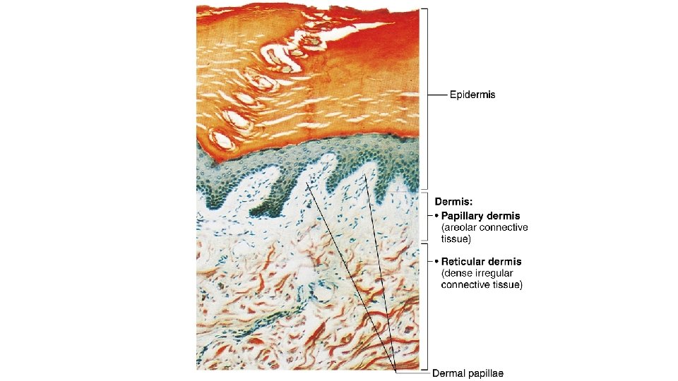
Dermis • • • Strong, flexible connective tissue Cells include fibroblasts, macrophages, and occasionally mast cells and white blood cells Fibers in matrix bind body together Contains nerves, blood vessels, and lymphatic vessels Contains epidermal hair follicles, oil glands, and sweat glands Two layers – – Papillary Reticular
Papillary Layer • • Superficial layer of areolar connective tissue consisting of loose, interlacing collagen and elastic fibers and blood vessels Loose fibers allow phagocytes to patrol for microorganisms
Papillary Layer • Dermal papillae: superficial region of dermis that sends fingerlike projections up into epidermis – Projections contains capillary loops, free nerve endings, and touch receptors (tactile corpuscles, also called Meissner’s corpuscles)
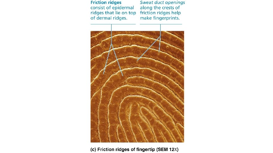
Papillary Layer • In thick skin, dermal papillae lie on top of dermal ridges, which give rise to epidermal ridges – Collectively ridges are called friction ridges • • • Enhance gripping ability Contribute to sense of touch Sweat pores in ridges leave unique fingerprint pattern
Reticular Layer • • Makes up ~80% of dermal thickness Consists of coarse, dense fibrous connective tissue – – Many elastic fibers provide stretch-recoil properties Collagen fibers provide strength and resiliency • Bind water, keeping skin hydrated
Reticular Layer • • Cutaneous plexus: network of blood vessels between reticular layer and hypodermis Extracellular matrix contains pockets of adipose cells
Reticular Layer • Cleavage (tension) lines in reticular layer are caused by many collagen fibers running parallel to skin surface – – Externally invisible Important to surgeons because incisions parallel to cleavage lines heal more readily
Cleavage lines in the
Reticular Layer • Flexure lines of reticular layer are dermal folds at or near joints – – – Dermis is tightly secured to deeper structures Skin’s inability to slide easily for joint movement causes deep creases Visible on hands, wrists, fingers, soles, toes
Flexure lines on digit Flexure lines on the palm Flexure lines of the hand
• Extreme stretching of skin cause dermal tears, leaving scars called striae – • Also known as “stretch marks” Acute, short-term traumas to skin cause blisters, fluid-filled pockets that separate epidermal and dermal layers
Skin Color • Three pigments contribute to skin color Melanin 1. • Only pigment made in skin; made by melanocytes – – • • • Packaged into melanosomes that are sent to keratinocytes to shield DNA from sunlight Sun exposure stimulates melanin production Two forms: reddish yellow to brownish black All humans have same number of keratinocytes, so color differences are due to amount and form of melanin Freckles and pigmented moles are local accumulations of melanin
• Excessive sun exposure damages skin – – – Elastic fibers clump, causing skin to become leathery Can depress immune system and cause alterations in DNA that may lead to skin cancer UV light destroys folic acid • – Necessary for DNA synthesis, so insufficient folic acid is especially dangerous for developing embryos Photosensitivity is increased reaction to sun • Some drugs (e. g. , antibiotics, antihistamines) and perfumes cause photosensitivity, leading to skin rashes
Skin Color Carotene 2. • • Yellow to orange pigment Most obvious in palms and soles Accumulates in stratum corneum and hypodermis Can be converted to vitamin A for vision and epidermal health Hemoglobin 3. • Pinkish hue of fair skin is due to lower levels of melanin – Skin of Caucasians is more transparent, so color of hemoglobin shows through
• Alterations in skin color can indicate disease – Cyanosis • – Erythema (redness) • – Blue skin color: low oxygenation of hemoglobin Fever, hypertension, inflammation, allergy Pallor (blanching or pale color) • Anemia, low blood pressure, fear, anger
• Alterations in skin color can indicate disease – Jaundice (yellow cast) • – Bronzing • – Liver disorders Inadequate steroid hormones (example: Addison’s disease) Bruises (black-and-blue marks) • Clotted blood beneath skin
Things to know: What are the layers of the skin? What are the layers of those layers? What is keratinization? What are the differences between papillary dermis and reticular dermis? What determines skin color? Where are these pigments found?