Ocular Anatomy Dr Ajai Agrawal Additional Professor Department


 cranial nerve, via the ophthalmic")


 Inferior Rectus (IR)")


 •")


- Slides: 52
Ocular Anatomy Dr. Ajai Agrawal Additional Professor Department of Ophthalmology A. I. I. M. S. , Rishikesh
Acknowledgement • Photographs included in the presentation are courtesy of Dr. Jonathan. J. Dutton and Dr. Thomas. G. Waldrop (Atlas of Clinical and Surgical Orbital Anatomy)
Learning Objectives At the end of this class students shall be able to • Identify various structures of the eye • Have a basic understanding of the structure and functions of various parts of the eye
• Orbit Lodges the eyeball • Eye lids – • Cover and protect • Lacrimal system. Secretes and drain tears • Extraocular musclesfor ocular movements • Layers of eyeballperform various functions
Bony Orbit • Made by 7 Bones: • Frontal • Zygomatic • Maxillary • Ethmoidal • Sphenoid • Lacrimal • Palatine
Frontal view of bony orbit
Outer eye: Eyelids The eyelids fulfill two main functions: • protection of the eyeball • secretion, distribution and drainage of tears
The eyelid muscles
Lid structure and movement ● The levator muscle extends from an attachment at the orbital apex to attachments at the tarsal plate and skin. ● The lids are securely attached at either end to the bony orbital margin by medial and lateral palpebral ligaments.
Innervation - Sensory innervation is from the trigeminal (fifth) cranial nerve, via the ophthalmic division (upper lid) and maxillary division (lower lid). - The orbicularis oculi is innervated by the facial (seventh) nerve. - The levator muscle in the upper lid is supplied by the oculomotor (third) nerve.
Blood supply Network of blood vessels which form an anastomosis between branches derived from the external carotid artery via the face and from the internal carotid artery via the orbit.
Lymphatics • Lymphatic fluid drains into the preauricular and submandibular nodes. • Preauricular lymphadenopathy is a useful sign of infective eyelid swelling (especially viral).
Lacrimal system • Lacrimal gland • Lacrimal passage • • Lacrimal puncta Lacrimal canaliculi Lacrimal sac Naso-lacrimal duct
Tear production • The lacrimal gland secretes most of the aqueous component of the tear film • Location: superotemporal part of the anterior orbit • Innervation : parasympathetic fibres carried by the facial nerve.
Lacrimal Apparatus/Drainage system
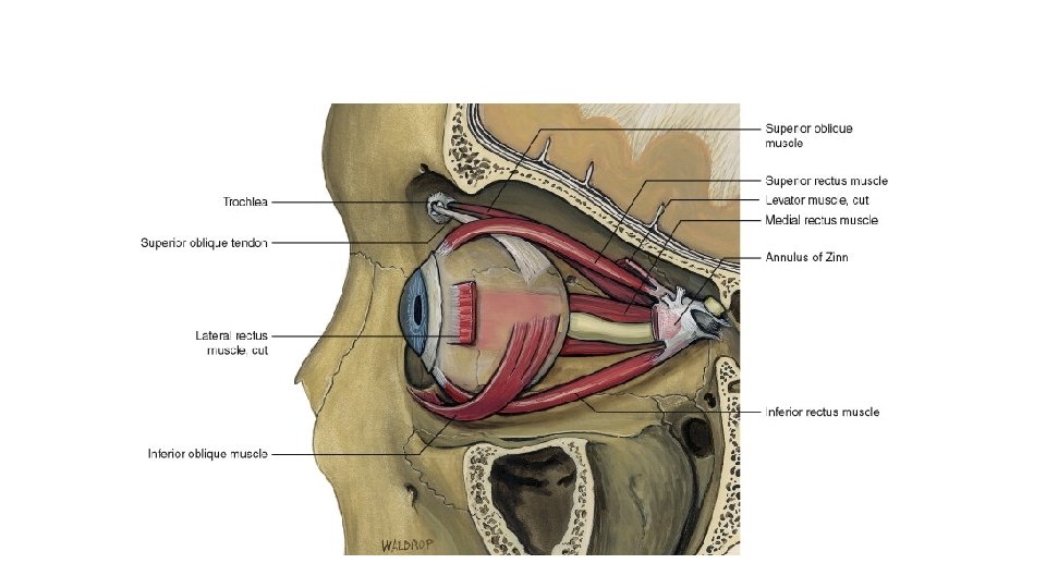
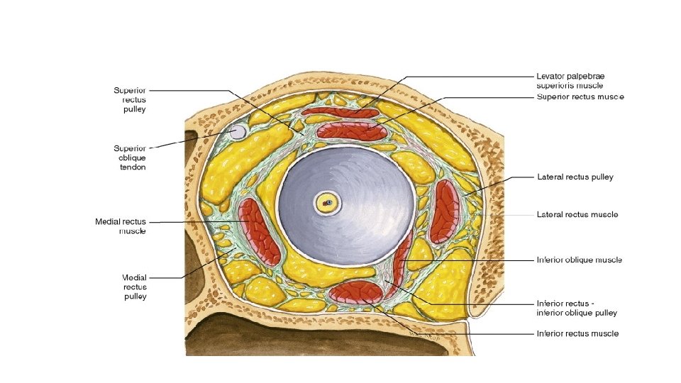
Extraocular muscles • 4 Recti • 2 Obliques Medial rectus (MR) Inferior Rectus (IR) Superior rectus (SR) Inferior oblique (IO) Superior Oblique(SO) Lateral Rectus (LR)
Extra-ocular muscles • Action results in eye movements • Medial Rectus --- Adduction • Inferior Rectus --Depression, Extorsion, Adduction • Superior Rectus --Elevation , Intorsion, Adduction • Inferior Oblique--Extorsion, Elevation, Abduction • Superior Oblique--Intorsion, Depression, Abduction • Lateral Rectus --- Abduction
EXTRAOCULAR MUSCLES
Nerve supply of Extra Ocular Muscles • Three Cranial Nerves supply the Extra Ocular Muscles • CN 3 rd-Oculomotor-MR, IR, SR, IO • CN 4 th-Trochlear—SO • CN 6 th- Abducens--- LR
Overview of ocular anatomy
Conjunctiva • The conjunctiva is a mucous membrane lining the eyelids. Covers the anterior eyeball up to the edge of the cornea. • At the upper and lower reflections between eyeball and eyelid, conjunctiva forms two sacs, the superior and inferior fornices.
Cornea and sclera The cornea and sclera form a spherical shell which makes up outer wall of eyeball.
Cornea and sclera • The sclera is : - principally collagenous, - avascular (apart from some vessels on its surface) - relatively acellular. It is perforated posteriorly by the optic nerve, and by sensory and motor nerves and blood vessels to the eyeball. The cornea and sclera merge at the corneal edge (the limbus).
Limbus • Juncture between the cornea and the sclera • Nourishes peripheral cornea • Assists in corneal wound healing • Pathway for aqueous outflow (contains trabecular meshwork and canal of schlemm)
Corneoscleral limbus • Transition zone: • anterior:bowman`s terminal-Descemet’s terminal • posterior:scleral spur/iris root • 1. 5~ 2. 0 mm width • Appearance • Semitransparant zone • White sclera • Angle of AC
The chief functions of the cornea • Protection against invasion of microorganisms into the eye • Transmission and focusing (refraction) of light.
Cornea • Made up of 5 layers • Specialized Transparent Tissue • No blood vessels • Primarily responsible for refracting light (43 -44 diopters) • More nerve endings than anywhere else in the body • Protection to the eye • The only part of the eye that is transplanted from one person to another
Cornea • Epithelium • Bowman membrane • Stroma • Descemet membrane (posterior limiting layer of cornea) • Endothelium
Cornea- 5 layered
Cornea • Oval • transverse: 11. 5~ 12 mm • vertical: 10. 5~ 11 mm • curvature:anterior: 7. 8 mm posterior: 6. 8 mm • thickness:centre: 0. 5~ 0. 55 mm periphery: 1 mm • Refractive index: 1. 377
Cornea 1. Epithelium : • 50μm • 5~ 6 layers • Squamous cell • Basal cell 2. Bowman’s membrane : • 12 μm • No cell • Collagen ,matrix • Renewing cycle≈7 days • Scar
Cornea 3. Stroma • 500 μm • 2~ 3% corneal cells • 1%matrix(GAGs) • Fibrous lamina 4. Descemet’s membrane – 10~ 12 μm – No structure – elasticity • 200~ 250 layers – Secreted by endothelial cells • Parallel to surface – Basal membrane of endothelial cell • Parallel collagen fibers • Ordered arrangement • Scar – Regeneration
Cornea 5. Endothelium : • 5 μm • Monolayer hexagon • 500 000 cells • Tight junctions • Pump
UVEA • The uvea comprises the • iris and ciliary body anteriorly • choroid posteriorly
Iris • Consists of connective tissue containing muscle fibres, blood vessels and pigment cells. Its posterior surface is lined by a layer of pigment cells. At its centre is an aperture, the pupil. The main function of pupil: Control light entry to the retina and to reduce intraocular light scatter.
Ciliary body • The ciliary body is a specialised structure uniting the iris with the choroid. • Secretes aqueous humour • Anchors the lens via the zonules, through which it modulates lens convexity. • The posterior part of the ciliary body merges into the retina at the ora serrata.
Choroid • The choroid, consisting of blood vessels, connective tissue and pigment cells, is sandwiched between the retina and the sclera. • It provides oxygen and nutrition to the outer retinal layers.
Lens • The discus-like lens comprises a mass of long cells known as fibres. Has a hard nucleus surrounded by less dense fibres, the cortex. • Relatively dehydrated • Transparent.
Aqueous humour • Fills the anterior and posterior chambers. • The anterior chamber is the space between the cornea and the iris. • Behind the iris and in front of the lens is the posterior chamber. They are connected by the pupil.
Formation • The ciliary body forms aqueous humour • By ultrafiltration and active secretion.
Drainage Aqueous circulates from the posterior to the anterior chamber through the pupil. Passes through the trabecular meshwork (a specialised tissue in the anterior chamber angle between the iris and the cornea). From here aqueous drains into Schlemm's canal.
Vitreous • A thick, transparent gel like substance that fills the center of eyeball, giving it form and shape • The vitreous body is 99% water but, vitally, also contains collagen fibrils and hyaluronan, which impart cohesion and a gel-like consistency. • The vitreous is adherent to the retina at certain points, particularly at the optic disc and at the ora serrata. • • Volume - 4. 5 ml RI: 1. 3360≈aqueous humor
Retina • Innermost layer of the eye. • Converts light energy electrical energy --->brain via the optic nerve • Composed of 10 layers. • Contains Photoreceptors • Cones-near Center (seeing detail and color) • Rods- in Periphery (seeing in low light and movement)
Retina • • • Transparent , thin Anterior rim:ora serrata 3 parts: Peripheral Retina Macula/fovea Optic disc
Retina • • Optic disc • Only optic nerve fibers • Physiological blind spot Macula • Thinnest • Point of sharpest vision is in the fovea located in the center of the macula
Histology of retina 1 RPE 2 Layers of Rods & Cones 3 External Limiting Membrane 4 Outer Nuclear layer 5 Outer Plexiform layer 6 Inner Nuclear layer 7 Inner Plexiform Layer 8 Ganglion Cell layer 9 Nerve Fibre layer 10 Internal limiting membrane
Retina • The blood supply of the retina is derived from the central retinal artery and vein, and from the choroid. • The retinal vessels enter and leave the eye through the optic nerve and run in the nerve fibre layer. A major arterial and venous branch, forming an 'arcade', supplies each of the retinal quadrants.
Optic nerve • The ganglion cell axons in the retinal nerve fibre layer make a rightangled turn into the optic nerve at the optic disc, which has no photoreceptors and corresponds to the physiological blind spot. • Behind the eyeball these axons become myelinated. • Here the optic nerve is surrounded by cerebrospinal fluid in an anterior extension of the subarachnoid space and is protected by the same membranous layers as the brain.
Optic Nerve Pathways/Visual Cortex • Message is carried down the optic nerve through pathways to occipital cortex; here vision becomes sight • At the optic chiasm, the nasal nerve fibers cross; temporal nerve fibers go straight back to cortex; this arrangement impacts on visual fields • Results in visual field losses can be predicted based on where damage is located on the optic nerve
Conclusion • The human eye is lodged inside bony orbit and protected by eyelids. • It has an outer protective scleral coat modified anteriorly to form cornea • The middle layer or uvea consists of the ciliary body and iris anteriorly and choroid posteriorly • The innermost layer is retina containing photoreceptors which lines the posterior two-thirds of choroid. • The optic nerve carries visual information to the visual cortex.