Shankarlal Agrawal Science College Salekasa Topic Cell Biology

Shankarlal Agrawal Science College, Salekasa Topic : - Cell Biology (for the session : - 2018 -19) by, Dr. Aparna S. khursel (Officiating Principal/ Head Department of Botany)

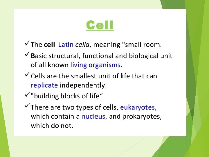
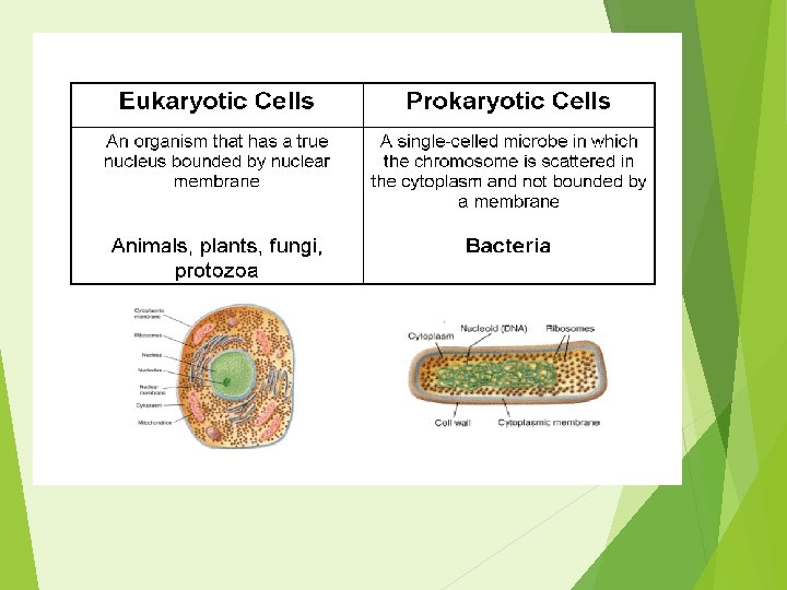
Cell Discovery v Robert Hooke: - Observed cork cells with a microscope and discovered cell walls. Named the structure “the cell” because it reminded him of the small rooms (cells) in which the monks lived. The study of cells is called Cell Biology
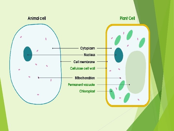

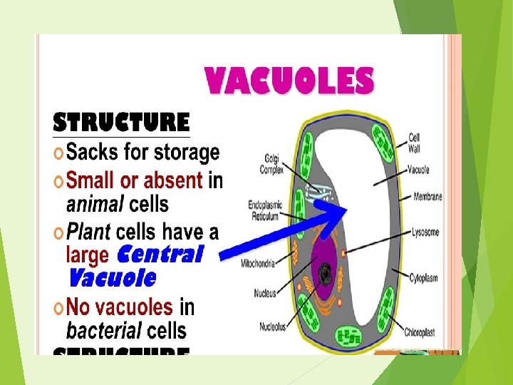
Inside the Cell : - Cell Covering & Cell Organelles embedded in Cytoplasm. Cell wall. Cell membrane. Nucleus. Mitochondria. Chloroplast. Ribosome. Endoplasmic reticulum. Golgi complex. Vacuole's. Lysosomes, peroxisomes, etc.

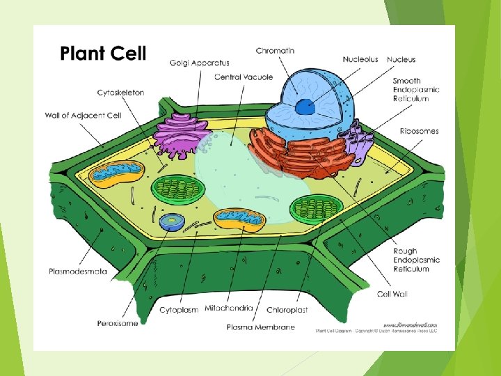
Structure Of Cell Wall A cell wall is a rigid, semi-permeable protective layer in some cell types. In plants, the cell wall is composed mainly of strong fibre's of the carbohydrate polymer cellulose. It is to form a framework for the cell to prevent over expansion and help to maintain the shape and form of the cell. The primary cell wall, generally a thin, flexible and extensible layer formed while the cell is growing. The secondary cell wall, a thick layer formed inside the primary cell wall after the cell is fully grown. It is not found in all cell types. Also containing lignin, which strengthens and waterproofs the wall. The middle lamella, a layer rich in pectins. This outermost layer forms the interface between adjacent plant cells and glues them together. A plasmodesmata is a channel through the cell wall that allows molecules and substances to move back and forth as needed, it also create junctions from cell to cell

Cell Walls

Function of cell wall Support: The cell wall provides mechanical strength and support. It also controls the direction of cell growth. Withstand turgor pressure: Turgor pressure is the force exerted against the cell wall as the contents of the cell push the plasma membrane against the cell wall. This pressure helps a plant to remain rigid and erect, but can also cause a cell to rupture. Regulate growth: The cell wall sends signals for the cell to enter the cell cycle in order to divide and grow. Regulate diffusion: The cell wall is porous allowing some substances, including proteins, to pass into the cell while keeping other substances out. Communication: Cells communicate with one another via Plasmodesmata (pores or channels between plant cell walls that allow molecules and communication signals to pass between individual plant cells). Protection: The cell wall provides a barrier to protect against plant viruses and other pathogens. It also helps to prevent water loss. Storage: The cell wall stores carbohydrates for use in plant growth, especially in seeds.

Structure of Plasma Membrane Plasma membrane can be defined as a biological membrane It is a thin semi permeable membrane layer, which surrounds the cytoplasm and other constituents of the cell. The plasma membrane (cell membrane) is made of two layers of phospholipids. The plasma membrane has many proteins embedded in it. The plasma membrane regulates the entry and exit of the cell. Many molecules cross the cell membrane by diffusion and osmosis. The fundamental structure of the membrane is phospholipid bilayer and it forms a stable barrier between two aqueous compartments. It is a fluid mosaic of lipids, proteins and carbohydrate. It is lipid bilayer, which contains -two layers of phospholipids, phosphate head is polar (water loving), fatty acid tails non-polar (water fearing) and the proteins embedded in membrane. The proteins present in the plasma membrane, act as pumps, channels, receptors, enzymes or structural components.

Plasma Membrane

Function of Plasma Membrane It separates the contents of the cell from its outside environment and it regulates what enters and exits the cell. Plasma membrane plays a vital role in protecting the integrity of the interior of the cell by allowing only selected substances into the cell and keeping other substances out. It also serves as a base of attachment for the cytoskeleton in some organisms and the cell wall in others. Thus the cell membrane supports the cell and helps in maintaining the shape of the cell. The cell membrane is primarily composed of proteins and lipids. While lipids help to give membranes their flexibility and proteins monitor and maintain the cell's chemical climate and assist in the transfer of molecules across the membrane. The lipid bilayer is semi-permeable, which allows only selected molecules to diffuse across the membrane. It helps in transporting molecules across cell membranes It helps in cell to cell communications

Fluid Mosaic Model

Plasma Membrane

Fluid Mosaic Model The fluid mosaic model explains various observations regarding the structure of functional cell membranes. According to this model, there is a lipid bilayer in which the protein molecules are embedded. The model, which was devised by SJ Singer and GL Nicolson in 1972, describes the cell membrane as a two-dimensional liquid that restricts the lateral diffusion of membrane components. Such domains are defined by the existence of regions within the membrane with special lipid and protein composition that promote the formation of lipid rafts or protein and glycoprotein complexes. Another way to define membrane domains is the association of the lipid membrane with the cytoskeleton filaments and the extracellular matrix through membrane proteins. The current model describes important features relevant to many cellular processes, including: cell-cell signaling, apoptosis, cell division, membrane budding, and cell fusion. The fluid mosaic model is the most acceptable model of plasma membrane.
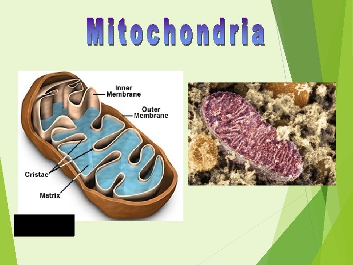

Structure of Mitochondria are rod shaped structure found in both animal and plant cells. It is a double membrane bound organelle, 3 to 4 u in length. It has the outer membrane and the inner membrane. The membranes are made up of phospholipids and proteins or lipoproteins. (protein-central lipid-protein) The outer membrane is Smooth & freely permeable to nutrient molecules, ions, energy molecules like the ATP and ADP molecules. The inner membrane of mitochondria is more complex in structure. It is folded into a number of folds many times and is known as the cristae. This folding help to increase the surface area inside the organelle. Cristae bears many particles called F 1 particles which contain ATP synthetase enzyme for the production of ATP molecules.

Structure of Mitochondria Various chemical reactions takes place in the inner membrane of the mitochondria. The double membrane of the mitochondria divides the organelle into two distinct parts - the Intermembrane Space And The Mitochondrial Matrix. The intermembrane space is the narrow space between the outer and the inner membrane. The mitochondrial matrix is the content enclosed by the inner membrane. The fluid inside the mitochondria is called the matrix. Mitochondria are independent organelles, they have their own DNA and ribosomes. They can replicate and multiply on their own and make their own proteins.

Structure of Mitochondria

Structure of Mitochondria

Function of Mitochondria The most important function of the mitochondria is to produce energy, store energy and supply energy. The simpler molecules of nutrition are sent to the mitochondria to be processed and to produce charged molecules. These charged molecules combine with oxygen and produce ATP molecules or ATP Synthesis. This process is known as oxidative phosphorylation. Mitochondria help the cells to maintain proper concentration of calcium ions within the compartments of the cell. The mitochondria also play important role in the process of Apoptosis or programmed cell death. Role in neurotransmitter metabolism& cholesterol metabolism Role as independent unit. Detoxification of ammonia. Power house of cell.

Structure of Mitochondria
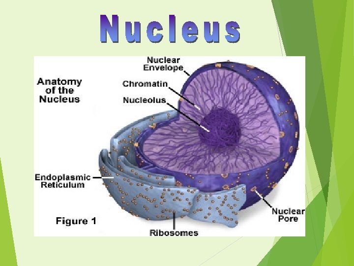

Structure of Nucleus The nucleus is the largest organelle of the cell. The nucleus appears to be dense, spherical organelle. It occupies about 10% of the total volume of the cell. The nuclear envelope is also known as the nuclear membrane. It is made up of two membranes the outer membrane and the inner membrane. The outer membrane of the nucleus is continuous with the membrane of the rough endoplasmic reticulum. The nuclear envelope is perforated with numerous pores called nuclear pores The nuclear pores regulate the passage of the molecules between the nucleus and cytoplasm The nucleus of the cell contains majority of the cells genetic material in the form of multiple linear DNA molecules. These DNA molecules are organized into structures called chromosomes. In the cell they are organized in a DNA-protein complex known as chromatin The nucleolus is not surrounded by a membrane, it is a densely stained structure found in the nucleus. It synthesizes and assembles ribosomes and r RNA During cell division, the nucleolus disappears

Nucleus

Functions of the Nucleus It Store & controls the heredity characteristics of an organism. It is responsible for Protein Synthesis, cell division, cell growth and cell differentiation. Stores heredity material in the form of deoxy-ribonucleic acid (DNA) strands. Also stores proteins and ribonucleic acid (RNA) in the nucleolus. It is a site for Transcription Process in which messenger RNA (m RNA) are produced for protein synthesis. Aids in exchange of DNA and RNA (heredity materials) between the nucleus and the rest of the cell. Nucleolus produces ribosomes and are known as protein factories. It also regulates the integrity of genes and gene expression.
")
DNA RNA PROTEIN (Ribosome)

Rough and Smooth ER

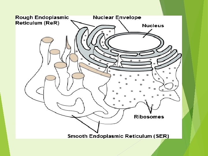
Structure of Endoplasmic Reticulum It can be defined as a eukaryotic organelle, which forms a network of tubules, vesicles and cisternae within the cells called as network of cisternae (saclike structures) It is a connecting link between plasma membrane, cytoplasm & nucleus. There are two regions of the Endoplasmic reticulum, which differ in both structure and function. One region is called as Rough Endoplasmic reticulum, as it contains ribosome attached to the cytoplasmic side of the membrane and they are the series of flattened sacs. The other region is called as Smooth Endoplasmic reticulum as it lacks the attached ribosome and they are tubule network This network increases the surface area for the storage of enzymes. Rough endoplasmic reticulum synthesizes proteins, while smooth endoplasmic reticulum synthesizes lipids and steroids. Thus, it extending from the cell or plasma membrane through the cytoplasm and forming a continuous connection with the nuclear envelope

Function of Endoplasmic Reticulum It is mainly responsible for the Transportation of proteins and other carbohydrates to another organelle, which includes lysosomes, Golgi apparatus, plasma membrane, etc. They play a vital role in the formation of the skeletal framework. They provide the increased surface area for cellular reactions. They help in the formation of nuclear membrane during cell division. They play a vital role in the synthesis, storing and transportation of proteins, lipids, glycogen and other steroids like cholesterol, progesterone, testosterone, etc.
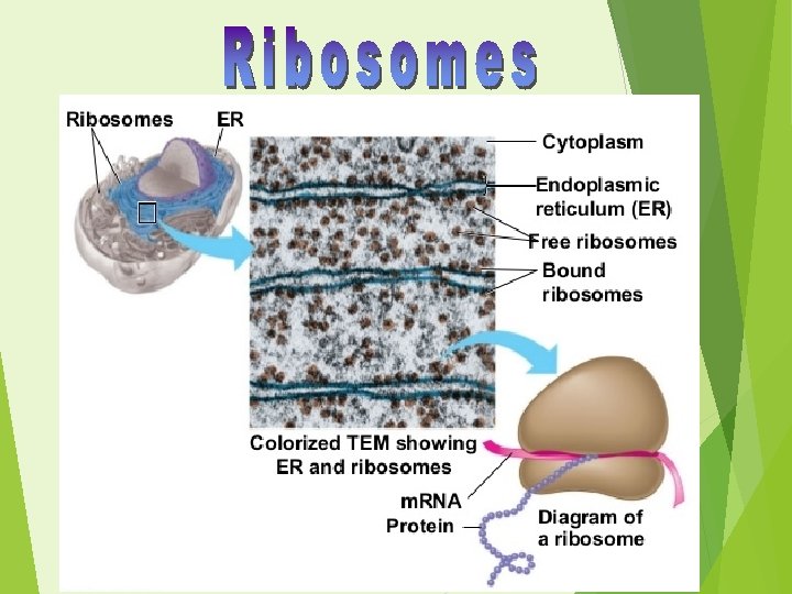

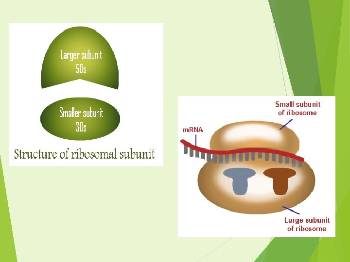
Structure of Ribosomes in a cell are located in two regions of the cytoplasm. They are found scattered in the cytoplasm and some are attached to the endoplasmic reticulum. When the ribosomes are bound to the ER, it is referred as rough endoplasmic reticulum. The bound and the free ribosomes are similar in structure and are invloved in protein synthesis. Ribosomes are tiny particles about 200 Ã…. Ribosomes are composed of both RNA and proteins Ribosome is made up of two subunits Prokaryotes have two subunit consisting of small subunit is of 30 S and the large subunit is of 50 S. Eukaryotes have two consisting of small (40 S) and large (60 S) subunit.

Ribosome Function They assemble amino acids to form specific proteins, proteins are essential to carry out cellular activities. The process of production of proteins, the deoxyribonucleic acid produces m. RNA by the process of DNA transcription. The genetic message from the m. RNA is translated into proteins during DNA translation. The sequences of protein assembly during protein synthesis are specified in the m. RNA. The m. RNA is synthesized in the nucleus and is transported to the cytoplasm for further process of protein synthesis. In the cytoplasm, the two subunits of ribosomes are bound around the polymers of m. RNA; proteins are then synthesized with the help of transfer RNA. The proteins that are synthesized by the free ribosomes present in the cytoplasm are used in the cytoplasm itself. The proteins produced by the bound ribosomes are transported outside the cell.

Chloroplasts

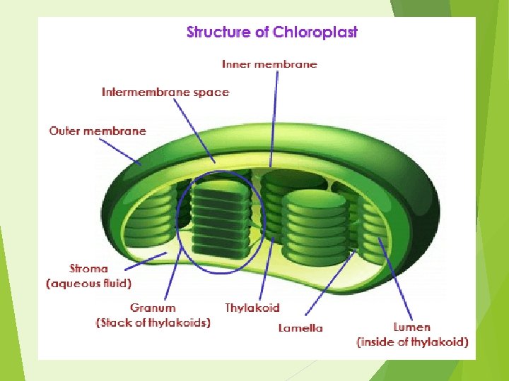
Structure of Chloroplasts vary from spheroid, filamentous saucer-shaped, discoid or ovoid shaped The chloroplast are double membrane bound organelles and are the site of photosynthesis The chloroplasts have a system of three membranes: the outer membrane, the inner membrane and the thylakoid system. The outer and the inner membrane of the chloroplast enclose a semi-gel-like fluid known as the Stroma. This stroma makes up much of the volume of the chloroplast, the thylakoids system floats in the stroma. A thin intermembrane space is present between the outer and the inner membrane of the chloroplast. The thylakoids are arranged in stacks known as Grana. Each granum contains around 10 -20 thylakoids. These thylakoids is the site for the light reactions of the photosynthesis to take place.

Chloroplast Function The most important function of chloroplast is to make food by the process of photosynthesis. Food is prepared in the form of sugars. During the process of photosynthesis sugar and oxygen are made using light energy, water, and carbon dioxide. Light reactions takes place on the membranes of the thylakoids. Chloroplasts, like the mitochondria use the potential energy of the H+ ions or the hydrogen ion gradient to generate energy in the form of ATP. The dark reactions also known as the Calvin cycle takes place in the stroma of chloroplast. Production of NADPH 2 molecules and oxygen as a result of photolysis of water. BY the utilization of assimilatory powers the 6 -carbon atom is broken into two molecules of phosphoglyceric acid.
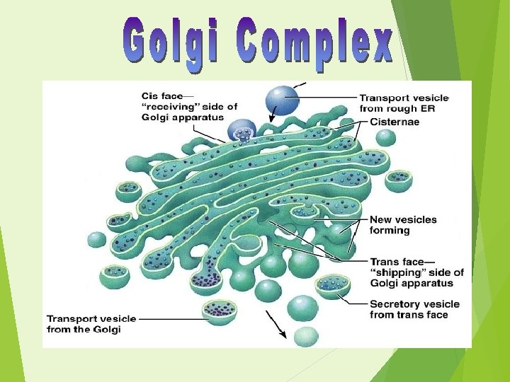

Structure of Golgi Apparatus They are membrane bound organelles, which are sac-like. They are found in the cytoplasm of plant and animal cells. The Golgi complex is composed of stacks of membrane-bound structures, these structures are known as the cisternae. . In a stack there about four to eight cisternae. Each cisternae is a disc enclosed in a membrane, it possess special enzymes of the Golgi which help to modify and transport of the modified proteins to their destination. The flat sacs of the cisternae are stacked and is bent and semicircular in shape. Each group of stacks is membrane bound and its insides are separated from the cytoplasm of the cell. The interaction in the Golgi membrane in responsible for the unique shape of the apparatus. One end of the stack is known as the cis face, it is the 'receiving department" while the other end is the trans face and is the "shipping department". The cis face of the Golgi apparatus is closely associated with the endoplasmic reticulum.
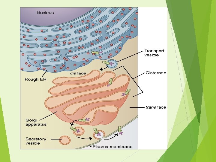

Golgi Apparatus Receives substances from ER, refines and packages them

Function of Golgi Apparatus It mainly modifies the proteins that are prepared by the rough endoplasmic reticulum. They are also involved in the transport of lipid molecules around the cell. The Golgi complex is thus referred as post office where the molecules are packaged, labelled and sent to different parts of the cell. The enzymes in the cisternae have the ability to modify proteins by the addition of carbohydrates and phosphate by the process of glycosylation and phoshphorylation respectively. It is also a major site of synthesis of carbohydrates. It play major role in transporting chemical substance in & out of cell.
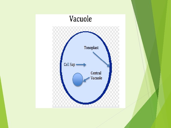

Structure of Vacuole The central vacuole comprises a large percentage of a plant cell's volume. The central vacuole itself is consists of two main bodies; the tonoplast and cell sap. The tonoplast is the membrane that encloses the vacuole and separates it from the cytoplasm. Like many other cell membranes, the tonoplast contains transport proteins that move substances into and out of the central vacuole. Cell sap is the solution of chemicals and molecules that fills the vacuole. Cell sap is made up of water, enzymes, inorganic ions, salts and also stores organic acids, sugars, amino acids, lipids, oligosaccharides, storage proteins, etc. It could also include pigments, which determine the colour of flowers. Some plant cells also store waste in the central vacuole.

Function of Vacuole The primary function of the central vacuole in plant cells is to maintain the turgor of the cell. Turgor is the pressure exerted on the cell wall by the contents of the cell. The central vacuole determines the amount of turgor based on it water content as a result of osmotic pressure. This is why when plants are not watered, they wilt; the reduction in turgor causes the plant cell to lose its rigidity. They act both as temporary stores for reserve materials and as final stores for waste products. Also help in intracellular digestion of complex molecules.

Thank you & Give your feedback
- Slides: 50