Neurons are nerve cells that are specialised for

 Cell Body")


 position once the resting")


- Slides: 38
Neurons are nerve cells that are specialised for the transmission of impulses to other neurons, muscle cells and gland cells Within the mammalian nervous system, there are three functional types of neurons – these are: • • • SENSORY (AFFERENT) NEURONS that transmit impulses from receptors towards the central nervous system (brain and spinal cord) MOTOR (EFFERENT) NEURONS that transmit impulses away from the central nervous system to effectors (muscles and glands) RELAY NEURONS that transmit impulses between sensory and motor neurons and are located within the central nervous system N. B. Two different spellings can be used for nerve cells, i. e. neurons or neurones; the spelling neuron has been used throughout this presentation
The Motor Neuron Dendrites (increase the surface area of the cell body) Cell Body surrounding the nucleus Axon Hillock Axon (containing axoplasm) Direction of Impulse Myelin Sheath Non-myelinated axon terminals Synaptic Knob Effector
Nerves and Neurons Nerves are bundles of nerve fibres and each nerve fibre is the long extension of a single neuron, e. g. the axon of a motor neuron Bundles of nerve fibres Myelinated nerve fibres Connective tissue surrounds the entire nerve, the bundles of nerve fibres and the nerve fibres within the bundles T. S. Whole Nerves may contain entirely motor fibres or sensory fibres or a mixture of both (mixed nerves)
Myelinated neurons within a nerve
The Electrical Nature of the Neuron Resting neurons, like all other human cells, are positive on the outside relative to the inside as a consequence of the unequal distribution of ions and charged organic molecules across the membrane The opposite charges are insulated by the high lipid content of the plasma membrane such that a voltage, called the membrane potential, exists across the membrane Cells that exhibit a membrane potential are said to be POLARISED The resting potential, or electrical difference across a neuron cell membrane when the neuron is inactive, is equal to -70 millivolts (for most neurons)
The Action Potential Depolarisation The presence of an appropriate stimulus, at a point on the neuron cell membrane, triggers an action potential at the point of stimulation Repolarisation Hyperpolarisation An action potential is a temporary reversal of the membrane potential; the inside of the neuron membrane becomes positive relative to the outside and then returns to the resting potential This temporary reversal of charge across the membrane of a ‘resting’ neuron is described as DEPOLARISATION The restoration of the resting membrane potential is described as REPOLARISATION, and the slight overshoot of the membrane potential to a value that is more negative than the resting potential is described as HYPERPOLARISATION
Neurons and Ions There are many different charged particles in the fluids both inside and outside neurons Sodium ions (Na+) and potassium ions (K+) are present in the highest concentrations The distribution of sodium and potassium ions across the neuronal cell membrane exerts the greatest effect on the membrane potential
Ion Channels Leak channels for potassium ions • • The passage of charged particles across the lipid bilayer of membranes is achieved only when there are specific channel and carrier proteins present Voltage-gated channels for both potassium and sodium ions There are TWO types of ion channel in the neuron membrane: VOLTAGE-GATED channels which open and close in response to changes in the voltage (membrane potential) across the membrane; these channels are CLOSED when the neuron is at rest LEAK channels; protein channels that remain permanently open There are Leak Channels for potassium ions within the neuron cell membrane but NO Leak Channels for sodium ions (however, a few sodium ions do leak into the neuron along the potassium channels)
Generating and Maintaining the Resting Potential Leak channels for potassium ions The potential difference across the unstimulated neuron membrane (i. e. the resting potential) is due to the activity of many Na+/K+ pumps within the membrane together with the existence of numerous K+ leak channels Na+/K+ pump AXOPLASM The permanently operating sodium-potassium pumps maintain chemical concentration gradients for sodium and potassium ions, by actively transporting sodium ions out of the neuronal axoplasm and potassium ions into the axoplasm
The ATP driven Na+/K+ pumps transport sodium ions out of the axoplasm and potassium ions into the axoplasm thereby creating a high internal potassium ion concentration and high external sodium ion concentration K+ Na+ K+ K+ K+ Na+ Na+ Na+ K+ Na+ Na+ The operation of the Na+/K+ pumps creates steep concentration gradients for sodium and potassium ions but leak channels in the membrane exist only for the potassium ions The permeability of the neuron membrane to potassium ions is therefore significantly greater than that for sodium ions Potassium ions begin to diffuse OUT of the axoplasm along their chemical concentration gradient through the leak channels
Potassium ions begin to diffuse out of the axoplasm along their chemical concentration gradient through K+ leak channels Na+ K+ Na+ Na+ K+ K+ K+ Na+ Na+ Na+ K+ K+ K+ Na+ Na+ K+ K+ K+ Na+ Within the axoplasm of the neuron there a number of negatively charged ions and proteins As the positively charged potassium ions leak out of the neuron and very few sodium ions leak in, the inside of the neuron becomes increasingly electrically negative relative to the outside
The development of an internal negative charge slows the OUTWARD movement of potassium ions through the leak channels; this electrical force attracts the positively charged potassium ions INTO the neuron Na+ chemical K+ concentration K+ gradient K+ K+ electrical gradient Na+ As the membrane potential approaches -70 millivolts, the electrical force attracting the positive potassium ions into the neuron is equal to the chemical concentration force driving potassium ions out of the neuron At the equilibrium potential of -70 millivolts, this electrochemical gradient for potassium ions disappears and there is NO NET MOVEMENT of K+; as many potassium ions enter the neuron as leave it The resting potential is thus maintained at a steady value of -70 millivolts and the membrane is described as being POLARISED (internally negative, externally positive)
The Generation of The Action Potential An action potential is a membrane potential that temporarily reverses in response to a stimulus and then returns to its original resting level Changes in the permeability of the axon membrane to sodium and potassium ions are responsible for the depolarisation and repolarisation phases of the action potential Changes in the permeability of the axon membrane during the action potential are due to the opening and closing of voltage-gated ion channels that control the movement of sodium and potassium ions through the membrane
The ‘unstimulated’ neuron is polarised and the resting membrane potential is -70 millivolts; voltage-gated sodium channels are in a closed position Extracellular fluid Voltage-gated sodium channel neuronal membrane Axoplasm
When an area of neuronal membrane is stimulated, depolarisation begins and the shift in membrane potential (to around -55 millivolts) triggers the opening of a few voltage-gated sodium channels; sodium ions rapidly enter the neuron along their steep concentration gradient Extracellular fluid Axoplasm
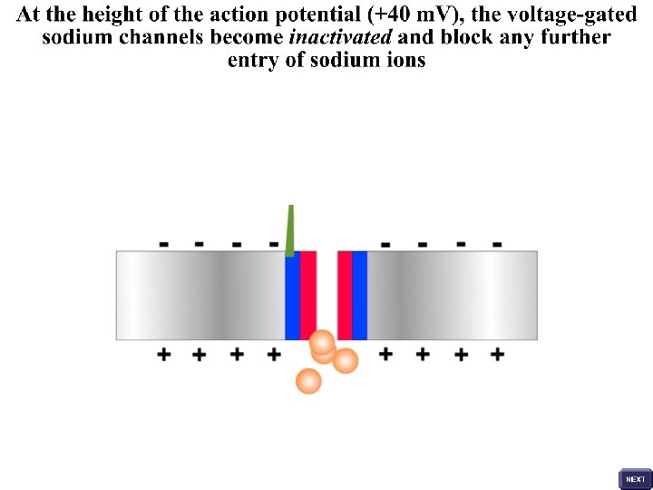
The influx of sodium ions results in further depolarisation of the membrane and the opening of more and more voltage-gated sodium channels (positive feedback) in the region of the stimulus Entry of sodium ions continues until the membrane potential reaches a value of around +40 millivolts; this is the height of the action potential Depolarised membrane
Voltage-gated potassium channels now open relatively slowly allowing potassium ions to move outwards across the neuronal membrane and re-establish the resting membrane potential during the process of repolarisation K+ Inactivated sodium channel The membrane is unresponsive to any further stimuli whilst the voltagegated sodium channels are inactivated; this refractory period lasts until the relatively slow, outward flow of potassium ions returns the membrane potential to its resting value (-70 millivolts)
Voltage-gated sodium channels return to a closed (rather than inactivated) position once the resting potential has been restored and this area of membrane can now respond to further stimulation Voltage-gated sodium channels in a closed position
The rapid influx of sodium ions Sodium channels leads to further depolarisation return to a closed position once the Voltage-gated sodium and the opening of more and resting potential channels are closed in more voltage-gated sodium channels (positive feedback) has been restored the resting neuron The membrane potential rapidly reaches a positive value and, at the height of the action potential, sodium gates are inactivated Voltage-gated potassium channels now open relatively slowly allowing potassium ions to move outwards across the neuronal membrane and repolarise the membrane The Na+/K+ pumps re-establish the resting potential following hyperpolarisation Hyperpolarisation A stimulus causes local depolarisation of the membrane If the change in membrane potential reaches the threshold value then a few voltage-gated sodium channels open
Summary of Changes in the Membrane Potential
Transmission of the Action Potential along an Non-myelinated Neuron All action potentials are propagated as self-generating waves of depolarisation along the length of the neuron These self-propagating action potentials travelling along the axon describe the nerve impulse When an action potential occurs on a segment of axon, it produces local currents which stimulate the adjacent area of neuron membrane; this area of membrane depolarises, and thus initiates another action potential ahead of the previous one As action potentials are propagated along the axon, the local currents produced spread out in all directions, but the nerve impulse travels only in one direction – it is unidirectional The unidirectional nature of the nerve impulse arises as the area of axon membrane that has recently depolarised is undergoing repolarisation and voltage-gated sodium channels are inactivated; the membrane is in a refractory state and temporarily incapable of producing an action potential
Inactivated sodium channels in previously stimulated area; membrane in refractory period Direction of propagation in relation to TIME
Direction of propagation in relation to TIME
Direction of propagation in relation to TIME
When an action potential occurs on a segment of the non-myelinated axon, it produces local currents that act as the stimulus for an adjacent segment of membrane which, in turn depolarises and generates an action potential This process continues unidirectionally along the axon such that an impulse travels down its length as a self-propagated wave of depolarisation
Refractory Periods The refractory period is the time during which a neuron membrane is unresponsive to stimuli • The Absolute Refractory Period coincides with the process of repolarisation in a previously active region and lasts for one millisecond; during this period the area of neuron membrane that has just been activated is totally unresponsive to any further stimuli • The Relative Refractory Period lasts for 4 to 10 milliseconds and is a period, following the absolute refractory period, when the membrane will respond only to stimuli of high intensity • The Excitability of the neuron membrane is below that of resting neuron during these refractory periods
A new stimulus, applied to an area of membrane that has just been active, will NOT initiate another action potential; this is the absolute refractory period and lasts for about 1 ms During the relative refractory period (4 to 10 ms), a stimulus of HIGH intensity MAY depolarise the region that has just been active
Features of Action Potentials • The All or Nothing Law; each neuron has a threshold of stimulation below which it will not fire an action potential; above this threshold all stimuli, regardless of strength, bring about an action potential whose magnitude is independent of the strength of the stimulus • Coding of Nervous Information; as the action potential is always of a fixed amplitude, then the code of the nerve impulse is a frequency code such that the stronger the stimulus, the greater the frequency of action potentials generated and the greater the amount of neurotransmitter released at the neuron terminals • Speed of Transmission; the speed of transmission of action potentials is affected by properties of the neurons themselves • Large Diameter Axons conduct impulses at greater speed due to the larger area of membrane available for ion movements and sodium/potassium pumps and the lower resistance to local intracellular current flow • Myelinated Neurons conduct impulses at greater speed due to the presence of the myelin sheath
Myelinated Neurons Due to the insulating properties of the myelin sheath, depolarisation of the axon membrane occurs only at the nodes In myelinated neurons the axon membrane is insulated by a fatty myelin sheath, formed by by the Schwann cells Myelin sheath AXOPLASM OF AXON Nodes of Ranvier The fatty myelin sheath insulates the axon against electrical interference from neighbouring cells This insulation is however incomplete and there are regularly spaced, uninsulated gaps called the nodes of Ranvier
Saltatory Conduction When an action potential occurs at a node along the axon, it produces electrical currents which depolarise the next node The action potential generated at this second node produces electric currents which depolarise the next adjacent node In myelinated neurons, nerve impulses are propagated by saltatory conduction and the action potential appears to jump from node to node
Speed of Transmission In myelinated neurons the speed of transmission of action potentials is greater than that observed in non-myelinated neurons of similar diameter The speed of transmission of action potentials along neurons is also affected by the diameter of the axon Large diameter axons conduct impulses at a greater speed
The ventral nerve cord of the earthworm contains many nerve fibres that include three giant axons Giant axons of the nerve cord These giant axons conduct impulses at very high speed and are responsible for the ESCAPE RESPONSE of the earthworm
Giant Axons in the Squid The escape response of the squid involves contraction of the muscles that surround its water-filled mantle cavity The giant axons that innervate these muscles have been studied extensively and provided us with much of our early knowledge of neuron function
Stimulation of Axons and Nerves The giant axon of the squid and the sciatic nerve of the frog were subjected to electrical stimulation of differing intensity Traces were obtained from the two preparations (axon and nerve) and these showed different responses to stimuli of increasing strength Consider the results obtained from stimulating axons and nerves and provide an explanation for the observed differences
• What do the voltages marked A represent? • Explain why the traces resulting from stimulation of the giant axon and the sciatic nerve are different
Acknowledgements Copyright © 2003 SSER Ltd. and its licensors. All rights reserved.