Neurons Neurons Brain Cells Discovered by Santiago Ramon

Neurons

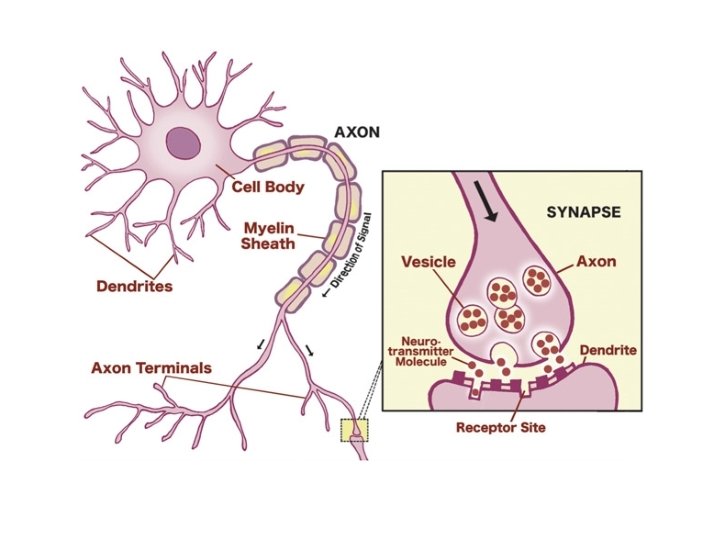
Neurons • Brain Cells • Discovered by Santiago Ramon y Cajal – Neurons are “distinguishable processing units” • Main components of a neuron – Dendrites – Soma – Axon

Dendrites • • • They extend from the Soma They branch several times They receive messages from on neurons They are the “input” for neurons At the end of each dendrite are spines which begin the message sending process in a neuron

Soma • This is the cell body • It contains the nucleus • The nucleus, indirectly the soma, has the following functions: – Genetic information – Directs protein synthesis – Supplies and directs energy needed for a neuron to function • Think of it as the control center

Axon • This is the output, it sends messages • The axon is covered in the myelin sheath – This myelin sheath “protects” the message • The end of each axon branches out and at the end of each branch is a terminal button (the end of the axon branch)

Other Terms • Synapse – where the axon and dendrites transfer messages from one neuron to another • Presynaptic – neuron sending the message • Postsynaptic – neuron receiving the message • The small gap that forms between the dendrites and axon is called the synaptic gap • Synaptic vesicles – package the message in chemical groupings • Neurotransmitters – these are the chemicals

Types of Cells in the Brain • Sensory neurons – receive information about the world around us • Motor neurons – movement and behavior. They get things going • Interneurons – takes input and processes it into meaningful representations – They take the input and define the action

Types of Cells in the Brain • Number of dendrites also defines cells of the brain – Unipolar: one dendrite – Bipolar: two dendrites – Multipolar: more then two dendrites • Most common is the pyramidal neuron – It is multipolar and the soma is a triangle shape

Types of Cells in the Brain • Glia Cells – another cell in the brain • Oligodendroglia – form the myelin sheaths – One cell can form the myelin sheaths on several axons • Microglia and astrocytes – digest debris of dead neurons, carry nutritional support from blood to the neurons, and regulate ionic composition of the extracellular fluids • Glia Cells do not carry the message

Communication Within and Between Neurons • Has two stages: 1. Electrical conduction of dendritic input to the initiation action of an action potential within a neuron 2. Chemical transmission across the synaptic gap
 fluid and extracellular (outside the cell)")
Resting Membrane Potential • Intracellular (inside the cell) fluid and extracellular (outside the cell) fluid of a neuron is composed of ions (it has an electric charge) • Cations = positively charge ions • Anions = negatively charged ions • Fluid make up: Sodium, Potassium, Chloride, and Anions

Resting Membrane Potential • Cell membrane – is composed of fat tissue, separates the cell from the extracellular fluids • There are channels, ion channels, that span the membrane – They allow extracellular and intracellular fluids to move back and forth

Resting Membrane Potential • Because there are different charges and amounts of ions in and outside the cell two forces are at work to maintain balance – Diffusion – Electrostatic pressure

Forces at Work • Diffusion – The force that moves molecules from places of high concentration to low

Forces at Work • Electrostatic Pressure – ions with the same charge will repel each other. Ions with opposite charges will attract to one another

Forces at Work • Equilibrium potential – the force of diffusion is equal and opposite the force of electrostatic pressure – The result: NO ION FLOW • The baseline electrical charge (think all things at rest) inside the cell compared to the outside is known as Resting Membrane Potential

Resting Membrane Potential • The net charge of a neuron is negative compared to the extracellular fluids
 • High concentration inside the cell,")
Forces on Groups of Ions • Anions (A-) • High concentration inside the cell, hence the negative charge. – Anions are not effected by these forces because it cannot flow through the membrane • It will effect both forces
 • Found in high concentrations inside")
Forces on Groups of Ions • Potassium (K+) • Found in high concentrations inside a cell • Diffusion will push Potassium out of the cell because there is a high concentration within • Electrostatic pressure will push Potassium in because of it positive charge and will attract to the negative field in the cell • Flows easily through the membrane • Opposite reactions for both forces
 • In high concentration outside the")
Forces on Groups of Ions • Chloride (Cl-) • In high concentration outside the cell • So… – What will diffusion do? – What will electrostatic pressure do? • Flows easily through the membrane • Opposite reactions to both forces
 • High concentration outside the cell")
Forces on Groups of Ions • Sodium (Na+) • High concentration outside the cell • So… – What will diffusion do? – What will electrostatic pressure do? • Does not flow easily through the membrane • The forces push Na+ into the cell • Sodium should not be in the cell so special pumps exist to remove it called sodiumpotassium pumps. 3 out for 2 in exchange

Message Sent • Hodgkin and Huxley discovered these actions in the 1930 s • Experimented on squids, they have axons that are 100 times the size of mammals • Hodgkin and Huxley would introduce a transient electrical charge down the axon • This is known as the action potential

Message Sent • The action potential is an all-or-nothing response that occurs when there is a change in electrical charge or potential in a positive direction – This action is known as depolarization • All-or-nothing – there are only two outcomes to the action potential, Do or Do Not
 that must be")
Message Sent • There is a certain membrane potential (think charge) that must be reached to initiate action potential – Threshold of excitation – It is a movement towards positive charge inside the cell

Message Received • Initiation of action potential – Each neurons is receiving hundreds of messages from synapsed neurons – Two basic actions can occur: – Excitatory postsynaptic potentials (EPSP) • This will cause depolarization – Inhibitory postsynaptic potentials (IPSP) • This will cause hyperpolaration

Message Received • Summate – add together over time and space – If a neuron receives two small EPSPs in the same synapse at the same time then it will create all large EPSP in the neuron – If a synapse receives an EPSP and IPSP they cancel out

Message Received • EPSPs and IPSPs change the charge by as little as 0. 1 to 40 m. V • The change in action potential is around 100 m. V • Remember: the EPSPs and IPSPs come in different strengths and all them are measured off the resting membrane potential – So, it could be one big one or lots of small ones to create the action potential

Message Received • If the EPSP is strong enough to reach the threshold of excitation then in initiates an action potential • Action potential travels down the axon until it hits the terminal buttons, which • Release neurotransmitters into the synapse gap • They connect with ionotropic receptors on the dentrites

Change in Membrane Potential During an Action Potential • As you can well imagine everything starts at resting membrane potential • At this point all channels are closed • When action potential occurs these channels open allowing ions to flow in or out • Na+ will rise (depolarize) the action potential – The inside of the cell becomes positive – Na+ channels will close until the cell returns to resting membrane potential (refractory)

Action Potential: All-or-nothing potential within a neuron EPSPs and IPSPs: Graded potential within a neuron that can be depolarizing or hyperpolarizing Release of Neurotransmitters: Chemical signals between neurons
- Slides: 31