Midyear Review 11 Organ Systems 1 Integumentary System


 Variable/ Stimuli Receptor Control Center (Nervous – Brain)")
 A. B. C. Depress Increase D. F. (output)")


 Copyright © 2003 Pearson Education, Inc.")


 Cells undergoing mitosis, Lies next to dermis,")


 Arranged in concentric rings")

 2. Cartilaginous joints: Immovable")


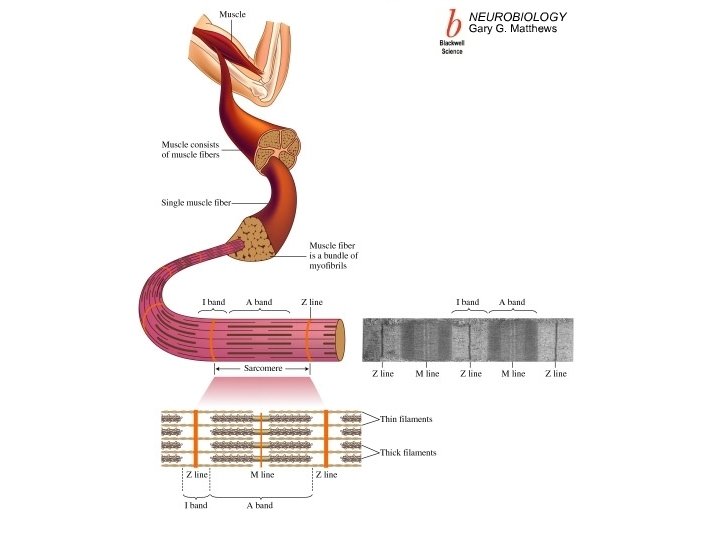


 Figure 6.")


 is generated which moves along the muscle fiber")


 Central nervous system: Brain & Spinal cord (PNS) Peripheral nervous system: Nerves outside")

 Engulf and dispose of debris, dead")


- Slides: 85
Midyear Review
11 Organ Systems 1. Integumentary System 2. Skeletal System 3. Muscular System 4. Nervous System 5. Endocrine System 6. Cardiovascular System 7. Lymphatic System 8. Respiratory System 9. Digestive System 10. Urinary System 11. Reproductive System
Organ System Overview • Integumentary • Forms the external body covering • Protects deeper tissue from injury • Synthesizes vitamin D • Location of cutaneous nerve receptors Figure 1. 2 a Copyright © 2003 Pearson Education, Inc. publishing as Benjamin Cummings Slide 1. 4
Organ System Overview • Skeletal • Protects and supports body organs • Provides muscle attachment for movement • Site of blood cell formation • Stores minerals Figure 1. 2 b Copyright © 2003 Pearson Education, Inc. publishing as Benjamin Cummings Slide 1. 5
Organ System Overview • Muscular • Allows locomotion • Maintains posture • Produces heat Figure 1. 2 c Copyright © 2003 Pearson Education, Inc. publishing as Benjamin Cummings Slide 1. 6
Organ System Overview • Nervous • Fast-acting control system • Responds to internal and external change • Activates muscles and glands Figure 1. 2 d Copyright © 2003 Pearson Education, Inc. publishing as Benjamin Cummings Slide 1. 7
Organ System Overview • Endocrine • Secretes regulatory hormones • Growth • Reproduction • Metabolism Figure 1. 2 e Copyright © 2003 Pearson Education, Inc. publishing as Benjamin Cummings Slide 1. 8
Organ System Overview • Cardiovascular • Transports materials in body via blood pumped by heart • Oxygen • Carbon dioxide • Nutrients • Wastes Figure 1. 2 f Copyright © 2003 Pearson Education, Inc. publishing as Benjamin Cummings Slide 1. 9
Organ System Overview • Lymphatic • Returns fluids to blood vessels • Disposes of debris • Involved in immunity Figure 1. 2 g Copyright © 2003 Pearson Education, Inc. publishing as Benjamin Cummings Slide 1. 10
Organ System Overview • Respiratory • Keeps blood supplied with oxygen • Removes carbon dioxide Figure 1. 2 h Copyright © 2003 Pearson Education, Inc. publishing as Benjamin Cummings Slide 1. 11
Organ System Overview • Digestive • Breaks down food • Allows for nutrient absorption into blood • Eliminates indigestible material Figure 1. 2 i Copyright © 2003 Pearson Education, Inc. publishing as Benjamin Cummings Slide 1. 12
Organ System Overview • Urinary • Eliminates nitrogenous wastes • Maintains acid – base balance • Regulation of materials • Water • Electrolytes Figure 1. 2 j Copyright © 2003 Pearson Education, Inc. publishing as Benjamin Cummings Slide 1. 13
Organ System Overview • Reproductive • Production of offspring Figure 1. 2 k Copyright © 2003 Pearson Education, Inc. publishing as Benjamin Cummings Slide 1. 14
Survival Needs 1. Nutrients: carbohydrates, proteins, lipids, vitamins, and minerals 2. Oxygen: 20 % in air, too much is flammable 3. Water: 60– 80% of body weight 4. Body temperature: 37 °C (98 °F) 5. Atmospheric pressure: force exerted by the weight of air; certain pressure is necessary for gas exchange
Homeostasis • Maintenance of a stable internal environment = a dynamic state of equilibrium Examples: -steady level of CO 2 and O 2 -blood sugar level -Blood pressure -body temperature
Homeostatic Control Mechanisms: Afferent Pathway (input) Variable/ Stimuli Receptor Control Center (Nervous – Brain) (Endocrine- Glands) Determines the set point. Depress Increase Effector Efferent Pathway (output)
Homeostatic Control Mechanisms: E. (input) A. B. C. Depress Increase D. F. (output)
Which is the right side of the heart? Side A or Side B? Side A Side B
Orientation and Directional Terms Table 1. 1 Copyright © 2003 Pearson Education, Inc. publishing as Benjamin Cummings Slide 1. 22
Orientation and Directional Terms Table 1. 1 (cont) Copyright © 2003 Pearson Education, Inc. publishing as Benjamin Cummings Slide 1. 23
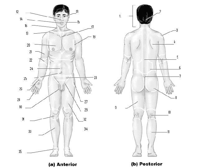
Body Landmarks • Anterior Figure 1. 5 a Copyright © 2003 Pearson Education, Inc. publishing as Benjamin Cummings Slide 1. 24
Body Landmarks • Posterior Figure 1. 5 b Copyright © 2003 Pearson Education, Inc. publishing as Benjamin Cummings Slide 1. 25
Body Planes Figure 1. 6 Copyright © 2003 Pearson Education, Inc. publishing as Benjamin Cummings Slide 1. 26
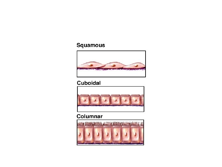
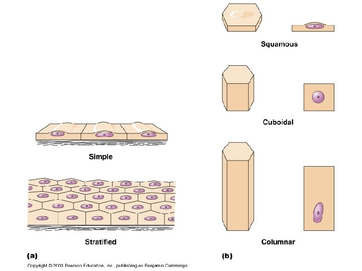
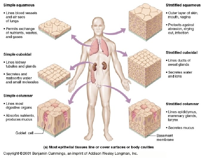
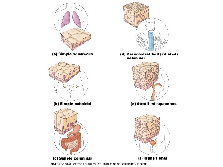
Quiz!! E Can You Identify the Classes of Epithelium? D A B C
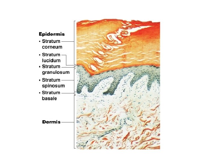
Skin Structure Figure 4. 4
Layers/Strata of Epidermis 1. Stratum basale (germinativum) Cells undergoing mitosis, Lies next to dermis, contains melanocytes 2. Stratum spinosum Most superficial layer that still receives adequate nourishment 3. Stratum granulosum Contains granules with a precursor to keratin and a waterproofing glycolipid 4. Stratum lucidum Occurs only in thick skin (palms, soles of feet) 5. Stratum corneum Shingle-like dead cells, filled with keratin (protein), 20 -30 cell layers thick
Functions of Bones 1. Support 2. Protection: skull/brain, vertebrae/spinal cord, ribs/soft organs 3. Movement: due to attached skeletal muscles 4. Storage: fat, minerals (calcium, phosphorus) 5. Blood cell formation: within the bone marrow of certain bones (aka. Hematopoiesis) Copyright © 2003 Pearson Education, Inc. publishing as Benjamin Cummings Slide 5. 2
Subdivisions of the Skeleton – Axial skeleton: bones of the longitudinal axis (skull, spine, ribs) – Appendicular skeleton: bones of the appendages (limbs and girdles)
Classification of Bones on the Basis of Shape Figure 5. 1 Copyright © 2003 Pearson Education, Inc. publishing as Benjamin Cummings Slide 5. 4 c
Gross Anatomy of a Long Bone Diaphysis Shaft compact bone Epiphysis Ends of long bone spongy bone surrounded by a thin layer of compact bone Copyright © 2003 Pearson Education, Inc. publishing as Benjamin Cummings Figure 5. 2 a Slide 5. 6
Microscopic Anatomy of Bone Figure 5. 3 Copyright © 2003 Pearson Education, Inc. publishing as Benjamin Cummings Slide 5. 10 b
Microscopic Anatomy of Bone Lacunae Cavities containing bone cells (osteocytes) Arranged in concentric rings Lamellae Rings around the central canal Sites of lacunae Copyright © 2003 Pearson Education, Inc. publishing as Benjamin Cummings Figure 5. 3 Slide 5. 11 a
• . . U 9 Endocrine SystemCh 09_JPGs�909_Hormonal. Contr ols_1. JPG
Structural Classification of Joints 1. Fibrous joints: Generally immovable (synarthroses) 2. Cartilaginous joints: Immovable or slightly moveable (amphiarthroses) 3. Synovial joints: Freely moveable (diarthroses) Q. Where might it be important to have immovable joints? Why? A. Immovable joints offer greater protection. B. Example: sutures in the skull Slide 5. 45
Fibrous Joints Bones united by fibrous tissue Sutures: -irregular edges interlock and are connected by fibrous connective tissue Example: bones of the skull Syndesmoses: - Allows more movement than sutures Example: distal end of tibia and fibula Figure 5. 27 d, e Slide 5. 46
Cartilaginous Joints Bones connected by cartilage Examples Pubic symphysis Intervertebral joints Cartilage between the ribs and sternum Figure 5. 27 b, c Slide 5. 47
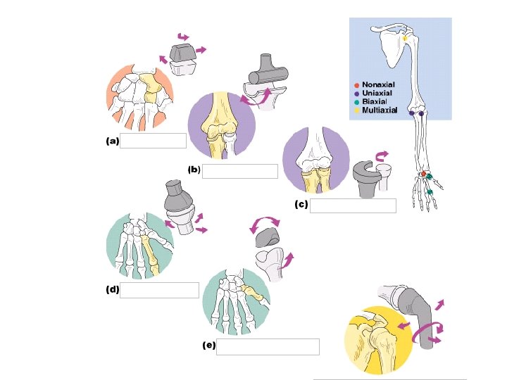
Types of Synovial Joints Based on Shape Figure 5. 29 a–c Copyright © 2003 Pearson Education, Inc. publishing as Benjamin Cummings Slide 5. 52 a
Types of Synovial Joints Based on Shape Figure 5. 29 d–f Copyright © 2003 Pearson Education, Inc. publishing as Benjamin Cummings Slide 5. 52 b
The Appendicular Skeleton Figure 5. 6 c Copyright © 2003 Pearson Education, Inc. publishing as Benjamin Cummings Slide 5. 32 b
The Muscular System Three Types of Muscle: 1. Skeletal muscle 2. Cardiac muscle 3. Smooth muscle Copyright © 2003 Pearson Education, Inc. publishing as Benjamin Cummings Slide 6. 1
Microscopic Anatomy of Skeletal Muscle Cells are multinucleate Nuclei are just beneath the sarcolemma Figure 6. 3 a Copyright © 2003 Pearson Education, Inc. publishing as Benjamin Cummings Slide 6. 9 a
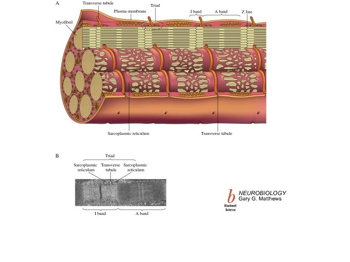
Microscopic Anatomy of Skeletal Muscle Sarcolemma: plasma membrane or cell membrane of a muscle cell Sarcoplasmic reticulum (SR)– stores calcium, smooth endoplasmic reticulum of a muscle cell Figure 6. 3 a Copyright © 2003 Pearson Education, Inc. publishing as Benjamin Cummings Slide 6. 9 b
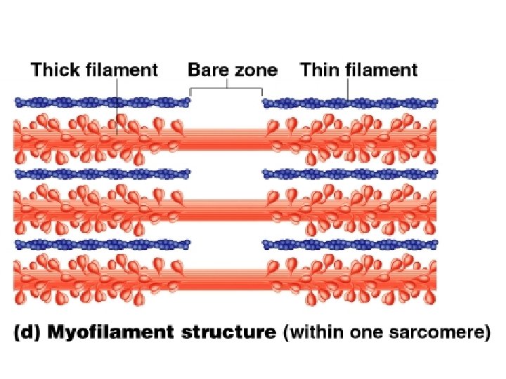
Microscopic Anatomy of Skeletal Muscle Myofibril: Bundles of myofilaments (actin and myosin) Figure 6. 3 b Copyright © 2003 Pearson Education, Inc. publishing as Benjamin Cummings Slide 6. 10 a
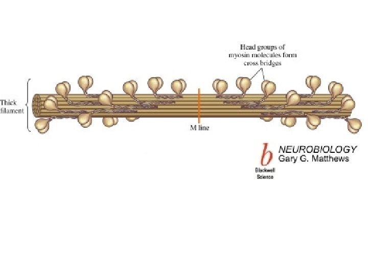
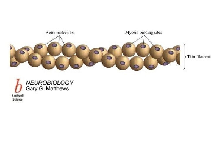
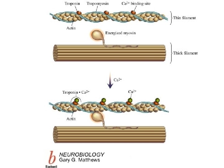
Sarcomere: Contractile unit of a muscle fiber between two 2 z-lines Myosin: Thick filaments that have heads (extensions, or cross bridges) Actin: Thin filaments that have binding sites where myosin heads form crossbridges Figure 6. 3 b Copyright © 2003 Pearson Education, Inc. publishing as Benjamin Cummings Slide 6. 10 b
The Sliding Filament Theory of Muscle Contraction 1. Action potential arrives at the end of the motor neuron 2. ACh (neurotransmitter) diffuses across the synaptic cleft and attaches to receptors on the sacrolemma of the muscle cell Copyright © 2003 Pearson Education, Inc. publishing as Benjamin Cummings Figure 6. 7 Slide 6. 17 a
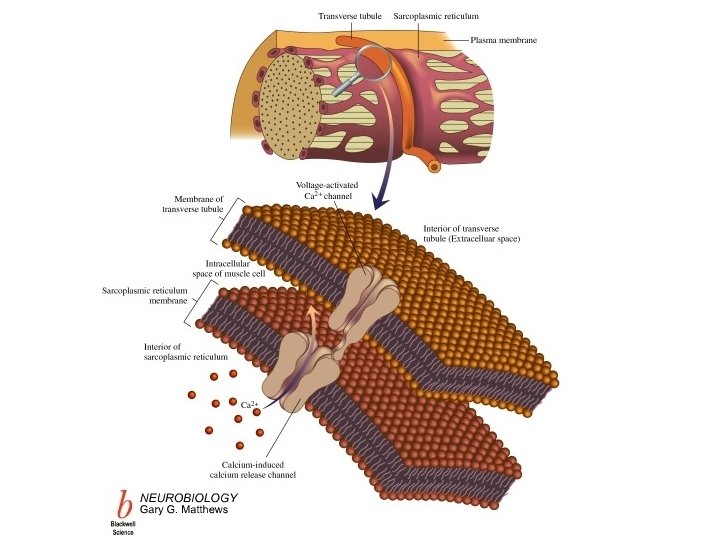
3. A muscle action potential (depolarization) is generated which moves along the muscle fiber surface 4. Depolarization spreads to the t-tubule 5. Ca 2+ is released from the sacroplasmic reticulum
6. Ca 2+ binds to troponin 7. Tropomyosin moves exposing the myosin binding sites on actin
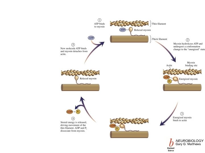
8. Myosin heads bind to the binding sites on actin and the heads pull on the actin causing the filaments to slide past each other
9. ATP binds to Myosin and is converted to ADP + Pi which re-energizes the myosin heads
10. Cross bridge cycling continues while Ca 2+ and ATP are present 11. Ca 2+ is pumped back into the sacroplasmic reticulum causing myosin binding sites to be covered and muscle activity to cease
Body Movements Figure 6. 13 Copyright © 2003 Pearson Education, Inc. publishing as Benjamin Cummings Slide 6. 33
Sensory Input: you see the red light Integration: red means stop Motor output: contract your muscles to step on the brake
(CNS) Central nervous system: Brain & Spinal cord (PNS) Peripheral nervous system: Nerves outside the brain and spinal cord PNS: -Afferent/Sensory division: Nerve fibers that carry information to the central nervous system -Efferent/Motor division: Nerve fibers that carry impulses away from the central nervous system Efferent: +Somatic nervous system = voluntar +(ANS) Autonomic nervous system = involuntary ANS: --Sympathetic --Parasympathetic Slide 7. 2
Astrocytes • Most numerous glial cell in the CNS • Brace/support neurons • star-shaped cells with many extensions • Fill spaces with scar tissue following an injury to the nervous system • have many extensions that are associated with and form a barrier between neurons and capillaries Slide 7. 5
Microglia Small spider-like phagocytes (the clean up crew) Engulf and dispose of debris, dead brain cells and bacteria Slide 7. 6
Ependymal cells • Line cavities of the brain and spinal cord • Have cilia that circulate cerebrospinal fluid in the CNS
Oligodendrocytes Produce myelin sheath around nerve fibers in the CNS don’t have the neurilemmal sheath that Schwann cells have The same oligodendrocyte can form myelin around many neurons, wheareas Schwann cells in the PNS form myelin only around part of one neuron. Figure 7. 3 d Copyright © 2003 Pearson Education, Inc. publishing as Benjamin Cummings Slide 7. 7 a
Satellite cells Protect and cushion neuron cell bodies in the PNS Figure 7. 3 e Copyright © 2003 Pearson Education, Inc. publishing as Benjamin Cummings Slide 7. 7 b
Schwann Cells • Form myelin sheath in the PNS • the Neurilemma is the outer part of the cell where the cytoplasm is located Figure 7. 5 Slide 7. 12
Nodes of Ranvier • gaps in myelin sheath along the axon where two Schwann cells meet (Myelinated nerve fiber x 540)
Multipolar neurons • many extensions from the cell body • majority of neurons Figure 7. 8 a Copyright © 2003 Pearson Education, Inc. publishing as Benjamin Cummings Slide 7. 16 a
Conjunctivitis: • inflammation of the conjunctiva • symptoms: eyes are red and inflamed Pinkeye: • infectious form of conjunctivitis • very contagious caused by bacteria/viruses
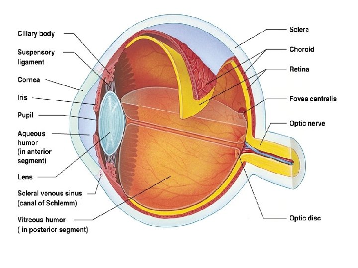
Neurons of the Retina Figure 8. 4 Copyright © 2003 Pearson Education, Inc. publishing as Benjamin Cummings Slide 8. 11