Meiosis Heredity and Variation Heredity Is the transmission


 Key Haploid (n) Diploid (2 n) Egg (n) Sperm (n)")


 Diploid (2 n) n Gametes n Mitosis n")


 Sister chromatids Chiasmata Spindle Telophase I and Cytokinesis")


- Slides: 43
Meiosis
Heredity and Variation • Heredity – Is the transmission of traits from one generation to the next • Variation – Shows that offspring differ somewhat in appearance and other traits from parents and siblings • Genetics – Is the scientific study of heredity and hereditary variation
Breeding in English Shepherds Mother Father
Breeding in English Shepherds - Offspring
Genes – A simple definition • Genes – Are the units of heredity – Are segments of DNA • Each gene in an organism’s DNA has a specific locus on a certain chromosome • Sexual organisms inherit one set of chromosomes from the mother and one set from the father
The Shallow End of the Gene Pool
Asexual Reproduction • For example – budding in Hydra is asexual reproduction that forms clones
Asexual Reproduction • Strawberry reproducing by putting out asexual runners – also forming clones
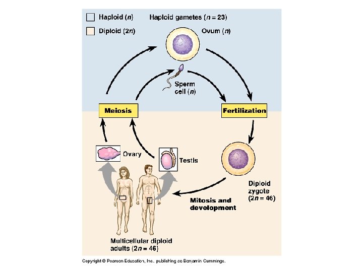
Haploid gametes (n 23) Key Haploid (n) Diploid (2 n) Egg (n) Sperm (n) MEIOSIS Ovary FERTILIZATION Testis Diploid zygote (2 n 46) Mitosis and development Multicellular diploid adults (2 n 46)
Chromosomes • Homologous chromosomes – Are the two chromosomes composing a pair – Have the same characteristics – Autosomes are the non-sex determining chromosomes • Sex chromosomes – Are distinct from each other in their characteristics – Are represented as X and Y in mammals – Determine the sex of the individual, XX being female, XY being male; X and X are homologous in female • A diploid cell – Has two sets of each of its chromosomes – In a human has 46 chromosomes (2 n = 46)
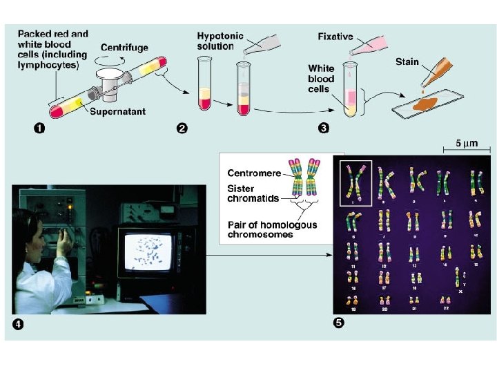
Pair of homologous duplicated chromosomes Centromere Sister chromatids Metaphase chromosome 5 m
Figure 13. x 3 Human female karyotype shown by bright field G-banding of chromosomes
Figure 13. x 5 Human male karyotype shown by bright field G-banding of chromosomes
Hairy Ears – a sex linked trait • Square symbol = male • Round symbol = female • Dark square = hairy ear trait
Chromosome number in humans if there was no meiosis prior to reproduction • First Generation - Sperm and Egg with 46 chromosomes each • Second generation 92 chromosomes • Third generation 184 chromosomes • Fourth generation 368 chromosomes • Etc.
Sexual life cycles Key Haploid (n) Diploid (2 n) n Gametes n Mitosis n MEIOSIS n FERTILIZATION n Diploid multicellular organism (a) Animals Zygote 2 n Mitosis n 2 n Diploid multicellular organism (sporophyte) Mitosis n Spores Gametes MEIOSIS 2 n Haploid unicellular or multicellular organism Haploid multicellular organism (gametophyte) n n Gametes FERTILIZATION (b) Plants and some algae n FERTILIZATION MEIOSIS 2 n Zygote Mitosis 2 n Zygote (c) Most fungi and some protists
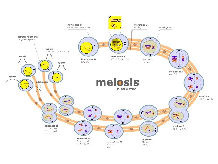
Interphase Pair of homologous chromosomes in diploid parent cell Duplicated pair of homologous chromosomes Sister chromatids Chromosomes duplicate Diploid cell with duplicated chromosomes
Interphase Pair of homologous chromosomes in diploid parent cell Duplicated pair of homologous chromosomes Sister chromatids Chromosomes duplicate Diploid cell with duplicated chromosomes Meiosis I 1 Homologous chromosomes separate Haploid cells with duplicated chromosomes
Interphase Pair of homologous chromosomes in diploid parent cell Duplicated pair of homologous chromosomes Sister chromatids Chromosomes duplicate Diploid cell with duplicated chromosomes Meiosis I 1 Homologous chromosomes separate Haploid cells with duplicated chromosomes Meiosis II 2 Sister chromatids separate Haploid cells with unduplicated chromosomes
MEIOSIS I: Separates sister chromatids MEIOSIS I: Separates homologous chromosomes Prophase I Metaphase I Centrosome (with centriole pair) Sister chromatids Chiasmata Fragments of nuclear envelope Duplicated homologous chromosomes (red and blue) pair and exchange segments; 2 n 6 in this example. Prophase II Metaphase II Anaphase II Telophase II and Cytokinesis Sister chromatids remain attached Centromere (with kinetochore) Spindle Homologous chromosomes Telophase I and Cytokinesis Anaphase I Metaphase plate Homologous chromosomes separate Microtubule attached to kinetochore Chromosomes line up by homologous pairs. Cleavage furrow Each pair of homologous chromosomes separates. During another round of cell division, the sister chromatids finally separate; four haploid daughter cells result, containing unduplicated chromosomes. Sister chromatids separate Two haploid cells form; each chromosome still consists of two sister chromatids. Haploid daughter cells forming
Prophase I Centrosome (with centriole pair) Sister chromatids Chiasmata Spindle Telophase I and Cytokinesis Anaphase I Metaphase I Sister chromatids remain attached Centromere (with kinetochore) Metaphase plate Fragments Homologous chromosomes of nuclear envelope Homologous chromosomes separate Microtubule attached to kinetochore Cleavage furrow Each pair of homologous chromosomes separates. Chromosomes line up Duplicated homologous chromosomes (red and blue) by homologous pairs. pair and exchange segments; 2 n 6 in this example. Two haploid cells form; each chromosome still consists of two sister chromatids.
Prophase II Metaphase II Anaphase II Telophase II and Cytokinesis During another round of cell division, the sister chromatids finally separate; four haploid daughter cells result, containing unduplicated chromosomes. Sister chromatids separate Haploid daughter cells forming
MEIOSIS MITOSIS Parent cell MEIOSIS I Chiasma Prophase I Duplicated chromosome Chromosome duplication 2 n 6 Chromosome duplication Homologous chromosome pair Metaphase I Anaphase Telophase Anaphase I Telophase I Daughter cells of meiosis I 2 n Daughter cells of mitosis 2 n Haploid n 3 MEIOSIS II n n Daughter cells of meiosis II
SUMMARY Property Mitosis Meiosis DNA replication Occurs during interphase before mitosis begins Occurs during interphase before meiosis I begins Number of divisions One, including prophase, metaphase, and telophase Two, each including prophase, metaphase, and telophase Synapsis of homologous chromosomes Does not occur Occurs during prophase I along with crossing over between nonsister chromatids; resulting chiasmata hold pairs together due to sister chromatid cohesion Number of daughter cells and genetic composition Two, each diploid (2 n) and genetically identical to the parent cell Four, each haploid (n), containing half as many chromosomes as the parent cell; genetically different from the parent cell and from each other Role in the animal body Enables multicellular adult to arise from zygote; produces cells for growth, repair, and, in some species, asexual reproduction Produces gametes; reduces number of chromosomes by half and introduces genetic variability among the gametes
Independent Assortment Possibility 2 Possibility 1 Two equally probable arrangements of chromosomes at metaphase I
Independent Assortment Possibility 2 Possibility 1 Two equally probable arrangements of chromosomes at metaphase I Metaphase II
Independent Assortment Possibility 2 Possibility 1 Two equally probable arrangements of chromosomes at metaphase I Metaphase II Daughter cells Combination 1 Combination 2 Combination 3 Combination 4
• Mitosis and meiosis have several key differences. – The chromosome number is reduced by half in meiosis, but not in mitosis. – Mitosis produces daughter cells that are genetically identical to the parent and to each other. – Meiosis produces cells that differ from the parent and each other.
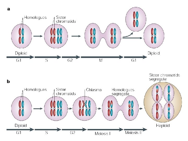
• Three events, unique to meiosis, occur during the first division cycle. 1. During prophase I, homologous chromosomes pair up in a process called synapsis. – A protein zipper, the synaptonemal complex, holds homologous chromosomes together tightly. – Later in prophase I, the joined homologous chromosomes are visible as a tetrad. – At X-shaped regions called chiasmata, sections of nonsister chromatids are exchanged. – Chiasmata is the physical manifestation of crossing over, a form of genetic rearrangement. – Prophase I is longest and most important phase
2. At metaphase I homologous pairs of chromosomes, not individual chromosomes are aligned along the metaphase plate. • In humans, you would see 23 tetrads. 3. At anaphase I, it is homologous chromosomes, not sister chromatids, that separate and are carried to opposite poles of the cell. – Sister chromatids remain attached at the centromere until anaphase II. • The processes during the second meiotic division are virtually identical to those of mitosis.
The synaptonemal complex binding together four homologous chromosomes
Chiasmata and crossing over
Prophase I of meiosis Pair of homologs Nonsister chromatids held together during synapsis
Prophase I of meiosis Pair of homologs Chiasma Centromere TEM Nonsister chromatids held together during synapsis
Prophase I of meiosis Pair of homologs Chiasma Centromere TEM Anaphase I Nonsister chromatids held together during synapsis
Prophase I of meiosis Pair of homologs Chiasma Centromere TEM Anaphase II Nonsister chromatids held together during synapsis
Prophase I of meiosis Pair of homologs Nonsister chromatids held together during synapsis Chiasma Centromere TEM Anaphase II Daughter cells Recombinant chromosomes
Human Female vs Male Meiotic Timelines
Sexual reproduction leads to genetic variation via: • Independent assortment during meiosis • Crossing over during meiosis • Random mixing of gametes (sperm and egg)