MECHANISMS OF CELL INJURY Manar Hajeer MD FRCPath


 � Chemical species with")

 during inflammation. � In phagosomes")
. � Glutathione (GSH)")


")


- Slides: 28
MECHANISMS OF CELL INJURY Manar Hajeer, MD, FRCPath.
MECHANISMS OF CELL INJURY � � Principles The cellular response to injury depends on: duration severity The consequences of injury depend on: type, status, adaptability, and genetic makeup of the injured cell Cell injury results from functional and biochemical abnormalities in one or more of several essential cellular components
The principal biochemical mechanisms and sites of damage in cell injury
Hypoxia and Ischemia � Defective oxidative phosphorylation >>Failure of ATP generation>>>depletion of ATP in cells � Failure of energy dependent pathways (membrane transport, protein synthesis, lipogenesis and phospholipid turnover) � Anaerobic glycolysis. � Liver cells Vs brain.
Hypoxia effects: � Reduced activity of membrane ATP dependent sodium pumps>> cell swelling � Lactic acid accumulation >> decreased PH>> failure of enzymes. � Disruption of the ribosomes>> decreased protein synthesis. � Accumulation of ROS � Damage to mitochondrial and lysosomal membranes. � Necrosis is the end result.
Ischemia-Reperfusion Injury � Paradoxical cell injury after restoration of blood flow to ischemic but viable tissues. � After myocardial and cerebral ischemia. � Increased generation of ROS from: � Injured cells with damaged mitochondria & defective antioxidant mechanisms. � Infiltrating new leukocytes. � Inflammation induced by influx of leukocytes, plasma proteins and complement
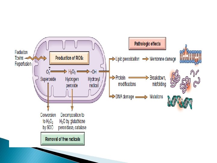
Oxidative Stress � Cellular abnormalities induced by ROS (free radicals) � Chemical species with single unpaired electron (extremely unstable) � ROS generated in: � Chemical injury (CCL 4) � Radiation injury (UV, Xray) � Hypoxia � Cellular aging � Inflammation � Ischemia-reperfusion injury.
Generation and Removal of Reactive Oxygen Species � 1 -Normally produced in small amounts in all cells during the redox reactions. � Oxygen is reduced to produce water. � Small amounts of highly reactive but short-lived toxic intermediates are generated. � Superoxide (O 2 • ), hydrogen peroxide (H 2 O 2), hydroxyl radical • OH.
� 2 -Produced in phagocytic leukocytes (neutrophils and macrophages) during inflammation. � In phagosomes and phagolysosomes. � O 2 >> superoxide >> H 2 O 2 >> hypochlorite. � Myeloperoxidase.
Removal of free radicals � Decay spontaneously � Superoxide dismutase (SOD). � Glutathione (GSH) peroxidases. � Catalase (one of most active enzymes known) � Endogenous or exogenous anti-oxidants (e. g. , vitamins E, A, and C and β-carotene)
Effects or ROS: � 1 -Lipid peroxidation of membranes. � (plasma, lysosomal & mitochondrial membranes) � 2 -Crosslinking and other changes in proteins. � ( degradation, fragmentation, loss of enzymatic activity & misfolding). � 3 -DNA damage. � Single strand breaks � 4 -Killing of microbes,
Cell Injury Caused by Toxins � Environmental chemicals |& substances produced by infectious pathogens. � Direct-acting toxins � Latent toxins.
Direct-acting toxins � Act directly by combining with a critical molecular component or cellular organelle. � Mercuric chloride poisoning � Contaminated seafood � Mercury binds to sulfhydryl groups of membrane proteins>>inhibit ATP-dependent transport and increase permeability. � Chemotherapeutic agents
Latent toxins � Not intrinsically active � Must be converted to reactive metabolites, then act on target cells. � Via cytochrome P-450 in SER of the liver. � Damage by formation of free radicals>>membrane phospholipid peroxidation. � CCl 4 and acetaminophen. � ER membranes >> decline in synthesis of enzymes and proteins +decreased synthesis of apoproteins >> fatty liver � Mitochondrial injury>> decreased ATP >> cell swelling >> cell death.
CCL 4 toxicity
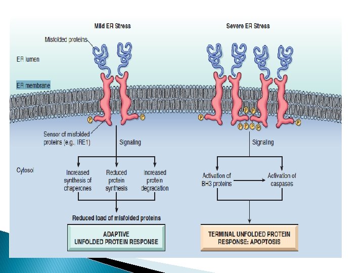
Endoplasmic Reticulum Stress � Chaperones � Misfolded in ER control proper protein folding proteins >> ubiquinated >> targeted to proteolysis � Unfolded protein response (adaptive response): increase chaperones production and decrease protein translation � If failed >> proapoptotic sensor activation (BH 3 -only family) + direct activation of caspases >>apoptosis by the mitochondrial pathway.
Causes of misfolding � Gene mutations � Aging � Infections, especially viral infections � Increased demand for secretory proteins such as insulin in insulin-resistant states � Changes in intracellular p. H in ischemia and hypoxia � Neurodegenerative diseases
Protein misfolding causes disease by: � Deficiency of an essential protein due to degradation � Cystic fibrosis � Inducing apoptosis of the affected cells � Neurodegenerative disorders (Alzheimer disease, Huntington disease & Parkinson disease) and type 2 diabetes � Improperly tissues � Amyloidosis folded proteins accumulation in extracellular
DNA Damage � Radiation � Chemotherapeutic agents � Intracellular generation of ROS � Mutations � DNA damage >> p 53 activation >>arrest cell cycle at G 1 phase for repair >> if repair is impossible >> apoptosis. � In P 53 mutations >> mutated cells replicate >> neoplastic change.
Inflammation � Pathogens � Necrotic cells, � Dysregulated immune responses (autoimmune diseases and allergies) � Inflammatory cells (neutrophils, macrophages, lymphocytes) secrete products that destroy microbes and damage host tissues.
Common Events in Cell Injury From Diverse Causes � Mitochondrial Dysfunction � Defects in Membrane Permeability
Mitochondrial Dysfunction � Energy factory � Hypoxia, toxins, radiation. � In necrosis and apoptosis. � Consequences: � Failure of oxidative phosphorylation, ATP depletion. � Abnormal oxidative phosphorylation, formation of ROS � Mitochondrial permeability transition pores, loss of membrane potential. � Release of cytochrome c >> apoptosis
Mitochondrial Damage and Dysfunction
Depletion of ATP
Influx of Calcium
Defects in Membrane Permeability � Mitochondrial membrane damage: decreased ATP � Plasma membrane damage: loss of osmotic balance, influx of fluids, leak of contents � Lysosomal digestion. membranes: leakage of enzymes >> cellular