Autophagy and intracellular accumulations Manar Hajeer MD FRCPath





- Slides: 20
Autophagy and intracellular accumulations Manar Hajeer, MD, FRCPath.
AUTOPHAGY auto, self; phagy, eating
Roles of autophagy Ø A survival mechanism in times of nutrient deprivation Ø Involved in the clearance of misfolded proteins (in neurons and hepatocytes) Ø With inflammatory bowel disease(? !) Ø Role in cancer
INTRACELLULAR ACCUMULATIONS Ø 1)Inadequate removal of a normal substance (fatty change in the liver) Ø 2)Accumulation of an abnormal endogenous substance (α 1 -antitrypsin) Ø 3)Failure to degrade a metabolite due to inherited enzyme deficiencies (storage diseases) Ø 4)Deposition and accumulation of an abnormal exogenous substance (carbon and selica)
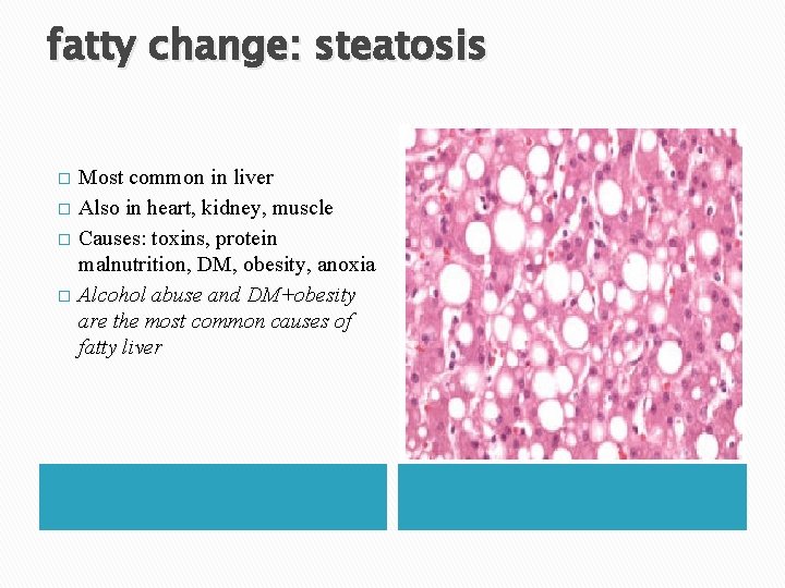
fatty change: steatosis � � Most common in liver Also in heart, kidney, muscle Causes: toxins, protein malnutrition, DM, obesity, anoxia Alcohol abuse and DM+obesity are the most common causes of fatty liver
Cholesterol and Cholesteryl Esters � Phagocytic cells become overloaded with lipid (triglycerides, cholesterol, and cholesteryl esters) � atherosclerosis is the most important example.
Proteins � � � Much less common than lipid accumulations Either excess external or internal synthesis Proximal renal tubules in nephrotic syndrome Russell bodies in plasma cells. Alcoholic hyaline in liver. Neurofibrillary tangles in neurons
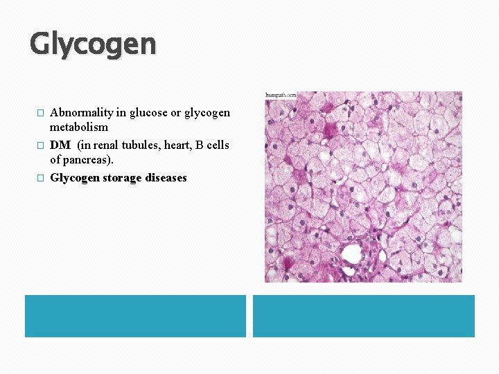
Glycogen � � � Abnormality in glucose or glycogen metabolism DM (in renal tubules, heart, B cells of pancreas). Glycogen storage diseases
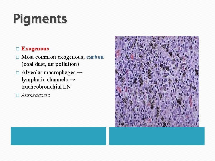
Pigments � � Exogenous Most common exogenous, carbon (coal dust, air pollution) Alveolar macrophages → lymphatic channels → tracheobronchial LN Anthracosis
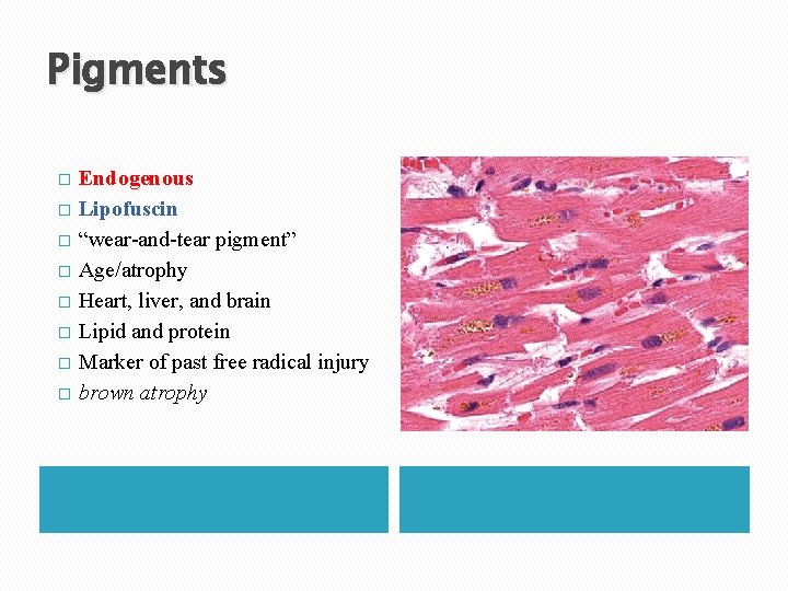
Pigments � � � � Endogenous Lipofuscin “wear-and-tear pigment” Age/atrophy Heart, liver, and brain Lipid and protein Marker of past free radical injury brown atrophy
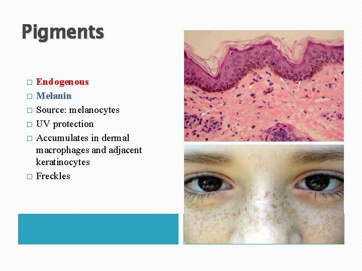
Pigments � � � Endogenous Melanin Source: melanocytes UV protection Accumulates in dermal macrophages and adjacent keratinocytes Freckles
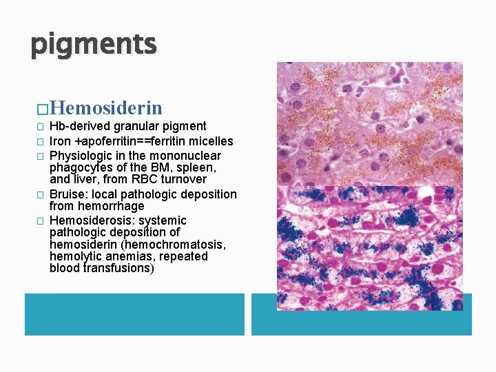
pigments �Hemosiderin � � � Hb-derived granular pigment Iron +apoferritin==ferritin micelles Physiologic in the mononuclear phagocytes of the BM, spleen, and liver, from RBC turnover Bruise: local pathologic deposition from hemorrhage Hemosiderosis: systemic pathologic deposition of hemosiderin (hemochromatosis, hemolytic anemias, repeated blood transfusions)
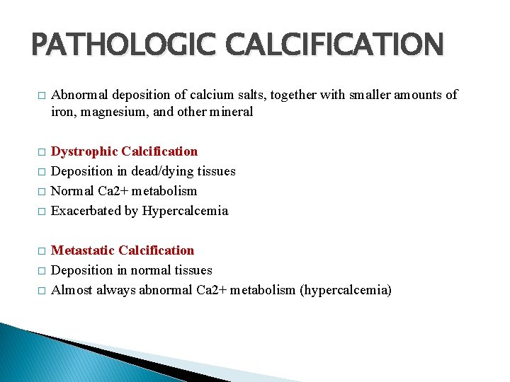
PATHOLOGIC CALCIFICATION � Abnormal deposition of calcium salts, together with smaller amounts of iron, magnesium, and other mineral � Dystrophic Calcification Deposition in dead/dying tissues Normal Ca 2+ metabolism Exacerbated by Hypercalcemia � � � Metastatic Calcification Deposition in normal tissues Almost always abnormal Ca 2+ metabolism (hypercalcemia)
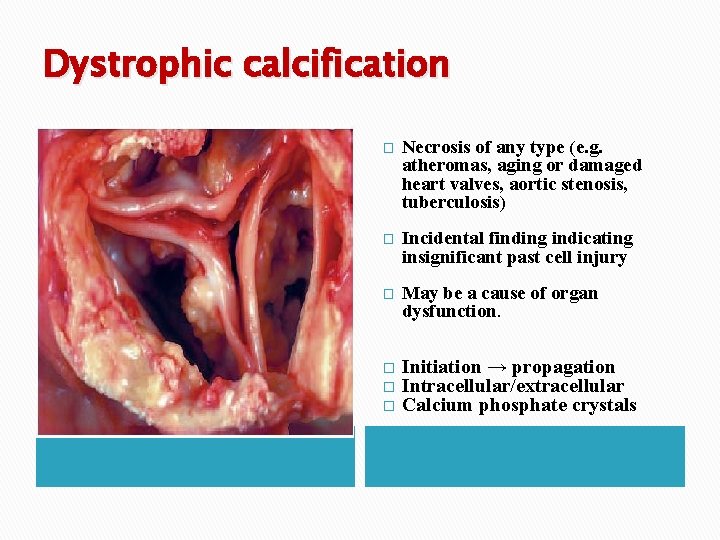
Dystrophic calcification � Necrosis of any type (e. g. atheromas, aging or damaged heart valves, aortic stenosis, tuberculosis) � Incidental finding indicating insignificant past cell injury � May be a cause of organ dysfunction. � � � Initiation → propagation Intracellular/extracellular Calcium phosphate crystals
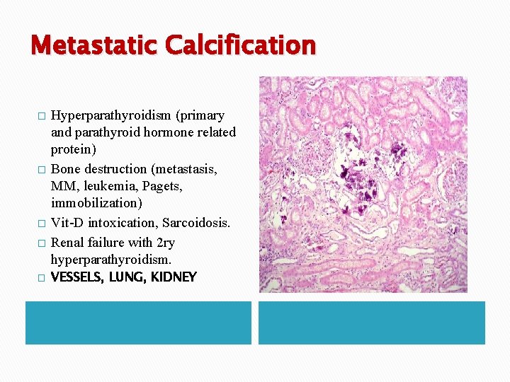
Metastatic Calcification � � � Hyperparathyroidism (primary and parathyroid hormone related protein) Bone destruction (metastasis, MM, leukemia, Pagets, immobilization) Vit-D intoxication, Sarcoidosis. Renal failure with 2 ry hyperparathyroidism. VESSELS, LUNG, KIDNEY
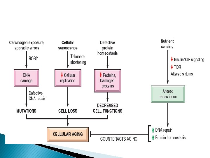
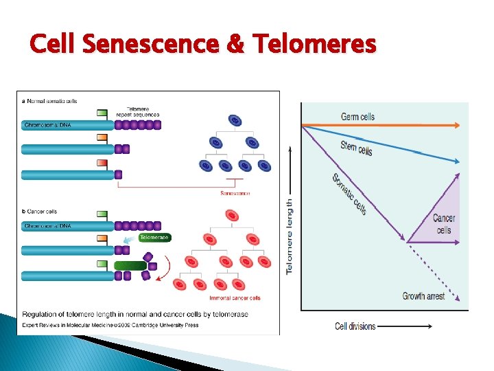
CELLULAR AGING � Age is one of the strongest independent risk factors for many chronic diseases, such as cancer, Alzheimer disease, and ischemic heart disease � Cellular aging is the result of a progressive decline in the life span and functional capacity of cells. � Several mechanisms (cumulative DNA damage, decreased cellular replication capacity, Defective protein homeostasis
Cell Senescence & Telomeres
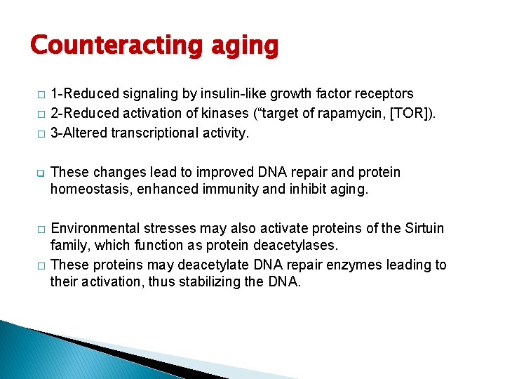
Counteracting aging � � � 1 -Reduced signaling by insulin-like growth factor receptors 2 -Reduced activation of kinases (“target of rapamycin, [TOR]). 3 -Altered transcriptional activity. q These changes lead to improved DNA repair and protein homeostasis, enhanced immunity and inhibit aging. � Environmental stresses may also activate proteins of the Sirtuin family, which function as protein deacetylases. These proteins may deacetylate DNA repair enzymes leading to their activation, thus stabilizing the DNA. �