Cellular responses to stress Adaptations injury and death
 (1 of 5) Ali Al Khader,")

…always reversible")


 -- exercise physiologic")


 ﺑﺎﻟﺘﺎﻟﻲ nutrient/supply ﻣﺎ ﻓﻲ ﻋﻨﺎ ﺑﺪﻧﺎ ﻧﻌﻤﻞ")


- Slides: 24
Cellular responses to stress (Adaptations, injury and death) (1 of 5) Ali Al Khader, M. D. Faculty of Medicine Al-Balqa’ Applied University
Before we talk about cellular adaptations: • Homeostasis: The steady state of cell structure and function (narrow range of changes in cell function according to external and internal environments) • In homeostasis: -Supplies: Available -Injurious (noxious) agents: Absent • Stress = demands (physiologic or pathologic), attack by noxious (injurious) factors, or abnormal DNA
Before we talk about cellular adaptations: Stress *Homeostasis Adaptations (new state of homeostasis)…always reversible *If the stress is more severe These are the cellular responses to stress (in general) Cell injury Reversible injury More severe Irreversible injury Cell death Apoptosis Necrosis
Cellular adaptations 1 - Hypertrophy 2 - Hyperplasia 3 - Atrophy 4 - Metaplasia troph (growth )ﻧﻤﻮ increase in the size not in the number.
Hypertrophy cell size, no change in number of cells ﺑﻴﺰﺩﺍﺩ ﺍﻟﺤﺠﻢ ﻭ ﻟﻴﺲ ﺍﻟﻌﺪﺩ ﻋﻠﻰ ﻣﺴﺘﻮﻯ ﺍﻟﺨﻠﻴﺔ ﺑﺴﺒﺐ ﺯﻳﺎﺩﺓ ﻋﺪﺩ ﻭ ﺣﺠﻢ ﺍﻟﻌﻀﻴﺎﺕ ﺩﺍﺧﻞ ﺍﻟﺨﻠﻴﺔ ﺍﻭﺯﻳﺎﺩﺓ ﺍﻟﺒﺮﻭﺗﻴﻨﺎﺕ ﺍﻧﺘﺎﺟﺎ ﻭ ﻧﻮﻋﻴﺔ the structural component. ﺍﻣﺎ ﺍﻟﻌﻀﻮ ﺑﺴﺒﺐ ﺯﻳﺎﺩﺓ ﺣﺠﻢ ﻭ ﻋﺪﺩ ﺍﻟﺨﻼﻳﺎ *Mechanism: (size + number) of organelles *May be physiologic or pathologic *Pathologic: The cause is abnormal *Physiologic: No abnormal cause **”Hypertrophy” of any thing = in its size *Cellular hypertrophy : increase in the size of the cell not in the number. *Organ hypertrophy : increase in the size of the organ by increasing in the size of the cell or in the number of the cell in the organ. The adaptation is either PHYSIOLOGICAL ���� OR PATHOLOGICAL ����
Physiologic hypertrophy, an example: • Skeletal muscle hypertrophy in weight lifters (because of mechanical stretch) • Increased workload (physiologic stress) • Skeletal muscle cells respond to this by Myocyte ��� Skeletal muscle �� ����� �� Skeletal muscle fiber increasing their size not number because they ���� Regeneration �� ��� have limited capacity to divide ����� ������� �� �����
• What is this organ? uterus • What did happen to it? Physiological hypertrophy • What is the difference between the lower two images? Left pic is smooth muscle in normal condition, right pic smooth muscle in pregnancy • What is the name of this condition? • Did the condition in this example occur alone or with another condition? • Is this condition physiologic or pathologic? Physiologic • What is the cause? Hormonal effect (estrogen and progesterone)
Smooth muscle cells are able to regenerate. hypertrophy and hyperplasia ������ smooth muscle cell in pregnancy�� ��� ����� *. Glandular epithelial cells in the breast in pregnancy and puberty��� �� ��� hypertrophy and hyperplasia �������
Pathologic hypertrophy, an example: *Cardiac myocytes in hypertension *Increased workload, but the cause here is pathological (not a physiologic stress) *Limited capacity to divide, so it respond by hypertrophy. *in systemic hypertension the cardiac muscle pump the blood against resistance *pathological hypertrophy ﻷﻨﻪ ﺍﻟﻤﺴﺒﺐ ﻣﺮﺿﻲ . ﻭﺍﻟﻲ ﻫﻮﻩ ﻣﺮﺽ ﺍﺭﺗﻔﺎﻉ ﺍﻟﻀﻐﻂ ﺍﻟﺸﺮﻳﺎﻧﻲ
Some mechanisms in hypertrophy • Mechanical triggers (e. g. , stretch) -- exercise physiologic - heart pathologic. Major physiologic hypertrophy. • Trophic triggers has larger rule in pathologic hypertrophy (e. g. , growth factors-hormones) -- in myometrium in the uterus. • Increased proteins, e. g. , contractile proteins (myofilaments: actin and myosin) -- in the skeletal and cardiac muscle. • Switch in protein types synthesized: - Example: Alpha form of myosin heavy chain is replaced by beta form…slower and more energetically economical contractions - ﻧﻮﻗﻒ ﺻﻨﺎﻋﺔ ﻧﻮﻉ ﻣﻦ ﺍﻟﺒﺮﻭﺗﻴﻨﺎﺕ ﻭ ﻧﺼﻨﻊ Expression for protein �� ���� Gene ﺑﺮﻭﺗﻴﻨﺎﺕ ﻣﺎ ﻛﺎﻧﺖ ﺗﺘﺼﻨﻊ ﻳﻌﻨﻲ ﻧﺘﺤﻜﻢ ﻋﻠﻰ ﺍﻝ
Hyperplasia: number = in number of cells that are capable to divide *May be physiologic or pathologic *May occur concurrently with hypertrophy…in this case the stimulus is usually the same • myometrium ﺑﻨﻔﺲ ﺍﻟﻮﻗﺖ ﻣﺜﻞ ﻓﻲ ﺍﻝ Hypertrophy ﺗﺤﺪﺙ ﻣﻊ ﺍﻝ Hyperplasia ﻣﻤﻜﻦ ﺍﻝ ﻭ ﻓﻲ ﻫﺬﻩ ﺍﻟﺤﺎﻟﺔ ﺑﻜﻮﻥ ﺍﻟﻤﺴﺒﺐ ﻭﺍﺣﺪ puberty & pregnancy ﻓﻲ ﺣﺎﻟﺘﻲ ﺍﻝ breast ﻭ ﻓﻲ ﺍﻝ )ﻧﻔﺴﻪ( ﺑﺎﻟﻌﺎﺩﺓ hormonal.
Physiologic hyperplasia, examples: *Glandular epithelium of the female breast at: -Puberty -Pregnancy Hormonal stimulation �� ����� �� physiologic process. �� ����� ��� ������� *Compensatory hyperplasia of the liver: After resection of a part of the liver, the remaining cells start to divide as early as 12 hours after …triggered by growth factors from: -Uninjured hepatocytes ﺍﻟﺨﻼﻳﺎ ﻧﻔﺴﻬﺎ ﺗﺆﺜﺮ ﻋﻠﻰ ﺑﻌﻀﻬﺎ ﺍﻭ ﻣﻦ ﺧﻼﻝ -Non-parenchymal (mesenchymal/stromal) cells …eventually: normal weight is restored and the hyperplasia is turned off by growth inhibitors
Pathologic hyperplasia abnormal condition • Mostly due to in hormones or growth factors, but the increase here is abnormal and not physiologic • Examples: *A disease called “Endometrial hyperplasia”: -It causes abnormal menstrual bleeding (menorrhagia) -Due to imbalance between estrogen and progesterone *Blood vessels and fibroblasts in wound healing: -Growth factors from leukocytes (WBCs) and connective tissue cells �� ������ prefoliation �� ���� inflammation
Endometrial hyperplasia >>. ﺍﻻﺳﺘﺮﻭﺟﻴﻦ ﻭ ﺍﻟﺒﺮﻭﺟﺴﺘﺮﻭﻥ ﺍﻻﺗﺰﺍﻥ ﺑﻴﻦ ﺑﺴﺒﺐ ﻋﺪﻡ uterus ﻣﺮﺽ ﻳﺤﺪﺙ ﻓﻲ ﺍﻝ • Menstrual cycle has 2 phases : 1 - proliferative phase > Estrogen ﻟﺤﺘﻰ ﺗﻔﺮﺯ ﺍﻻﺳﺘﺮﻭﺟﻴﻦ ovary ﻭ ﺑﺘﺄﺜﺮ ﻋﻠﻰ ﺍﻝ pituitary ﻓﻲ ﻫﺬﻩ ﺍﻟﻤﺮﺣﻠﺔ ﺑﺘﻄﻠﻊ ﻫﺮﻣﻮﻧﺎﺕ ﻣﻦ ﺍﻝ 2 - ovulation 3 - secretory phase > Progesterone In normal condition Estrogen do hyperplasia in the endometrial cell , *Progesterone control Estrogen effect. In case of imbalance between (estrogen and progesterone) then we will have Endometrial hyperplasia.
Pathologic hyperplasia, another example ﺑﺸﻜﻞ ﻏﻴﺮ ﻃﺒﻴﻌﻲ GF & HORMONEs ﺳﺒﺒﻪ ﺍﻓﺮﺍﺯ • Skin warts: ﺍﻟﺜآﻠﻴﻞ -Etiology: Human papilloma virus (HPV) -Mechanism: Epithelial hyperplasia due to growth factors …the GFs are encoded either by viral genes or host cell genes induced by the virus ﺍﻣﺎ ﺗﻔﺮﺯ ﻣﺒﺎﺷﺮﺓ ﻣﻦ ﺍﻟﻔﺎﻳﺮﻭﺱ ﺍﻭ ﻳﺘﺤﻔﺰ ﺍﻧﺘﺎﺟﻬﺎ GF ﺍﻝ . Host cell by the virus ﻣﻦ ﺍﻝ ﻳﻌﻤﻞ Host cell ﻟﺠﻴﻦ ﻓﻲ ﺍﻝ activation ﻣﻦ ﺧﻼﻝ ﻋﻤﻞ Patho hyperplasia ﺣﺘﻰ ﺗﺴﺒﺐ GF ﺍﻓﺮﺍﺯ ﻝ Hyperplasia in the Epidermis ﺑﺼﻴﺮ ﻋﻨﺎ
ﺍﺫﺍ ﺗﻮﻗﻒ ﺍﻓﺮﺍﺯ ﺍﻻﺳﺘﺮﻭﺟﻴﻦ ﺑﺮﺟﻊ ﺍﻟﻮﺿﻊ ﻃﺒﻴﻌﻲ *Pathologic hyperplasia may be a precancerous condition, and the best example of this risk is: Endometrial hyperplasia *In sometimes pathologic hyperplasia consider as a fertile environment for Cancer of uterus (Endometrial adenocarcinoma) * ﻣﻦ ﺍﻫﻢ ﺍﻋﺮﺍﺽ ﺍﻝ Endometrial hyperplasia >> Menorrhagia ( heavy bleeding )
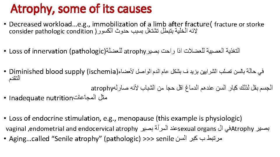
Atrophy = Shrinkage ( in cell size) ﺑﺎﻟﺘﺎﻟﻲ nutrient/supply ﻣﺎ ﻓﻲ ﻋﻨﺎ ﺑﺪﻧﺎ ﻧﻌﻤﻞ ﺗﻘﺸﻒ ﻭ ﻧﻘﻠﻞ ﺻﻨﺎﻋﺔ ﺍﻟﺒﺮﻭﺗﻴﻨﺎﺕ metabolism ﻭ ﻧﻘﻠﻞ ﺍﻝ ﻭ ﻧﺰﻳﺪ ﺗﺤﻄﻴﻢ ﺑﺮﻭﺗﻴﻨﺎﺕ ﻣﻌﻴﻨﺔ ﻋﺸﺎﻥ ﻧﻘﻠﻞ . ﺍﻟﻤﺼﺎﻧﻊ ﺍﻟﻲ ﻋﻨﺎ *Atrophy of any thing = in its size *organ trophy ﻋﻠﻰ ﻣﺴﺘﻮﻯ ﺍﻟﻌﻀﻮ *Mechanisms: - metabolic activity - synthesis (due to decreased metabolism) and degradation of proteins, so the substance of the cell is generally decreased
Atrophy, more about its mechanisms ﻧﻮﻗﻒ ﺍﻟﺰﻳﺎﺩﺓ ﻭ ﻧﺰﻳﺪ ﺍﻟﺘﺤﻄﻴﻢ • Degradation of proteins is mainly by: ubiquitin-proteasome pathway -Ubiquitin is a small peptide -Ubiquitin binds to the cellular protein and targets it for degradation in proteasomes -Ubiquitin ligase(enzyme) attaches ubiquitin to the protein that will be degraded **This pathway is also responsible for the accelerated proteolysis seen in cancer cachexia ( ﺍﻟﻌﻀﻼﺕ ﻭ ﺍﻟﺒﺮﻭﺗﻴﻦ ﻓﻲ ﺍﻟﺠﺴﻢ ﺗﺤﻄﻤﺖ. . ﺍﻟﺯﺍﻝ ) ﺑﺤﻴﺚ ﺑﺼﻴﺮ ﻣﺮﻳﺾ ﺍﻟﺴﺮﻃﺎﻥ ﺟﻠﺪ ﻭ ﻋﻈﻢ • Autophagy: ﺍﻟﺒﻠﻌﻤﺔ ﺍﻟﺬﺍﺗﻴﺔ - = Self-eating - Common in atrophy - Cell components are isolated in autophagic vacuoles
Metaplasia ﺑﺘﺤﻮﻝ ﻧﻮﻉ ﺍﻟﺨﻠﻴﺔ ﻝ ﻧﻮﻉ ﺍﺧﺮ • Definition: One adult cell type is replaced by another adult cell type in response to stress • Metaplasia occurs in epithelial or mesenchymal cell types…mesenchymal type is generally pathological • Epithelial metaplasia can be pathological or physiological ; ex >> from squamous to columnar • Mesenchymal metaplasia is pathological ; ex >> ﻓﻲ ﺍﻟﻌﻀﻠﺔ ﺍﻟﻲ bone ﺑﺘﻜﻮﻥ ﺷﻮﻳﺔ ( ﺗﻌﺮﺿﺖ ﻟﺠﺮﺡ ) ﺑﻤﻌﻨﻰ ﺍﺧﺮ ﺑﺼﻴﺮ ﺗﻜﺱ ﺍﻭ ﺗﻌ ﻡ ﻓﻲ ﺍﻟﻌﻀﻠﺔ • The new type is better in withstanding the new environment • The stem cells are reprogrammed…not trans-differentiation of already differentiated cells
• What is the cause of this condition? Smoking • Is it epithelial or mesenchymal metaplasia? epithelial • Mention 2 disadvantages of the condition in this example > 1 - we lose Cilia. 2 - Cancerogens. In normal condition the type of cells in trachea are ( pseudostratified ciliated columnar epithelium ) which transfer to ( squamous ) to bear the new chemical environment. normal abnormal
3 more examples of metaplasia • Columnar-type metaplasia in the lower esophagus due to gastroesophageal reflux disease (GERD)… What is the name of this condition? • Squamous metaplasia of respiratory epithelium in vitamin A deficiency …Vitamin A has a role in cell differentiation • Bone formation in foci of injury (a mesenchymal metaplasia)
Done by : RAGHDA AL-DROUBI MARIAM ABABNEH