
Inflammation1 Manar Hajeer MD FRCPath Definition Protective response
- Slides: 35
Inflammation-1 Manar Hajeer, MD, FRCPath
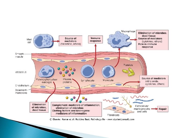
Definition � Protective response involving host cells, blood vessels, and proteins and other mediators that is intended to eliminate the initial cause of cell injury, as well as the necrotic cells and tissues resulting from the original insult, and to initiate the process of repair. � Without inflammation, infections would go unchecked and wounds would never heal. � However, can cause considerable harm (if strong, prolonged or inappropriate)
Components of inflammation � Leukocytes � Plasma proteins and mediators � Blood vessels � ECM
� Inflammation � The is normally controlled and self-limited. outcomes of acute inflammation � Elimination of the noxious stimulus. � Decline of the reaction and repair of the damaged tissue, � Persistent injury resulting in chronic inflammation.
Acute inflammation
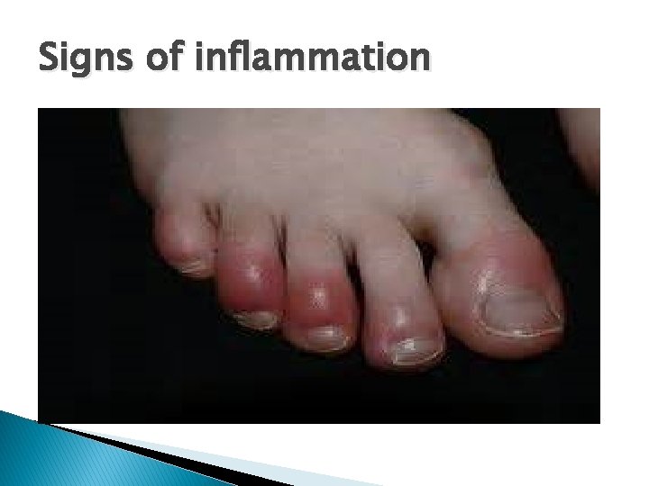
Signs of inflammation
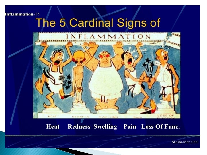
Hotness versus fever � Hotness : localized, due to vasodilation. � Fever : systemic, caused by mediators mainly prostaglandins. � Hotness is a cardinal sign of inflammation, fever is not!
Infection versus inflammation � Infection cause inflammation. � Infection is caused by a pathogen. � Inflammation is the protective response that is caused by infections among other causes.
Stimuli for Acute Inflammation � Infections � Trauma � Physical and chemical agents � Tissue necrosis � Foreign bodies � Immune reactions
Inflammation. . ITIS � Tonsillitis � Apendicitis � Pancreatitis � Liver…? � Lung. . ?
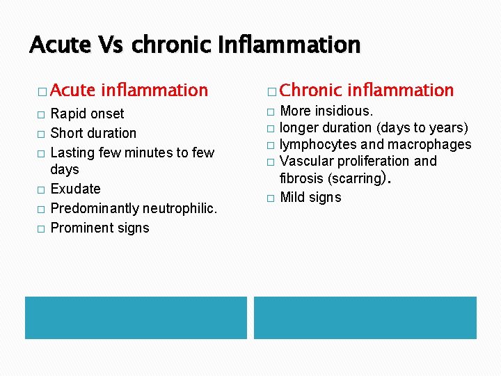
Acute Vs chronic Inflammation � Acute � � � inflammation Rapid onset Short duration Lasting few minutes to few days Exudate Predominantly neutrophilic. Prominent signs � Chronic � � � inflammation More insidious. longer duration (days to years) lymphocytes and macrophages Vascular proliferation and fibrosis (scarring). Mild signs
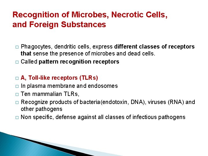
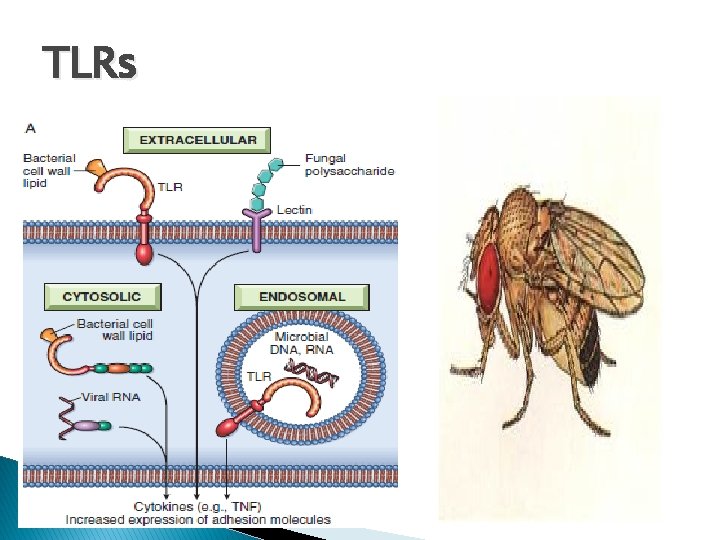
Recognition of Microbes, Necrotic Cells, and Foreign Substances � � � � Phagocytes, dendritic cells, express different classes of receptors that sense the presence of microbes and dead cells. Called pattern recognition receptors A, Toll-like receptors (TLRs) In plasma membrane and endosomes Ten mammalian TLRs, Recognize products of bacteria(endotoxin, DNA), viruses (RNA) and other pathogens Non specific, defense against all classes of infectious pathogens
TLRs
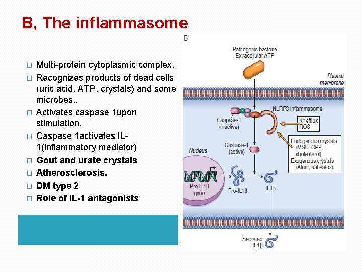
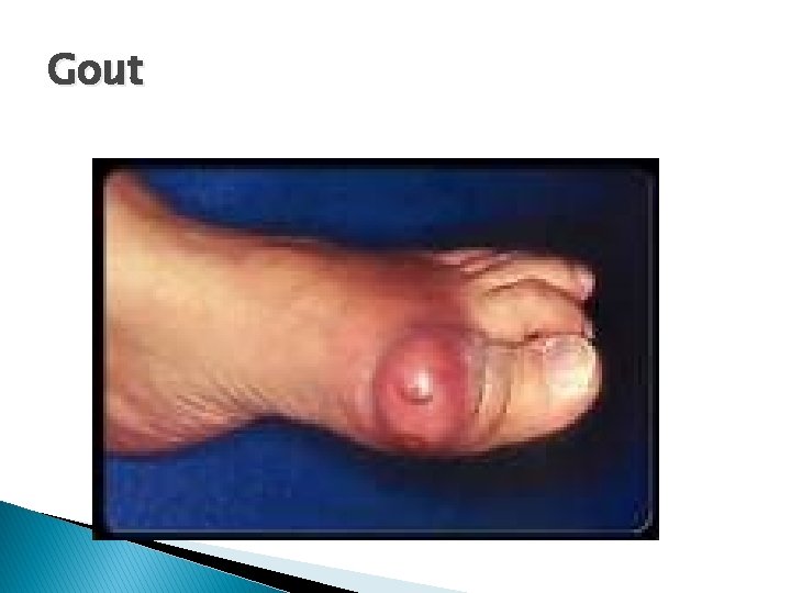
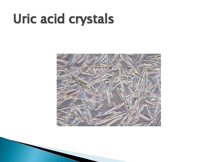
B, The inflammasome � � � � Multi-protein cytoplasmic complex. Recognizes products of dead cells (uric acid, ATP, crystals) and some microbes. . Activates caspase 1 upon stimulation. Caspase 1 activates IL 1(inflammatory mediator) Gout and urate crystals Atherosclerosis. DM type 2 Role of IL-1 antagonists
Gout
Uric acid crystals
Explain what happens in gout, atherosclerosis and DM type 2? ?
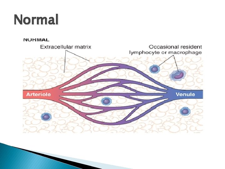
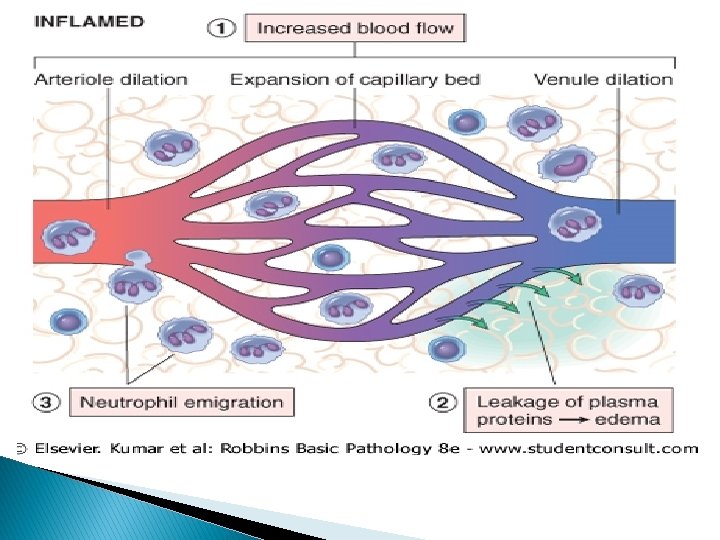
Components of acute inflammation � Vascular changes: Alterations in vessel caliber resulting in increased blood flow (vasodilatation) and structural changes that permit plasma proteins to leave the circulation (increased vascular permeability). � Cellular events: Emigration of the leukocytes from the microcirculation and accumulation in the focus of injury (cellular recruitment and activation). The principal leukocytes in acute inflammation are neutrophils.
Normal
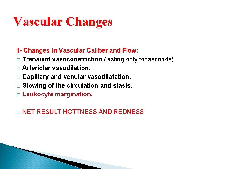
Vascular Changes 1 - Changes in Vascular Caliber and Flow: � Transient vasoconstriction (lasting only for seconds) � Arteriolar vasodilation. � Capillary and venular vasodilatation. � Slowing of the circulation and stasis. � Leukocyte margination. � NET RESULT HOTTNESS AND REDNESS.
Transient vasoconstriction � � Affects arterioles Due to neural reflex 3 -5 seconds in mild injuries. Several minutes in more severe injury (like burns).
Vasodilatation u u u First in arterioles Then capillaries and venules. Increasing blood flow. Hotness and reddness. Increased local hydrostatic pressure Transudation of protein poor fluid into the extravascular space
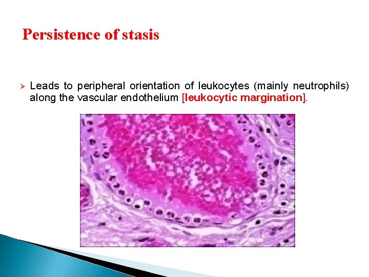
Persistence of stasis Ø Leads to peripheral orientation of leukocytes (mainly neutrophils) along the vascular endothelium [leukocytic margination].
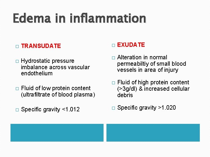
Vascular changes 2 - Increased Vascular Permeability: � increasing vascular permeability that allows the movement of protein -rich fluid and even cells (exudate)=====EDEMA. � Components of exudate ◦ Fluid ◦ Fibrin ◦ Cells: neutrophils predominate (6 -72 hours), also macrophages (involved slightly later: 48+ hours)
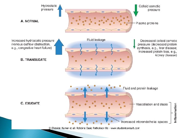
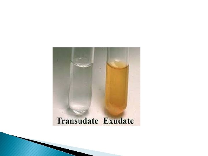
Edema in inflammation � TRANSUDATE � EXUDATE � Hydrostatic pressure imbalance across vascular endothelium � Alteration in normal permeabiltiy of small blood vessels in area of injury � Fluid of high protein content (>3 g/dl) & increased cellular debris � Specific gravity >1. 020 � Fluid of low protein content (ultrafiltrate of blood plasma) � Specific gravity <1. 012
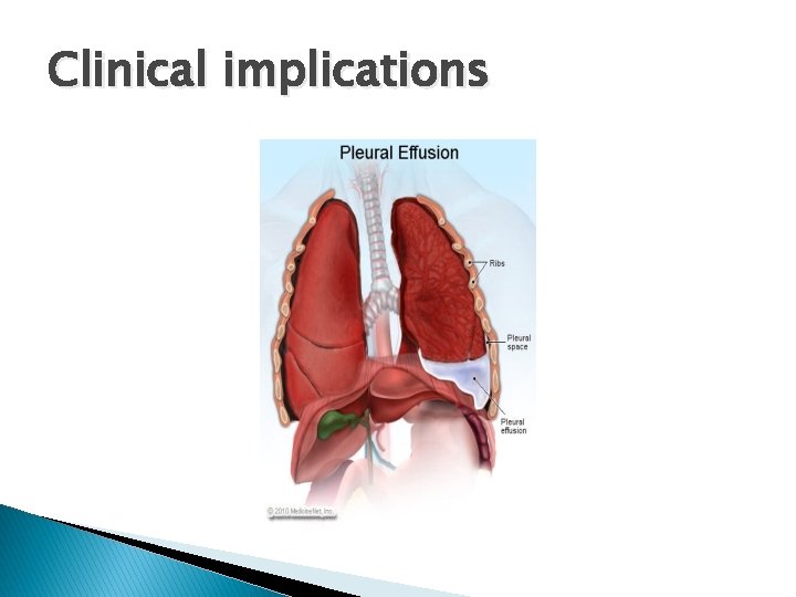
Clinical implications
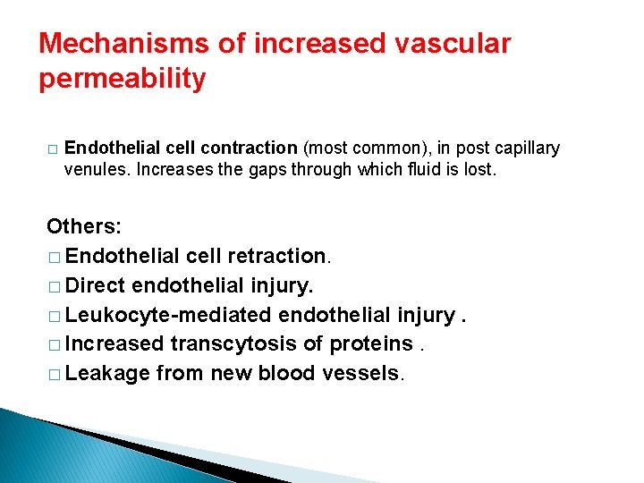
Mechanisms of increased vascular permeability � Endothelial cell contraction (most common), in post capillary venules. Increases the gaps through which fluid is lost. Others: � Endothelial cell retraction. � Direct endothelial injury. � Leukocyte-mediated endothelial injury. � Increased transcytosis of proteins. � Leakage from new blood vessels.
� Endothelial cell contraction ◦ Reversible ◦ Immediate transient response but short lived (15 -30 minutes) ◦ Induced by: histamine, bradykinin, leukotriens, neuropeptide substance P ◦ Mostly in postcapillary venules � Endothelial cell retraction ◦ Reversible ◦ Starts 4 -6 hours after injury and stays for 24 hours ◦ Induced by: IL-1 and TNF, IFN-g 33
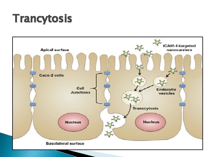
� Direct endothelial injury ◦ Severe injury. E: g burns or infections ◦ Immediate sustained response ◦ All microvessels can be affected � Delayed prolonged response: ◦ Begins after delay (2 -12 hours), lasts for hours or days ◦ Caused by mild thermal injury, UV radiation, bacterial toxins. � Increased transcytosis through vesiculovacuolar pathway ◦ Stimulated by VEGF Leakage from newly formed blood vessels � 34
Trancytosis