Cell injury Dr Heyam Awad FRCPath Causes of


 •")

 • (1) plasma membrane changes such as blebbing,")


- Slides: 20
Cell injury Dr Heyam Awad FRCPath
Causes of cell injury/ 1 • • Chemica agents Infections. Immunologic Genetic factors Nutritional imbalances Physical agents Aging.
Causes of cell injury/ 2 Oxygen deprivation. . Hypoxia and ischemia. Hypoxia= oxygen deficiency Ischemia = loss of blood supply due to impaired arterial flow or reduced venous return. -Ischemia is the most common cause of hypoxia. -Other causes of hypoxia: *reduced oxygen carrying capacity in anemia or carbon monoxide deficiency. *inadequate oxygenation of the blood as in pnumonia.
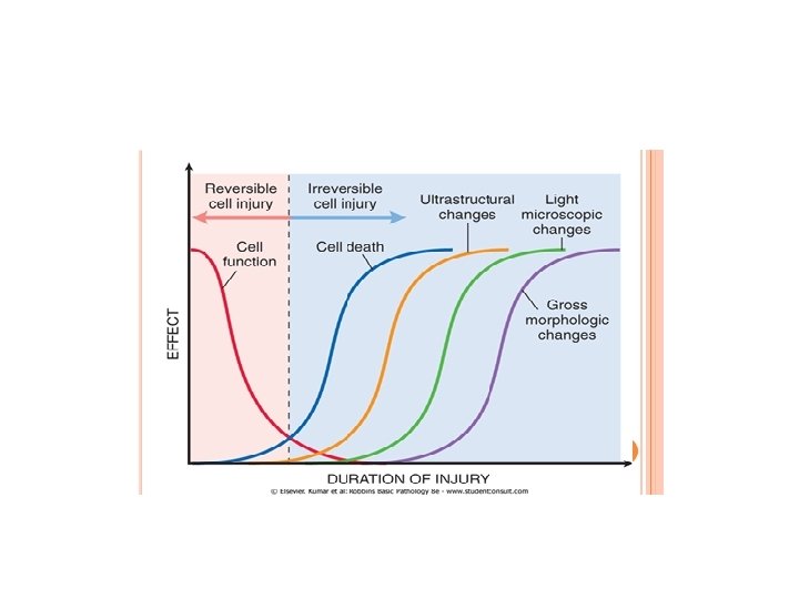
Rules and principles/ 1 • Cell response to injurious stimuli depend on type, duration and severity of the injury. • Example: low dose of a toxin cause reversible injury whereas larger dosed can cause cell death. • Short-lived ischemia. . Reversible • Ischemia of long duration… death
Rules and principles/ 2 • Response to injury also depends on type, status, adaptability and genetic makeup of the injured cell. • Example: skeletal muscle cells can stand 2 -3 hours of ischemia without irreversible injury but cardiac muscles die in 20 -30 minutes. • Glycogen content in hepatocytes can determine their response to injury. . How? • Genetic polymorphism in cytochrome P-450 influences response to toxins.
Rules and principles/ 3 Cell injury results from functional and biochemical changes in essential cellular components, mainly: • Mitochondrial function • Calcium homeostasis • Cell and organelle membranes • DNA • Protein synthesis and folding.
Rules and principles/ 4 • All injurious stimuli first affect the molecular or biochemical level. • Cellular functions lost before cell death occurs. • The morphologic changes of cell injury (or cell death) occur very late.
Rules/4 example • Ischemia of the heart… coronary artery occlusion. • Myocardial cells loose function ( become noncontractile) after 1 -2 minutes of ischemia. • They die 20 -30 minutes after ischemia. • It takes 2 -3 hours to recognise ultrastructural changes of death (EM) • 6 -12 hours by light microscope to appear dead.
Morphology of reversible cell injury • Cellular swelling : due to failure of energydependent ion pumps in the plasma membrane causing inability to maintain ion and fluid homeostasis. • Fatty change : small or large lipid vacuoles (hepatocytes and myocardial cells)
Cell swelling • The first manifestation of almost all forms of cell injury. • Reversible. • Grossly: organ affected becomes pale and gains weight. • Micro: small clear cytoplasmic vacuoles … which are distended endoplasmic reticulum.
Cell swelling
Cell swelling
Fatty change • In cells participating in fat metabolism: hepatocytes and myocardial cells) • Reversible • Fat droplets.
Fatty change
Ultrastructural changes of reversible injury (EM) • (1) plasma membrane changes such as blebbing, blunting or distortion of microvilli, and loosening of intercellular attachments. • (2) mitochondrial changes such as swelling and the appearance of phospholipid-rich amorphous densities. • (3) dilation of the ER with detachment of ribosomes and dissociation of polysomes. • (4) nuclear alterations, with clumping of chromatin.
EM changes
Mechanisms of cell injury • • • ATP depletion Mitochondrial damage Calcium influx Oxygen derived free radicals membrane defects Damage to DNA and protein
mechanisms
Mitochondrial damage