CAUSES AND MECHANISMS OF CELL INJURY Manar Hajeer

 Ø Chemical Agents Ø")


 Influx across plasma membrane(ATP")
")
 � Attack nucleic")


 Injury � Direct toxicity: combining molecular component or cellular organelle � Examples:")
- Slides: 28
CAUSES AND MECHANISMS OF CELL INJURY Manar Hajeer, MD, FRCPath.
Causes of cell injury Ø Oxygen Deprivation (Hypoxia Vs ischemia) Ø Chemical Agents Ø Infectious Agents Ø Immunologic Reactions Ø Genetic Factors Ø Nutritional Imbalances Ø Physical Agents Ø Aging
Oxygen Deprivation
Chemical Agents
Infectious Agents
Immunologic Reactions
Genetic Factors
Nutritional Imbalances
Physical Agents
MECHANISMS OF CELL INJURY � � Principles The cellular response to injury depends on: type of injury duration severity The consequences of injury depend on: type, status, adaptability, and genetic makeup of the injured cell Cell injury results from functional and biochemical abnormalities in one or more of several essential cellular components
The principal targets of cell injury are:
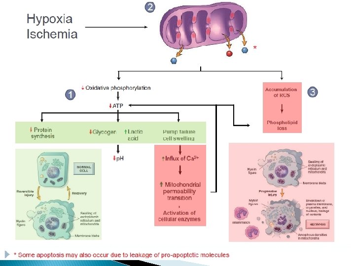
Depletion of ATP
Significant depletion of ATP has widespread effects on many critical cellular systems � Plasma membrane ATP-dependent sodium pumps � Increase in anaerobic glycolysis � Failure of ATP-dependent Ca pumps leads to influx of Ca. � Structural disruption of the protein synthetic apparatus. � Irreversible damage to mitochondrial and lysosomal membranes, leading to necrosis.
Mitochondrial Damage and Dysfunction
Mitochondrial damage may result in several biochemical abnormalities � Progressive depletion of ATP, � Formation of reactive oxygen species � Formation of a high-conductance channel in the mitochondrial membrane (mitochondrial permeability transition pore) � Released of certain proteins into the cytoplasm to activate apoptosis
Influx of Calcium
� Sources of intracellular calcium: Intracellular stores (mitochondria and ER) Influx across plasma membrane(ATP dependent). � Increased cytosolic Ca activates a number of enzymes, � Calcium induces apoptosis by Direct activation of caspases Increasing mitochondrial permeability
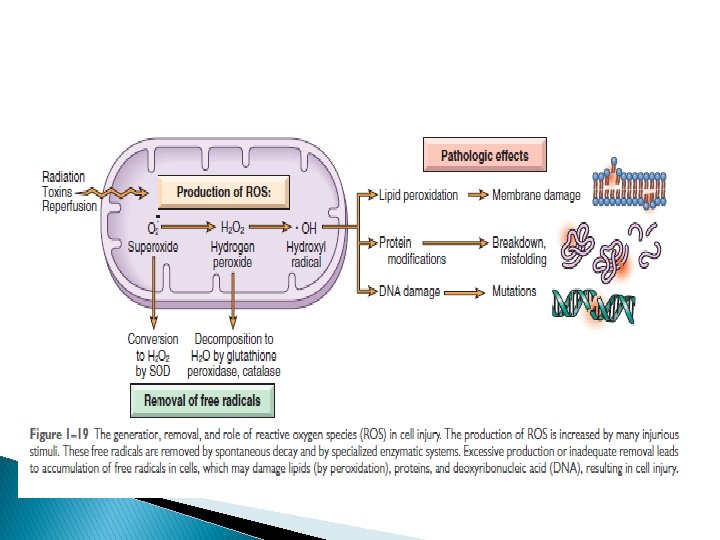
Accumulation of Oxygen-Derived Free Radicals (Oxidative Stress)
Free radicals � Chemical species with single unpaired electron (extremely unstable) � Attack nucleic acids, cellular proteins and lipids. � Molecules that react with free radicals are converted into free radicals. � Involved in: ischemia-reperfusion, chemical and radiation injury, toxicity from oxygen , cellular aging, microbial killing by phagocytes, and tissue injury in inflammation. � The damage caused by them is determined by their rates of production and removal
ROS are produced by two major pathways � Normally in small amounts in all cells during (redox) reactions in the mitochondria. � In phagocytic leukocytes (neutrophils and macrophages) for destroying microbes and inflammatory reactions.
� Generation of free radicals � Radiant energy � Exogenous chemicals � Inflammation � Removal of free radicals: � SPONTANIOUS DECAY � Superoxide dismutases (SODs) � Glutathione (GSH) peroxidases. � Catalase. � Endogenous or exogenous antioxidants (vitamins E, A, and C and β-carotene
Defects in Membrane Permeability Cell membranes, lysosomal membranes, mitochondrial membranes.
Damage to DNA and Proteins � Cells have mechanisms that repair damage to DNA. � If damage is too severe, trigger apoptosis. � Improperly folded proteins have similar effect
CLINICOPATHOLOGIC CORRELATIONS: EXAMPLES OF CELL INJURY AND NECROSIS � Ischemic � � � and Hypoxic Injury Ischemia injures tissues faster than hypoxia Reduced generation of ATP Functional consequences (heart muscle) If oxygen is restored, disturbances are reversible. If ischemia persists, irreversible injury and necrosis.
Ischemia-Reperfusion Injury � Restoration of blood flow to ischemic but viable tissues results, in the death of cells that are reversibly injured. � Mechanism: � By generation of ROS from parenchymal, endothelial cells and leukocytes � By influx of leukocytes and plasma proteins (complement)
Chemical (Toxic) Injury � Direct toxicity: combining molecular component or cellular organelle � Examples: mercuric chloride poisining (seafood), chemotherapeutic drugs. � Indirect toxicity: converted to reactive toxic metabolites, involves the formation of free radicals. (cytochrome P-450) � Examples: Carbon tetrachloride (CCl 4), acetaminophen