Cell injury Dr H Awad Mechanisms of cell


 • Leak of cytochrome c out of")

 Reduced (shrinkage) Pyknosis → karyorrhexis →")
- Slides: 32
Cell injury Dr H Awad
• Mechanisms of cell injury
Mechanisms of cell injury • • • ATP depletion Mitochondrial damage Calcium influx Oxygen derived free radicals membrane defects Damage to DNA and protein
ATP depletion • • ATP. . Importance? Sources of ATP? Causes of depletion? Effects of ATP depletion?
Effects of ATP depletion • Membrane permeability affected due to effects on Na- K pump. • Increased acidity due to increased non oxidative phosphorylation. • Failure of calcium pump. . Increased intracellular calcium. • Structural disruption in protein synthesis apparatus. . Detacment of ribosomes and dissociation of polysomes
Mitochondrial damage • Failure of oxidative phosphrylation. . ATP depletion. • Abnormal oxidative phosphorylation. . ROS • Mitochondrial permeability transition pores. . Loss of membrane potentional and p. H change • Release of proteins that initiate apoptosis
Calcium influx • Activates enzymes… phospholipases, proteases, endonucleases, ATPases… so cell destruction. • Calcium can directly activate caspases… apoptosis
Oxygen free readicals • Definition? ? • Effect? ? • Formation? ?
Oxygen free radicals • Lipid peroxidation • Cross linking of proteins • DNA damage
Membrane permeability defects Caused by: Decreased phospholipid synthesis due to ATP depletion. Increased phospholipid breakdown. . Due to increased calcium ROS. . Lipid breakdown products
Irreversible injury • Two types? • Define each. .
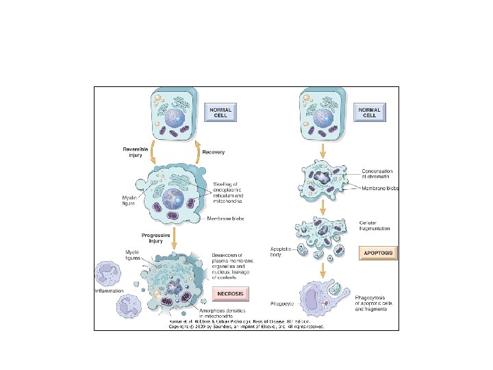
Necrosis • Morphologic changes that follow cell death in living tissues.
NECROSIS • Denaturation of intracellular proteins. • Digestion of cells by lysosomal enzymes of dying cells ( autolysis) and leukocytes (heterolysis).
Apoptosis
Apoptosis • cell death induced by a tightly regulated suicide program in which cells activate enzymes capable of degrading the cells' own nuclear DNA and nuclear and cytoplasmic proteins. • Fragments of the apoptotic cells then break off, giving the appearance that is responsible for the name (apoptosis, "falling off"). .
apoptosis • The plasma membrane remains intact. • Apoptotic bodies (contain portions of the cytoplasm and nucleus) become targets for phagocytosis before their contents leak out and so there would be no inflammatory reaction.
Causes of Apoptosis • Physiologic situations: To eliminate cells that are no longer needed OR to maintain a steady number of various cell populations in tissues.
• Think of examples of physiologic apoptosis…
Physiologic apoptosis • Embryogenesis. • involution of hormone-dependent tissues upon hormone withdrawal. (endometrium and breast after pregnancy) • Cell loss in proliferating cell populations. (GIT) • Death of host cells after serving their useful function. (neutrophils and lymphocytes in inflammation) • Elimination of potentially harmful self-reactive lymphocytes. • Cell death induced by cytotoxic T lymphocytes (tumor cells and viraly infected cells)
Pathologic situations Examples: DNA damaged cells, . Cells with accumulation of misfolded proteins, Certain infections (viral ones): may be induced by the virus (as in human immunodeficiency virus infections) or by the host immune response (as in viral hepatitis). • Pathologic atrophy in parenchymal organs after duct obstruction (pancreas, parotid and kidney) • •
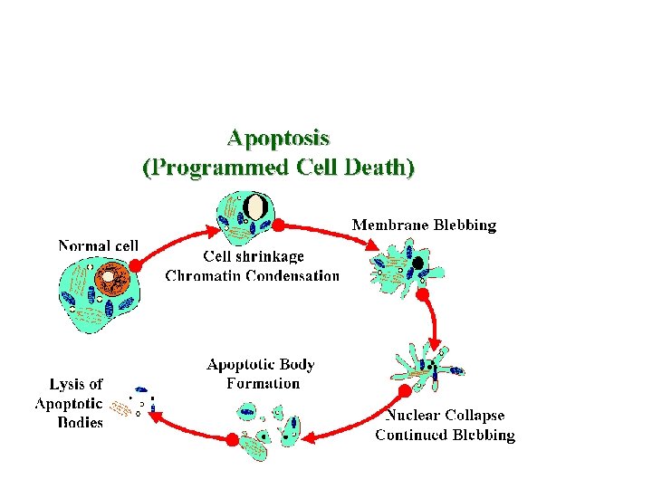
Morphology • Cell shrinkage: dense cytoplasm, tightly packed organelles. • Chromatin condensation: peripherally under the nuclear membrane. • Formation of cytoplasmic blebs • apoptotic bodies: blebbing then fragmentation into membrane bound apoptotic bodies composed of cytoplasm and tightly packed organelles with or without nuclear fragments.
Morphology • Phagocytosis of apoptotic cells or cell bodies by macrophages (quickly hence no inflammation).
• Draw apoptotic cell.
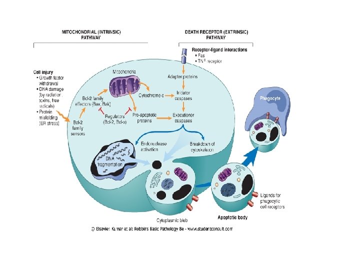
Mechanisms of Apoptosis • Activation of enzymes called caspases. • Two main pathways: • 1 - Mitochondrial pathway (intrinsic) • 2 - Death receptor pathway (extrinsic)
Mechanisms of Apoptosis • Activation of enzymes called caspases. • Two main pathways: • 1 - Mitochondrial pathway (intrinsic) • 2 - Death receptor pathway (extrinsic)
• 1 - mitochondrial pathway (intrinsic) • Leak of cytochrome c out of mitochondria and activation of caspase 9… • 2 - death receptor pathway (extrinsic) • Involved in elimination of self-reactive lymphocytes and in killing of target cells by some cytotoxic T lymphocytes. • Activation of caspase 8.
• What are the differences between necrosis and apoptosis? ? ?
Feature Necrosis Cell size Nucleus Enlarged (swelling) Reduced (shrinkage) Pyknosis → karyorrhexis → karyolysis Fragmentation into nucleosome-size fragments Plasma membrane Disrupted Intact; altered structure, especially orientation of lipids Cellular content Enzymatic digestion; may leak out of cell Adjacent inflammation Physiologic or pathologic role Frequent Intact; altered structure, especially orientation of lipids No Invariably pathologic (culmination of irreversible cell injury) Apoptosis Often physiologic, means of eliminating unwanted cells; may be pathologic after some forms of cell injury, especially DNA damage