Integumentary System Epidermis Dermis Hypodermis Functions of the

 UV")


 – each has keratinocytes – stratum basale mitosis layer")


 – Merocrine – most numerous water producing for coolness –")


- Slides: 56
Integumentary System Epidermis Dermis Hypodermis
Functions of the skin – Barrier Water Bactericidal conditions (acid mantle, dry, oil) UV Fat-soluble vitamins can be absorbed (A, D, E, K); poisons – – Vitamin D synthesis Sensory Heat/cold Pressure Touch Texture Injury Vibration
Functions of the skin – Thermoregulation Receptors tell hypothalamus to constrict blood vessels to cool; dilate to warm the skin – Social functions – Goosebumps – piloerector muscle contracts, pulling follicle; traps air
Epidermis
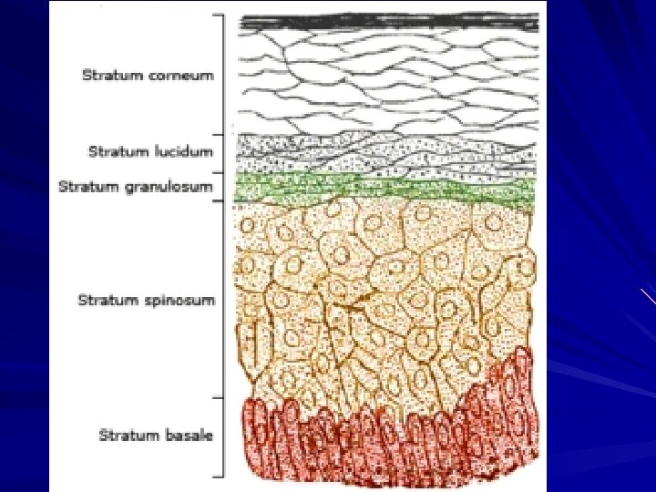
Epidermis Layers (deep to superficial) – each has keratinocytes – stratum basale mitosis layer – new cells melanocytes – melanin Merkel cells – touch – stratum spinosum keratinocytes – bridge to one another by desmosomes Langerhans cells – present pathogen to immune system
Epidermis stratum granulosum – 2 -5 keratinocyte layers & waterproofing layer – grainy cell layer stratum lucidum – eleidin – immature form of keratin – clear cell layer stratum corneum – < 30 layers of cells – exfoliation/desquamate
Dermis Structures
Layers Dermis – papillary – areolar tissue; immunity Meissner’s corpuscles – pain & touch – reticular – dense irregular connective tissue Pacinian corpuscles – pressure sensor Ruffini corpuscle – stretch, heavy touch stretch marks – tears in collagen fibers
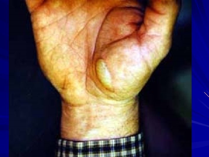
Pruney fingers The wrinkles that occur in skin after prolonged exposure to water are sometimes referred to as pruney fingers or water aging. This is a temporary skin condition where the skin on the palms of the hand or feet becomes wrinkly. In recent past the common explanation was based on water absorption in the keratin-laden epithelial skin when immersed in water; [6], this caused the skin to expand, resulting in a larger surface area and forcing it to wrinkle. Usually the tips of the fingers and toes are the first to wrinkle because of a thicker layer of keratin and an absence of hairs which secrete the protective oil called sebum. In 1935, Lewis and Pickering already found that the skin in the median nerve distribution failed to wrinkle in patients with median nerve palsy. This suggested a mechanism other than simple water absorption. Recent research shows that wrinkling is related to vasoconstriction [7][8]. Water probably initiates the wrinkling process by altering epidermal electrolyte homeostasis as it diffuses into the porous skin of the hands and soles via their many sweat ducts. Altered epidermal electrolyte homeostasis would lead to a change in membrane stability of the surrounding dense network of nerve fibers and trigger increased vasomotor firing with subsequent vasoconstriction. Vasoconstriction, through loss of volume, leads to negative digit pulp pressure resulting in a downward pull on the overlying skin, which wrinkles as it is distorted [9]. This insight resulted in bedside tests for nerve damage and vasoconstriction. For instance a patient with Diabetic neuropathy caused by Diabetes mellitus will have a different finger wrinkling pattern than a healthy subject. Wrinkling is often scored with immersion of the hands for 30 minutes in water or EMLA cream with measurements steps of 5 minutes, and counting the number of visible wrinkles in time. Not all healthy persons have finger wrinkling after immersion, so it would be safe to say that sympathetic function is preserved if finger wrinkling after immersion in water is observed, but if the fingers emerge smooth it cannot be assumed that there is a lesion to the autonomic supply or to the peripheral nerves of the hand[10].
Cutaneous Glands sebaceous - holocrine – oil ceruminous – wax of ear; cerumen – eardrum pliable, waterproofing; bactericidal mammary – modified apocrine sweat glands
Cutaneous Glands Sweat (sudoriferous) – Merocrine – most numerous water producing for coolness – Apocrine – sparse or absent in some ethnicities milky due to fatty acids scent glands responding to stress & sexual stimulation occur in groin, anal region, areola, beard in mature males still secrete by exocytosis, but larger lumen so called apocrine instead of merocrine
Dermis Structures
Hypodermis Connects dermis to muscles Subcutaneous fat
Skin coloration Normal color controlled by – hemoglobin – melanin – Carotene
Skin Discoloration 1. cyanosis – blue; low O 2 2. erythema – red; increased blood flow 3. jaundice – yellow; bilirubin buildup 4. 5. 6. 7. (hemoglobin destruction) & liver doesn’t dispose it bronzing – “tan”; Addison disease = adrenal cortex hormone deficiency pallor – pale; low blood flow albinism – white; lack of melanin hematoma - bruise
Integumentary System Review
Common Skin Disorders
Acne
Boil
Bacterial Intertrigo
Ringworm
Shingles
Warts
Tinea versicolor
Vitiligo
Psoriasis
Leprosy
Leprosy
Eczema
Poison Ivy
Uncommon skin disorders
Langerhans Cell Histiocytosis
Toxic Epidermal Necrolysis
Scleroderma
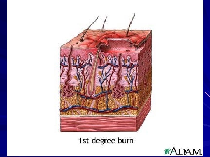
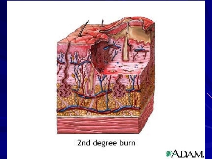
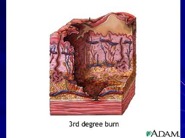
Other skin concerns Cancer – Basal cell – Squamous cell – Malignant melanoma Burns (old classification) – 1 st degree – 2 nd degree – 3 rd degree
Skin Cancers What is Cancer?
Learn the ABC’s! Asymmetry: half the mole looks different from the other half Border: the edges of the mole are irregular or ragged Color: moles are non-uniform in color, and Diameter: normal moles are typically smaller than six millimeters (a quarter inch) in diameter. Melanomas are generally bigger, although recently doctors have seen melanomas between three and six millimeters in diameter.
Basal cell carcinoma
Basal cell carcinoma
Basal cell carcinoma
Basal cell carcinoma
Squamous cell carcinoma
Squamous cell carcinoma
Malignant melanoma
Malignant melanoma
Burn Classification
1 st degree burn
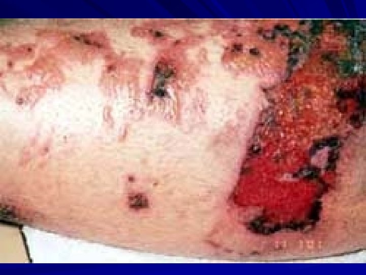
2 nd degree burn
3 rd degree burn