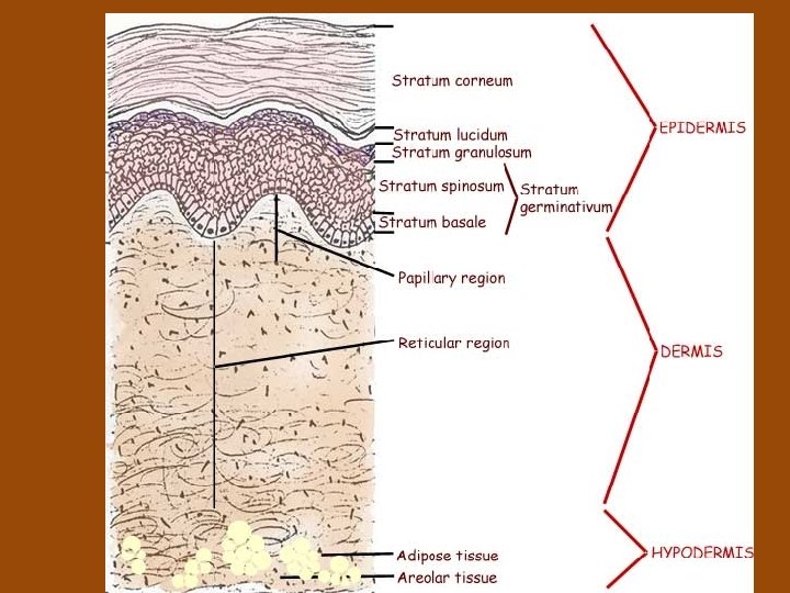
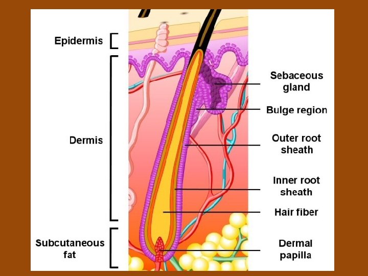
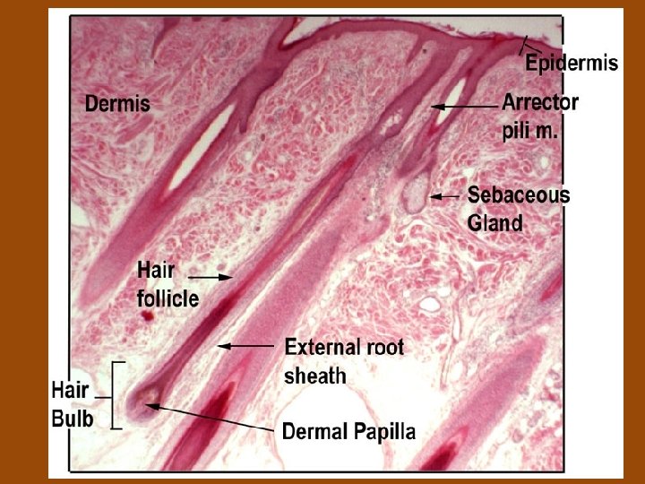
Integumentary System SKIN LAYERS Epidermis and Dermis EPIDERMIS

 • EPIDERMIS (outside to inside) – Stratum Corneum •")


 – Thicker than Epidermis")


 – Vitamin D production (hormone")

 • Heat Loss –Increases the flow of blood to")


")
 • All are exocrine glands (they release their")


- Slides: 35
Integumentary System
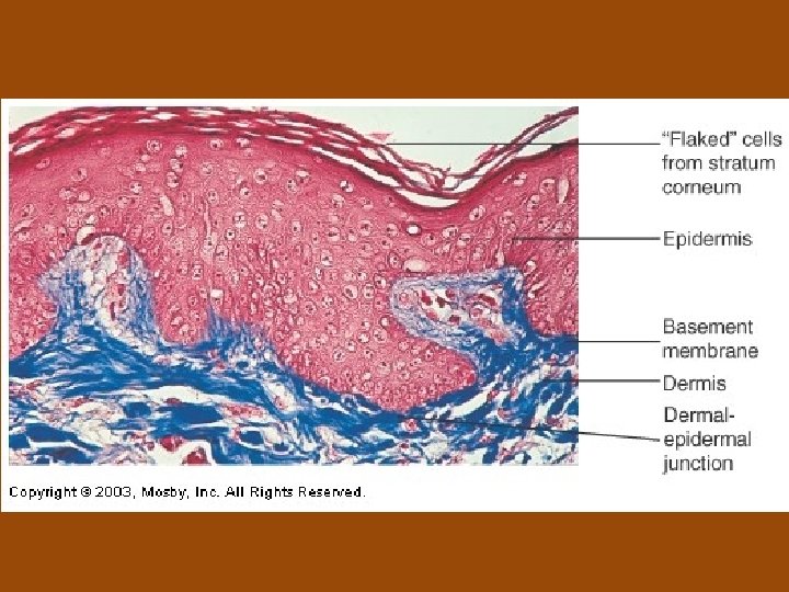
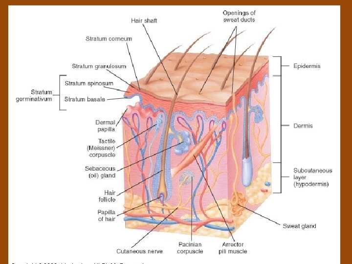
SKIN LAYERS (Epidermis and Dermis) • EPIDERMIS (outside to inside) – Stratum Corneum • • Thin and flat cells Dead Continually shed (35 days to new skin) Cytoplasm – full of protein keratin – Water repellent • Cells are very dry – If soak in water, cells act as sponge and absorb water – Skin puckers, bulges when in water a long time – Allowed to dry out wrinkles disappear • Barrier to water loss, outside chemicals, and outside bacteria
– Stratum Lucidum • • Clear layer Dead - No nuclei Prevents water loss Apparent in “thick layers” of skin – soles of feet and palms of hand – Stratum Granulosum • Keratinization begins in this layer • Intense staining granules
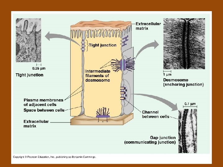
– Stratum spinosum • Irregular-shaped cells • Desmosomes – joining adjacent cells – Spot welds – Heat, friction, or irritants break bonds and results in blisters • Gives a spiny appearance • Heavy production of keratin (heavy RNA and protein synthesis)
– Stratum basale Single layer of columnar cells Deepest cells undergo mitosis Migrate from this layer to surface of skin Regeneration of cells about 3 -4 weeks Abrasions or constant pressure increases mitotic activity • Leads to a thickened corneum (calluses) • • •
• DERMIS – Corium (much thicker than the epidermis) – Thicker than Epidermis – Skin strength in the dermis – Storage area for water
– Papillary layer Dermis • Thin layer that projects into the epidermis – dermal papillae • Responsible for fingerprinting
Dermis – Reticular layer • Collagen fibers give strength to the skin – Commercial processing of animal skin – leather – Elastic fibers give skin its give • Point of attachment for muscle – Results in facial expressions and scalp movement • Hair follicles have arrector pili muscle attached – Hair stands on end in extreme fright (skin crawl) – Raises the skin around it – “goose bumps” • Doesn’t shed and regenerate • Contains sensory receptors (pain, pressure, touch, and temperature)
• SUBCUTANEOUS LAYER OR HYPODERMIS – Not really part of the skin itself • Lies between dermis and underlying tissues – Rich in fat or adipose tissue – Choice for subcutaneous injections and absorption of materials
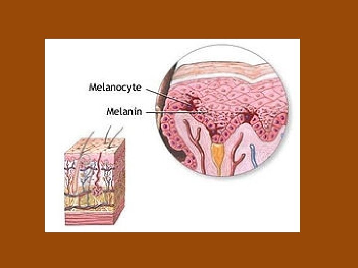
• SKIN COLOR –Determined by 3 factors • Amount of melanin • Carotene – yellow pigment • Hemoglobin at the surface
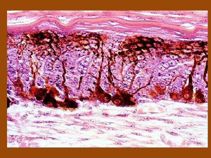
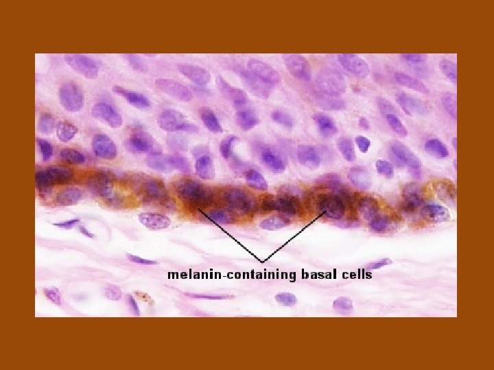
– Melanin – production occurs in Melanocytes • Located in the Stratum basale • Melanin – dark brown pigment • Tyrosine (aa) → Melanin – Requires enzyme tyrosinase • Absence of enzyme – results in albinism • Decreased enzyme – results in graying of hair and white skin of elderly • Controlled by genes • Environmental control – exposure to sun (increases melanin production) – Meant as protection to UV damaging rays (mutations)
–Carotene – yellowish color –Hemoglobin – reddish color • Depends on volume of blood in skin capillaries
• FUNCTIONS OF THE SKIN – Protection – • Microorganisms – mechanical and lysozymes • From water loss – keratin • Chemicals – keratin • Physical damage – Collagen strength • UV radiation - Melanin
– Sensation – sensory receptors puts us in contact with our external environment • Pressure, touch, pain, and temperature
– Excretion – salts and wastes (ammonia and urea) – Vitamin D production (hormone or endocrine function)– UV light causes a chemical reaction that allows vitamin D formation • Important in calcium absorption for strong bones
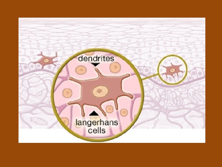
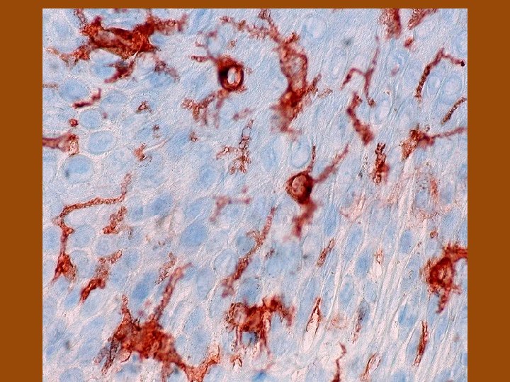
– Immunity • Macrophages are in the tissue to repel foreign invaders • Langerhans cells work with Helper T cells – trigger immune reaction to disease
– Body Temperature Homeostasis (balance) • Heat Loss –Increases the flow of blood to the skin dermis – Vasodilation –Sweating and evaporation allow heat to be removed • Heat Conservation –Decreases the flow of blood to the skin dermis – Vasoconstriction
• SKIN APPENDAGES • HAIR – Present all over body EXCEPT – lips, nipples, palms of hands and soles of feet – Parts of the hair • Shaft – hair seen above the surface of the skin • Root part of shaft below the surface – Follicle – tube that surrounds the hair below the surface of the skin » Epidermal layers spread down into the dermis » Contain mitotic layer for hair shaft formation » Melanocytes are present that give color to the hair – Papilla - Supplies blood capillaries for nourishment
– Hair color • Amount of melanin deposited in the middle of the hair determines color • Special melanin containing iron results in red hair • Gray and White hair decrease or absence of melanin – Straight or curly hair • Straight hair – round shaft • Curly hair – flat shaft
• NAILS – Nail body - visible part of nail (keratinized epithelial cells) – Root of nail – hidden by the cuticle • Lunula - crescent-shaped white area • Stratum germinativum below lunula – site of nail growth – Rich blood vessels below nail give a pink color to nail
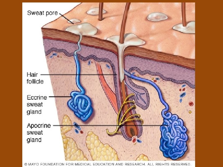
• SKIN GLANDS (Cutaneous glands) • All are exocrine glands (they release their secretions to the skin surface via ducts) – Sebaceous (Oil) Glands- found all over the skin, except on palms of the hands and soles of the feet. • Sebum- secreted by sebaceous glands – Made up of an oily substance and fragmented cells – A lubricant that keeps the skin soft and moist and prevents the hair from becoming brittle. – Contains chemicals to kill bacteria
– Sweat glands- “sudoriferous glands”more than 2. 5 million person • Eccrine glands- found all over the skin –Produce sweat (water plus some salts, vitamin C, traces of metabolic wastes (ammonia, urea, uric acid), and lactic acid. –The sweat glands are supplied with nerve endings
• Apocrine sweat glands- axillary and genital areas –Larger than eccrine glands –Secretions contain fatty acids and proteins as well as all substances present in eccrine secretions. –May be milky or yellow in color. –Odorless, but when bacteria begin to grow on the nutrients in the secretions, it takes on a musky smell. –Apocrine glands begin to function during puberty under the influence of androgen