HemostasisThrombosis I Normal HemostasisThrombosis Assessment of Clotting System

Hemostasis/Thrombosis I Normal Hemostasis/Thrombosis; Assessment of Clotting System

HEMOSTASIS & THROMBOSIS • Platelets • Coagulation Cascade • Regulation of Coagulation

CIRCULATORY SYSTEM • Low volume, high pressure system • Efficient for nutrient delivery to tissues • Prone to leakage 2º to endothelial surface damage • Small volume loss large decrease in nutrient delivery • Minimal extravasation in critical areas irreparable damage/death of organism

HEMOSTASIS Primary vs. Secondary vs. Tertiary • Primary Hemostasis – Platelet Plug Formation – Dependent on normal platelet number & function – Initial Manifestation of Clot Formation • Secondary Hemostasis – Activation of Clotting Cascade Deposition & Stabilization of Fibrin • Tertiary Hemostasis – Dissolution of Fibrin Clot – Dependent on Plasminogen Activation

HEMOSTATIC DISORDERS Suspicions • Spontaneous bleeding • Prolonged or excessive bleeding after procedures or trauma • Simultaneous bleeding from multiple sites

HEMOSTATIC DEFECTS
 • • • Dental Extractions Prior Surgery/Biopsy Easy bruising")
HEMOSTATIC ABNORMALITIES "Screening Tests" (History) • • • Dental Extractions Prior Surgery/Biopsy Easy bruising Heavy menses Deliveries Blood transfusion

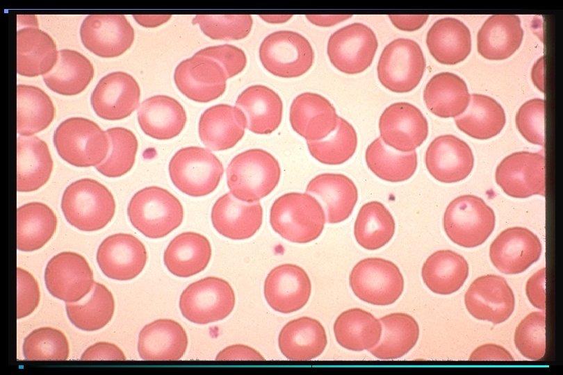
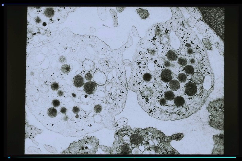
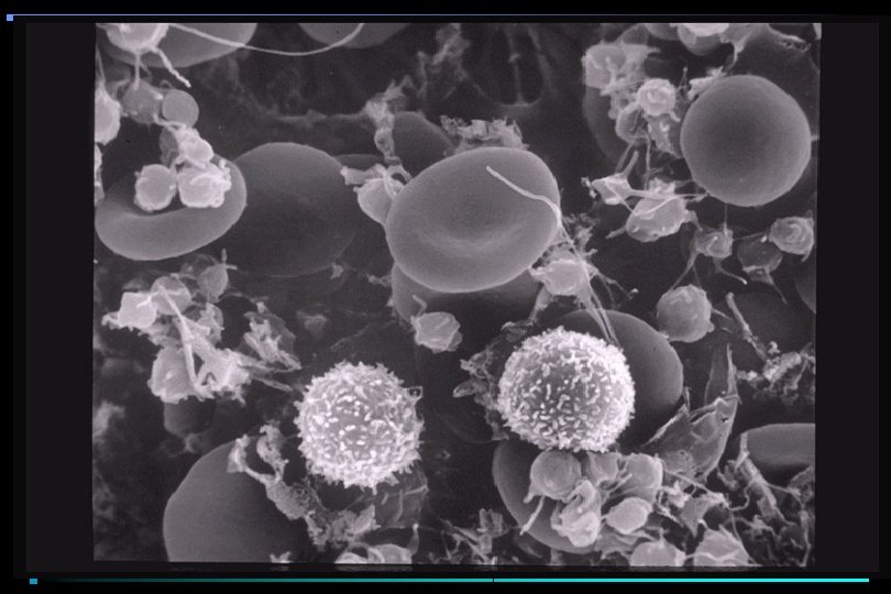
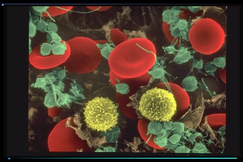
PLATELETS • Anucleate cellular fragments; multiple granules, multiple organelles • Synthesis controlled by IL-6, IL-3, IL-11, & thrombopoietin • Circulate as inactive, non-binding concave discs • On stimulation, undergo major shape change • Develop receptors for clotting factors • Develop ability to bind to each other & subendothelium

PLATELET ACTIVATION Resting Platelets v. WF Platelet Adhesion

Initial Thrombin Formation IIa TR II X Xa VIIa -TF IIa

PLATELET ACTIVATION II Platelet Activation X Xa VIIa -TF IIa

Platelet Release Tx. A 2, ADP, Serotonin, Fibrinogen, Thrombospondin II X Xa VIIa -TF IIa

PLATELET PLUG FORMATION

COAGULATION CASCADE General Features • Zymogens converted to enzymes by limited proteolysis • Complex formation requiring calcium, phospholipid surface, cofactors • Thrombin converts fibrinogen to fibrin monomer • Fibrin monomer crosslinked to fibrin • Forms "glue" for platelet plug

COAGULATION CASCADE

THROMBIN • • • Serine protease Cleaves fibrinogen to fibrin Activates V to Va, VIII to VIIIa Activates platelets-cleaves thrombin receptor Activates XIII to XIIIa In presence of thrombomodulin activates Protein C to APC

COAGULATION CASCADE

Platelet Release Tx. A 2, ADP, Serotonin, Fibrinogen, Thrombospondin IX IXa VIIa -TF VIII V II X Xa VIIa -TF VIIIa Va

Tenase/Prothrombinase complex assembly X VIIIa/IXa VIIIa R Xa II IIa Va/Xa Va R

FIBRIN FORMATION A A B B T F XIIIa

VITAMIN K DEPENDENT CARBOXYLASE • Post-translational modification • Factors II, VII, IX, X; proteins C & S • Converts 1 st 7 -12 glutamic acids to γ-carboxyglutamic acid • Confers calcium binding and lipid binding on these proteins • Without vitamin K, secrete desγ-carboxyglutamic acid containing proteins (inactive in coagulation)

γ -CARBOXYGLUTAMIC ACID COO- COOCH 2 CH O 2, CO 2 CH 2 NH 3+ CH COOGlu COO- Vit K Carboxylase NH 3+ CH COO- γ-carboxy Glu

PLATELET FUNCTION STUDIES • Bleeding Time • Platelet Count • Platelet Aggregation Studies

BLEEDING TIME vs. PLATELET COUNT

COAGULATION CASCADE

Clotting Tests • Recalcification times • Blood collected in sodium citrate, a weak calcium chelator • Amount designed to drop calcium concentration to < 1 m. M • At that level, blood won’t clot spontaneously • RBC’s & platelets centrifuged off, leaving plasma (unclotted liquid portion of blood)

Prothrombin Time • Mixture of: – 50% patient’s platelet poor plasma – 25% Mixture of Tissue Factor & phospholipid species – 25% Calcium chloride (to bring final calcium concentration to c. 3 -5 m. M) – Time to clot formation measured
 FVII TF FVIIa Ca+2 VIIa/TF FX Middle")
COAGULATION CASCADE EXTRINSIC PATHWAY Prothrombin Time (PT) FVII TF FVIIa Ca+2 VIIa/TF FX Middle Components Ca+2 FXa Ca+2 T V Va Va/Xa/PL PT Common Pathway Ca+2 T FG F
 • 50% patient’s platelet-poor plasma • 25% mixture")
a. PTT (activated partial thromboplastin time) • 50% patient’s platelet-poor plasma • 25% mixture of phospholipid & surface active agent (Celite, Kaolin) • 25% Calcium Chloride to bring calcium concentration to 3 -5 m. M • Time to clot formation measured

COAGULATION CASCADE INTRINSIC PATHWAY FXIIa FXI a. PTT Surface Active Components HMWK FXIa FIX Ca+2 T VIII FIXa Ca+2 VIIIa/IXa/PL FX Middle Components Ca+2 FXa Ca+2 T V Va Va/Xa/PL PT Common Pathway Ca+2 T FG F

COAGULATION CASCADE Contact Phase Prekallikrein Kallikrein F XII HMWK F XIIa F XIa

COAGULATION CASCADE Intermediate Phase Ca+2 F VII/TF F VIIa/TF XIa VIIa/TF/PL F IX Ca T VIIIa + F IXa +2 Ca+2 IXa/VIIIa/PL FX Ca+2 F Xa Tenase Complex

COAGULATION CASCADE Common Pathway IXa/VIIIa/PL FX V VIIa/TF/PL Ca+2 Va + FX F Xa Xa/Va/PL PT Prothrombinase Complex Ca+2 T Fibrinogen Fibrin monomer

CLOTTING ASSAYS Specific factors/Inhibitors • Deficiency states of every clotting protein described • 50% of any clotting factor will yield normal PT and/or a. PTT • Mix 1/2 patient plasma with 1/2 factor deficient plasma; then perform PT and/or a. PTT • Factor deficient plasma with missing factor will prolonged clotting time; all others WNL • If more than one clotting assay prolonged, implies inhibitor to clotting protein
- Slides: 38