BCH 471 Experiment 10 COGULATION PROFILE Clotting time
 COGULATION PROFILE Clotting time, Bleeding time, and Prothrombin time")
+ BCH 471 Experiment (10) COGULATION PROFILE Clotting time, Bleeding time, and Prothrombin time

+ Coagulation �Coagulation is a complex process by which blood forms clots. �It is an important part of haemostasis (the cessation of blood loss from a damaged vessel). � Disorders of coagulation can lead to an increased risk of bleeding (hemorrhage) or clotting (thrombosis).

+ Hemostasis is maintained in the body via three mechanisms : n Vascular spasm - Damaged blood vessels constrict n Platelet plug Platelats adhere formation to - damaged endothelium to form platelet plug (primary hemostasis) n Blood Coagulation - Clots form upon the conversion of fibrinogen to Fibrin (secondary hemostasis).

+ Clotting Cascade mechanism in which a is cascade A • enzymes activate other enzymes sequentially usually leading to an amplification of an initial signal. Pathways • Initially independent, then they converge on common pathway leading Extrinsic • a fibrin clot to the formation of Intrinsic • pathways leads to these of Each • conversion of factor X (inactive) to factor
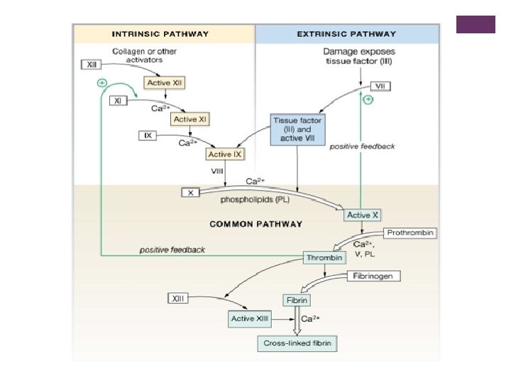

+ What triggers extrinsic and intrinsic pathways n Extrinsic—Release of biochemicals from broken blood vessels/damaged tissue. n Intrinsic—No tissue damage, blood contacts damaged endothelial layer of blood vessel walls.

+ Clotting time n Test for intrinsic system n Simple test but takes time and rarely done now n Method: n Venous blood is taken and placed on glass test tube at 37°C and it observed at time intervals until clotting occurs n Normal blood takes 5 -10 min to clot n Longer periods Coagulation defects (e. g. Hemophilia)

+ Clotting time - capillary method

BLEEDING TIME n Provides assessment of platelet count and function n Method: n It is determined by noting time at which blood coming out a small cut, no longer forms a spot on a piece of filter paper placed in contact with cut surface n The normal range from 2 -4 min
 § Measures effectiveness of the extrinsic pathway Method: An excess of")
PROTHROMBIN TIME (PT) § Measures effectiveness of the extrinsic pathway Method: An excess of tissue factor and Ca 2+ ions are added to diluted § § plasma containing citrate (anticoagulant) and then the time taken for the mixture to clot is measured § Normal value 10 -15 secs High PT low levels of thrombin § Results from liver disease due to deficiency of prothrombin, fibrinogen, § V, VII and X factors
- Slides: 10