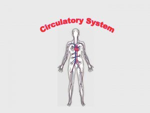
The Human Circulatory System Page 1 of 101


- Slides: 101

The Human Circulatory System Page 1 of 101

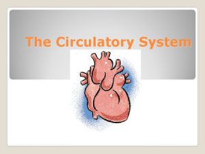
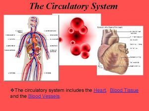
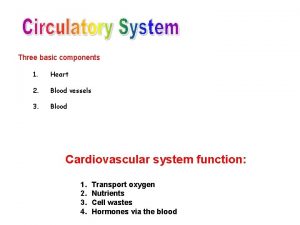
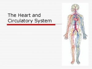
Introduction • Humans and other vertebrates have a closed circulatory system: – This means that circulating blood is pumped through a system of vessels – This system consists of the heart (pump), series of blood vessels and the blood that flows through them. Page 2 of 101

Heart Anatomy Page 3 of 101

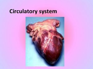
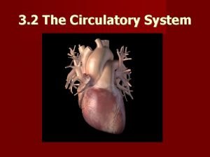
The Heart • • • Located near the center of your chest Hollow structure Composed almost entirely of muscle About the size of your clenched fist Enclosed in a protective sac called the pericardium Page 4 of 101

The Heart • In the walls of the heart, two layers of tissue form a sandwich around a thick layer of muscle called the myocardium. • Contractions of the myocardium pump blood through the circulatory system. • The heart contracts about 72 times per minute • Pumps about 70 m. L of blood with each contraction. • The right and left sides of the heart are separated by a septum, or wall. • The septum prevents the mixing of oxygen rich and oxygen poor blood. Page 5 of 101

The Heart • • • On each side of the septum are two chambers. The upper chamber (receives blood) is the atrium. The lower chamber (pumps blood out of heart) is the ventricle. The heart has a total of 4 chambers: 2 atriums 2 ventricles Page 6 of 101

Pathway of Blood • Deoxygenated blood passes from the right atrium into the right ventricle and then goes to the lungs. • From the lungs, blood moves back toward the heart into the left atrium to the left ventricle and then passes into the aorta to go to the rest of the body Page 7 of 101

Valves • As the heart contracts, blood flows into the ventricles and then out through the ventricles. • Flaps of connective tissue, called valves, are located between the atria and ventricles. • Blood moving keeps the valves open. • When the ventricles contract, the valves close which prevent blood from flowing back into the atria. • There also valves that stop blood from re-entering the ventricles after the blood has left. • This system of valves keeps blood moving in one direction which increases the pumping efficiency of the heart. Page 8 of 101

Heart Beat • Heart muscles are composed of individual fibers • Each atrium and ventricle contracts as a unit. • Each contraction begins with a group of cardiac muscle cells in the right atrium known as the senatorial node (SA node) • Because the SA node paces the heart it is known as the pacemaker. • The impulse spreads from the pacemaker to the rest of the atria. • From the atria, a signal is sent to the atrioventricular node and then to a bundle of fibers in the ventricle. • When the ventricle contracts, blood flows out. Page 9 of 101

Blood Vessels • As blood moves through the circulatory system it moves through 3 types of blood vessels: • Arteries • Capillaries • Veins Page 10 of 101

Arteries • Large vessels • Carry blood from heart to tissues of body • Carry oxygen rich blood, with the exception of pulmonary arteries. • Thick walls-need to withstand pressure produced when heart pushes blood into them. Page 11 of 101

Capillaries • Smallest blood vessels • Walls are only one cell thick and very narrow. • Important for bringing nutrients and oxygen to tissues and absorbing CO 2 and other waste products. Page 12 of 101

Veins • Once blood has passed through the capillary systems it must be returned to the heart. • Done by veins • Walls contains connective tissue and smooth muscle. • Largest veins contain one way valves that keep blood flowing toward heart. • Many found near skeletal muscles. When muscles contract, blood is forced through veins. Page 13 of 101

Blood Pressure • The heart produces pressure • The force of blood on the wall of the arteries is known as blood pressure. • Blood pressure decreases as the heart relaxes, but the rest of the circulatory system is still under pressure. Page 14 of 101

Blood Pressure • When blood pressure is taken, the cuff is wrapped around the upper portion of the arm and pumped with air until blood flow in the artery is blocked. • As the pressure in the cuff is relaxed, 2 numbers are recorded. – Systolic pressure- the first number taken, is the force felt in the arteries when the ventricles contract. – Diastolic pressure- the second number taken, is the force of the blood on the arteries when the ventricles relax. Page 15 of 101

Disorders of Circulatory System • Atherosclerosis – Fatty deposits (plaque) in walls of arteries – Deposits can obstruct flow of blood which can raise blood pressure – Increases risk of blood clots – If clot breaks free it can obstruct blood flow to tissues. Page 16 of 101

Disorders of Circulatory System • Heart Attack – Due to atherosclerosis, coronary arteries may become blocked (blood can’t get to heart muscle) – Heart muscle begins to die due to lack of O 2 Page 17 of 101

Disorders of Circulatory System • Stroke – Blood clot may break free and block a vessel leading to the brain. – Brain cells are starved of oxygen and nutrients – Loss of function may occur – Can cause paralysis, loss of ability to speak or death. Page 18 of 101

Blood • Composed of plasma and blood cells • Types of Cells are: – Red Blood Cells – White Blood Cells – Platelets Page 19 of 101

Blood • Plasma – Straw colored – 90% water – 10% dissolved gases, salts, nutrients, enzymes, hormones, wastes, and proteins. Page 20 of 101

Blood • Plasma proteins – 3 Types: Albumins, globulins and fibrinogen. – Albumins and Globulins- transport substances such as fatty acids, hormones and vitamins. – Fibrinogen- Responsible for blood’s ability to clot Page 21 of 101

Blood • Red Blood Cells – – – Page 22 of 101 Most numerous type Transport oxygen Get color from hemoglobin Disk shaped Made in red bone marrow Circulate for 120 days

Blood • White Blood Cells – Guard against infection, fight parasites, and attack bacteria – Number of WBC’s increases when body is fighting – Lymphocytes produce antibodies which fight pathogens and remember them Page 23 of 101

Blood • Platelets – Aid the body in clotting – Small fragments – Stick to edges of broken blood cell and secrete clotting factor to help form clot. Page 24 of 101

Blood Clotting Problems • Hemophelia – Genetic disorder that disrupts clotting – People must be very careful to avoid injury – Can be treated by injecting extracts that contain the missing clotting factor. Page 25 of 101

Introduction to the Human Cardiovascular System Page 26 of 101

INTRODUCTION The cardiovascular system is transport system of body It comprises blood, heart and blood vessels. The system supplies nutrients to and remove waste products from various tissue of body. The conveying media is liquid in form of blood which flows in close tubular system. Page 27 of 101

FUNCTION OF CARDIOVASCULAR SYSTEM Transport nutrients, hormones Remove waste products Gaseous exchange Immunity Blood vessels transport blood ◦Carries oxygen and carbon dioxide ◦Also carries nutrients and wastes Heart pumps blood through blood vessels Page 28 of 101

COMPONENTS OF CARDIOVASCULAR SYSTEM • BLOOD • HEART • BLOOD VESSELS Page 29 of 101

BLOOD • The Blood: Blood cells & Plasma • Blood cells 1 - Erythrocytes - Red Blood Cells 2 - Leucocytes 3 - Thrombocytes • Plasma is fluid portion Page 30 of 101

HEART • Heart is a four chambered, hollow muscular organ approximately the size of your fist • Location: – Superior surface of diaphragm – Left of the midline – Anterior to the vertebral column, posterior to the sternum Page 31 of 101

FUNCTIONS OF THE HEART • Generating blood pressure • Routing blood Heart separates pulmonary and systemic circulations • Ensuring one-way blood flow Heart valves ensure one-way flow • Regulating blood supply Changes in contraction rate and force match blood delivery to changing metabolic needs Page 32 of 101

BLOOD VESSELS • Blood Vessels -A closed network of tubes • These includes: Ø Arteries Ø Capillaries Ø Veins Page 33 of 101

BLOOD VESSELS Arteries(Distributing channel) • Thick walled tubes • Elastic Fibers • Circular Smooth Muscle • Capillaries (microscopic vessels) • One cell thick • Serves the Respiratory System Veins (draining channel) • General structure 1. Tunica intima 2. Tunica media 3. Tunica adventitia Page 34 of 101

CLASSIFICATION OF BLOOD VESSELS • Conducting Vessels • Distributing Vessels • Resistance Vessels • Exchange Vessels • Capacitance / Reservoir Vessels Page 35 of 101

ARTERIES • Blood vessels that carry blood away from the heart are called arteries. • They are thickest blood vessels and they carry blood high in oxygen known as oxygenated blood (oxygen rich blood). • Accompanied by vein and nerves • Lumen is small • No valves • Repeated branching Page 36 of 101

CLASSIFICATION OF ARTEIES • Elastic- e. g. (Aorta & its Major branches) • Muscular -e. g. (Renal, Testicular, Radial, Tibial etc. ) • Arterioles (<0. 1 mm)Terminal arterioles, Meta-arterioles, Thoroughfare, channel/ preferred Page 37 of 101

CAPILLARIES (5 -8 micron) • The smallest blood vessels are capillaries and they connect the arteries and veins. • This is where the exchange of nutrients and gases occurs. BODY CONTAINS TWO KINDS OF CAPILLARIES • CONTINUOUS-SKIN, LUNG, SMMOTH MUSCLE, CONNECTIVE TISSUES • FENESTRATED- PANCREAS, ENDOCRINE GLANDS, SMALL INTESTINE, CHOROID PLEXUS, CILLIARY PROCESS etc. Page 38 of 101

SINUSOIDS • SINUSOIDS- Large irregular vascular space (30 -40 micron) eg. Liver, Spleen, Bone marrow, suprarenal, Parathyroid etc. Page 39 of 101

VEINS • Blood vessels that carry blood back to the heart are called veins. • They have one-way valves which prevent blood from flowing backwards. • They carry blood that is high in carbon dioxide known as deoxygenated blood (oxygen poor blood). Page 40 of 101

• • • Ø Ø Ø Ø Page 41 of 101 VEINS Thin Walled Large irregular lumen Have valves Dead space around Types: Large Medium Small Veins without valves: SVC & IVC Hepatic, Renal Uterine, Ovarian not Testicular Facial Pulmonary Umbilical Emissary Portal Veins <2 mm

VEINS • Ø Ø Ø • 1. 2. 3. 4. 5. Page 42 of 101 Veins without Muscular tissue: Dural venous sinuses Pial Veins Retinal Veins of erectile tissue of sex organs Veins of spongy bones Factors responsible for venous return: Muscle contraction Negative intrathoracic pressure Pulsation of arteries Gravity Valves

ANASTOMOSIS • Communication between vessels • ARTERIAL: Actual( end to end & convergent)-Palmar, plantar, Circle of Willis, Labial Intestinal arcade, etc. Potential-Coronary, around joints etc. Page 43 of 101

ANASTOMOSIS • 1. 2. 3. 4. 5. 6. 7. Page 44 of 101 ARTERIOVENOUS ANASTOMOSIS: Skin of nose Lips External Ear Mucus membrane of GI & nose Erectile tissue of sex organ Thyroid Tongue

END ARTERIES • 1. 2. 3. END ARTERIES: Central artery of retina Arteries of spleen, liver, kidneys, metaphyses of long bones Central branches of cerebral cortex Page 45 of 101

CIRCULATION – Coronary circulation – the circulation of blood within the heart. – Pulmonary circulation – the flow of blood between the heart and lungs. – Systemic circulation – the flow of blood between the heart and the cells of the body. – Fetal Circulation Page 46 of 101

SYSTEMIC AND PULMONARY CIRCULATION Pulmonary circulation The flow of blood between the heart and lungs. Systemic circulation The flow of blood between the heart and the cells of the body. Page 47 of 101

CORONARY CIRCULATION: ARTERIAL SUPPLY Page 48 of 101 48

PORTAL CIRCULATION Portal circulation - the flow of blood between tow set of capillaries before draining in systemic veins. Page 49 of 101

FETAL CIRCULATION Page 50 of 101

PLACENTA UMBILICAL ARTERY DESCENDING AORTA (Through Ductus Arteriosus) PULMONARY TRUNK RIGHT VENTRICLE ASCENDING AORTA UMBILICAL VEIN PORTAL VEIN (Through Ductus Venosus) INFERIOR VENA CAVA RIFHT ATRIUM (Through Foramen Ovale) LEFT ATRIUM Page 51 of 101

APPLIED § Diseases and Disorders § § § § Page 52 of 101 BLOOD PRESSURE HAEMORRHAGE/STROKE ARTERIOSCLEROSIS ANEURYSM CORONARY ARTERY DISEASE (CAD) HEART ATTACK CONGESTIVE HEART FAILURE (CHF) ANEMIA, HEMOPHILIA, AND LEUKEMIA

APPLIED • Problems with the cardiovascular system are common, but they don’t just affect older people. • Many heart problems affect children and teenagers. QUESTIONS All of the following are features of veins except: a)Thin walls b)Thin tunica media c)Thin tunica adventia d)Wide lumen Page 53 of 101

REFERENCES 1 - General Anatomy by Vishram Singh 2 - Clinical Anatomy by R. Snell 3 -Gray’s Anatomy Page 54 of 101

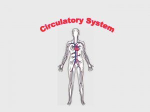
Circulatory system Transporting gases, nutrients, wastes, and hormones Page 55 of 101

Page 56 of 101

Features and Functions Features • Circulatory systems generally have three main features: – Fluid (blood or hemolymph) that transports materials – System of blood vessels – A heart to pump the fluid through the vessels Page 57 of 101

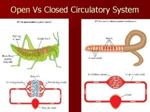
Types of circulatory systems • Animals that have a circulatory system have one of two kinds: – Open: fluid is circulated through an open body chamber. – Closed: fluid is circulated through blood vessels. Page 58 of 101

Open system • Arthropods and most mollusks have an open circulatory system. • Hemolymph is contained in a body cavity, the hemocoel. A series of hearts circulates the fluid. Page 59 of 101

Closed system • Vertebrates, annelid worms, and a few mollusks have a closed circulatory system. • Blood is moved through blood vessels by the heart’s action. It does not come in direct contact with body organs. Page 60 of 101

Why does an open circulatory system limit body size? 1. Hearts are too small for growth. 2. Too little blood to support a larger animal. 3. Less efficient in moving oxygen to body tissues. 4. Hemocoel must be shed for growth. Page 61 of 101

W O R K • Why did homeothermy (“warm. T bloodedness) only develop in organisms with. O G a closed circulatory system? E T H E R Page 62 of 101

Blood Page 63 of 101

Components • Blood is made up of four major components. What do each of these do? – Plasma: the liquid portion. – Red blood cells. – White cells. – Platelets. Page 64 of 101

Red blood cells • RBCs lose their nucleus at maturity. • Make up about 99% of the blood’s cellular component. • Red color is due to hemoglobin. Page 65 of 101

Hemoglobin • Hemoglobin is a complex protein made up of four protein strands, plus ironrich heme groups. • Each hemoglobin molecule can carry four oxygen atoms. The presence of oxygen turns hemoglobin bright red. Page 66 of 101

RBC lifespan • RBCs live about 4 months. Iron from hemoglobin is recycled in the liver and spleen. • The hormone erythropoeitin, made by the kidneys, stimulates the production of RBCs in red bone marrow. Page 67 of 101

If your diet is poor in iron, what will happen to your RBCs? 1. You will make fewer because there is less iron to make hemoglobin. 2. You will make more to make up for the lack of iron in hemoglobin. 3. You will make just as many. Page 68 of 101

W O R K • One of the illegal drugs that some top T Olympic athletes have been caught using is O G erythropoetin. What would this hormone do E T that would give athletes an edge in H E competitions? R Page 69 of 101

White cells • White blood cells defend against disease by recognizing proteins that do not belong to the body. • White cells are able to ooze through the walls of capillaries to patrol the tissues and reach the lymph system. Page 70 of 101

Platelets • Platelets are cell fragments used in blood clotting. • Platelets are derived from megakaryocites. Because they lack a nucleus, platelets have a short lifespan, usually about 10 days. Page 71 of 101

Blood clotting • Platelets aggregate at the site of a wound. • Broken cells and platelets release chemicals to stimulate thrombin production. • Thrombin converts the protein fibrinogen into sticky fibrin, which binds the clot. Page 72 of 101

Which blood cells transport oxygen? 1. 2. 3. 4. White cells Red cells Platelets All blood cells Page 73 of 101

• If a person had a defect in the gene for fibrinogen, what health problems could this cause? Page 74 of 101 W O R K T O G E T H E R

Blood Vessels Page 75 of 101

Classes of blood vessels • Blood vessels fall into three major classes: – Arteries and arterioles carry blood away from the heart. – Veins and venules carry blood to the heart. – Capillaries allow exchange of nutrients, wastes and gases. Page 76 of 101

Arteries • Arteries are thick-walled, and lined with smooth muscle. • How does the structure of an artery help with its function? Page 77 of 101

Arterioles • Arterioles branch off of arteries. • Arterioles can constrict to direct and control blood flow. They may, for example, increase or decrease blood supply to the skin. • How might arterioles be involved when: – Your skin turns red when you are hot. – A person’s face turns pale with fright. Page 78 of 101

Capillaries • Body tissues contain a vast network of thin capillaries. • Capillary walls are only one cell thick, allowing exchange of gases, nutrients, and wastes. • Capillaries are so fine that RBCs must line up single-file to go through them. Page 79 of 101

Venules • Venules are thin-walled collectors of blood. • Low pressure in the venules allows the capillary beds to drain into them. Page 80 of 101

Veins • Veins have thinner walls than arteries. • Veins have fewer smooth muscle cells, but do have valves. How do valves and the skeletal muscles help veins function? Page 81 of 101

W O R K • Besides the ability to contract and move T blood, why do arteries need to be so thick O G and strong? E T • Varicose veins are veins in the legs that are H E swollen, stretched, and painful. What factors. R could lead to this condition, and how can varicose veins be prevented? Page 82 of 101

Atherosclerosis • LDL cholesterol forms plaques in arteries, triggering inflammation. • The immune system forms a hard cap over the plaque, partially blocking the artery. Caps can rupture, creating clots that can close off an artery. Page 83 of 101

Preventing heart attacks • Both genetic and environmental factors contribute to atherosclerosis. • Blood LDL cholesterol can be reduced by a low-fat diet that emphasizes high-fiber foods, antioxidants, and “good” fats (monounsaturated fats, omega-3 oils), and reduce trans-fats. • Regular exercise also contributes significantly to LDL cholesterol reduction. Page 84 of 101

What is always true of arteries? 1. Always carry oxygenated blood. 2. Always carry deoxygenated blood. 3. Always carry blood to the heart. 4. Always carry blood away from the heart. Page 85 of 101

Besides having to constrict to move blood, why are artery walls so thick and strong? 1. Arteries must move oxygenated blood. 2. Arteries must withstand very high blood pressure when the heart contracts. 3. Arteries must move blood out to all parts of the body. Page 86 of 101

Why are capillary walls so thin? 1. Because capillaries are thin and narrow 2. To allow exchange of gases and nutrients. 3. To force RBCs to move through in single file. Page 87 of 101

W O R K • Some people who are at high risk for heart T attacks may be advised by their doctors to O G take low doses of aspirin daily. What effects E does aspirin have that would help prevent TH E heart attacks? R Page 88 of 101

Heart Page 89 of 101

The Vertebrate Heart • Vertebrate hearts are separated into two types of chambers – Atria (singular: atrium): receive blood from body or lungs. Contractions of the atria send blood through a valve to the ventricles. – Ventricles: receive blood from atria, contract to send blood to body or lungs. Page 90 of 101

Two-chambered heart • The simplest vertebrate heart is the two-chambered heart, seen in fishes. • A single atrium receives blood from the body cells. A ventricle sends blood to the gills to collect oxygen. Page 91 of 101

Three-chambered heart • Separate atria allow some separation of oxygenated and deoxygenated blood, which was an advantage for land organisms (reptiles, amphibians). • Though blood can mix in the ventricle, mixing is minimal. Some reptiles have partial separation of the ventricle. Page 92 of 101

Four-chambered heart • The four-chambered heart, seen in birds and mammals, allows complete separation of oxygenated and deoxygenated blood. • Complete separation is necessary to support a fast metabolism found in homeotherms. Page 93 of 101

“Dual pump” operation The four-chambered heart acts as two pumps. Page 94 of 101

Page 95 of 101

Keeping Time • The sinoatrial (SA) node is nervous tissue that times heart beats. • The SA node causes atria to contract, and sends the signal to the atrioventricular (AV) node to signal the ventricles to contract. Page 96 of 101

Blood pressure • Systolic pressure = pressure when the heart contracts. • Diastolic pressure = pressure between heart beats. Page 97 of 101

Which set of heart vessels moves deoxygenated blood from the body to the lungs? 1. Right atrium, right ventricle 2. Right atrium, left atrium 3. Left atrium, left ventricle 4. Right ventricle, left ventricle Page 98 of 101

If your blood pressure is 90/70, the 70 represents: 1. Systolic pressure – heart contracts 2. Systolic pressure – heart is relaxed 3. Diastolic pressure – heart contracts 4. Diastolic pressure – heart is relaxed Page 99 of 101

An electric pacemaker can be connected to the heart to replace a faulty: 1. 2. 3. 4. AV node Bicuspid valve SA node Tricuspid valve Page 100 of 101

• Hypertension (high blood pressure) puts people at risk for heart disease. What longterm effects would an increase in blood pressure have on the heart? • What other organ system is involved in hypertension? Page 101 of 101 W O R K T O G E T H E R
 Respiratory digestive and circulatory system
 Tiny air sacs at the end of the bronchioles
 Circulatory system and respiratory system work together
 Title page for apa format
 Circulatory system steps in order
 Superchp
 Heart fish
 Do clams have a closed circulatory system
 Horse heart
 Carbon dioxide levels in the blood
 Closed circulatory system
 Parts of circulatory system
 Smallest blood vessel
 Double circulatory system
 How circulatory system work
 What makes up the cardiovascular system
 Gas exchange lungs
 Single vs double circulatory system
 Phylum arthropoda characteristics
 Crustaceans characteristics
 Single vs double circulatory system
 Circulatory system foldable
 Invertebrate circulatory system
 Fetal pig circulatory system
 Unit 6:8 circulatory system
 Open circulatory system
 Open circulatory system
 Circulatory system function
 Function of the circulatory system
 Arthropods circulatory system
 Difference between open and closed circulatory system
 Chapter 34 section 1 the circulatory system
 Reptile circulatory system
 Excretory system of silkworm
 Function of the circulatory system
 Mass transport in animals
 Circulatory system interactions with other systems
 Vena cava function in circulatory system
 All parts of the heart
 Chapter 19 the cardiovascular system blood vessels
 Circulatory system
 Functions of the digestive system
 Reptile circulatory system
 Chapter 33 circulatory and respiratory systems
 Mollusca open circulatory system
 Root hair cells
 Blood vessels circulatory system
 Frog circulatory system open or closed
 Circulatory system
 Components of blood
 Circulatory system jeopardy
 Circulatory system trivia
 Circulatory system in a sentence
 The circulatory system
 Where is the femoral pulse located quizlet
 Excretory system quizizz
 Salamander circulatory system
 Circulatory system
 Cardiorespiratory system includes
 Polychaete respiration
 Chambered heart animals
 Types of circulatory system
 Double circulatory system
 3 main functions heart
 Circulatory system
 Blood vessels
 Parts of blood
 Pressure in circulatory system
 Diagram of circulatory system
 How to remember the circulatory system
 Circulatory system crash course
 Hình ảnh bộ gõ cơ thể búng tay
 Bổ thể
 Tỉ lệ cơ thể trẻ em
 Chó sói
 Glasgow thang điểm
 Hát lên người ơi
 Môn thể thao bắt đầu bằng chữ f
 Thế nào là hệ số cao nhất
 Các châu lục và đại dương trên thế giới
 Cong thức tính động năng
 Trời xanh đây là của chúng ta thể thơ
 Mật thư tọa độ 5x5
 Phép trừ bù
 độ dài liên kết
 Các châu lục và đại dương trên thế giới
 Thể thơ truyền thống
 Quá trình desamine hóa có thể tạo ra
 Một số thể thơ truyền thống
 Cái miệng xinh xinh thế chỉ nói điều hay thôi
 Vẽ hình chiếu vuông góc của vật thể sau
 Thế nào là sự mỏi cơ
 đặc điểm cơ thể của người tối cổ
 Thế nào là giọng cùng tên?
 Vẽ hình chiếu đứng bằng cạnh của vật thể
 Fecboak
 Thẻ vin
 đại từ thay thế
 điện thế nghỉ
 Tư thế ngồi viết
 Diễn thế sinh thái là

