Head injury Trauma is a type of injury


 Noncontrasted axial computed tomography (CT) scan • (B)")

 with not damaged brainstem")


- Slides: 32
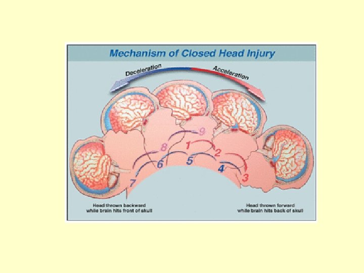
Head injury • Trauma is a type of injury which effects the body by external force being applied in a violent and sudden manner. When dealing with motorcycle accidents, it's important to understand the types of forces which a rider is subjected to, the body parts affected by these forces.
Head injury • First, let's define the different types of trauma. 1. Penetrating trauma 2. Blunt trauma 3. Acceleration/deceleration trauma
Head injury • Penetrating trauma is an object entering the body or head due to an object striking the body, or the body being placed in motion and striking an object which then penetrates the body. Blunt trauma occurs when an object strikes the body or head with force causing a compression of body tissues which results in injury.
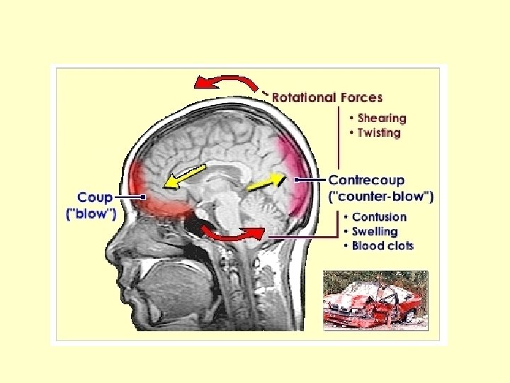
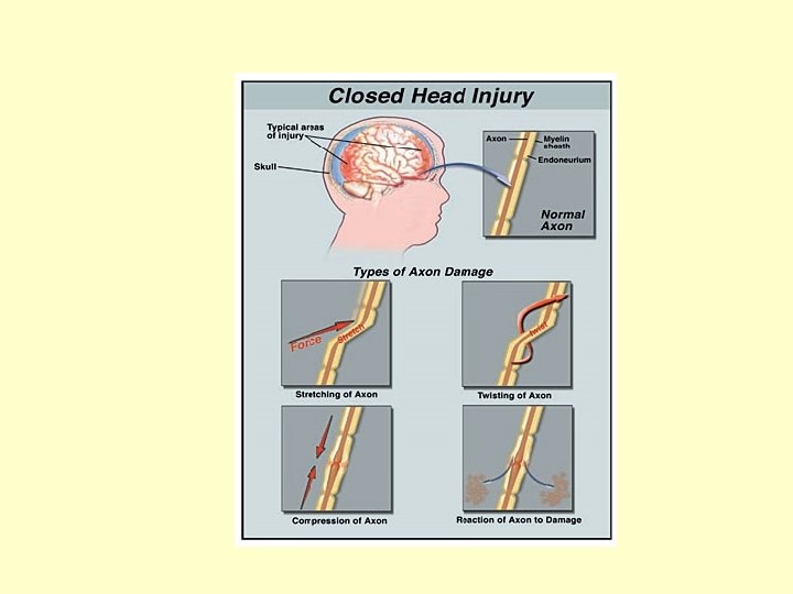
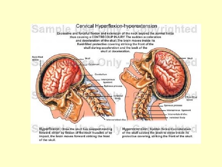
Head injury • Acceleration/deceleration trauma occurs when the body is moving and strikes another moving or stationary object. This results in • a change of motion for the body, or • a complete stop of motion, which causes stretching and tearing of body tissues • preťahovanie a trhanie telesných tkanív
Head injury • Concussion of brain – commotio cerebri • Compression of brain – subdural, epidural haematoma • Contussion of brain – intracerebral contussion with bleeding
Commotio cerebri • • • Reversible damage Generalised asynapsia Amnesia – retro or anterograde Unconsciousness +- short duration Desorientation Vomitus, headache
Commotio cerebri • Take care about patient !!! • Free interval after head injury • Development of subdural or epidural haematoma
• Bleeding from veins • Slower development than in epidural haematoma
Signs of subdural and epidural haematoma • • Anizokoria Bradykardia Hemiparesis Focal signs
Subdural haematoma
Brain CT Subdural haematoma Epidural haematoma
Isodense subdural haematoma • There is some midline shift to the left • The grey matter is apparently increased in thickness on the right due to the adjacent isodense blood • The sulcal pattern is effaced on the right • Where sulci are visible, they are displaced medially from the cranial vault by the thickness of the haematoma.
Epidural haematoma
Epidural haematoma • Bleeding from arteries • Quicker development • Bone fracture – often • Plane X-ray of the skull
Subdural, Epidural haematoma • Therapy • Epidural - emergency operation • Subdural - emergency operation or, in the small hematoma we can onitor the patient • After some time – chronic subdural hematoma
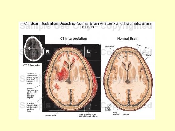
Traumatic intracerebral haemorrhage
Severe closed head injury • A) Noncontrasted axial computed tomography (CT) scan • (B) a fluid-attenuated inversion recovery (FLAIR) magnetic resonance (MR, right) image of a 10 -year-old boy 48 hours after he sustained a severe closed head injury. • The region of hyperintense signal in the brain stem visible on the MR image cannot be detected on the CT scan.
Consequences of head injury • • Posttraumatic parkinsonism Posttraumatic epilepsy Apalic syndrom Brain death
Apalic syndrom • Signs of damage of brain cortex (pallium) with not damaged brainstem • Brainstem reflexes are preserved, including spontaneous breathing and heart function • Patient is not able to be in verbal or visual contact with other people • Muscle hypertonia
Apalic syndrom • Patient is fed by nasogastric tube or by gastrostomia • Positive axial reflexes – grip, saccing reflexes • Patient is incontinent
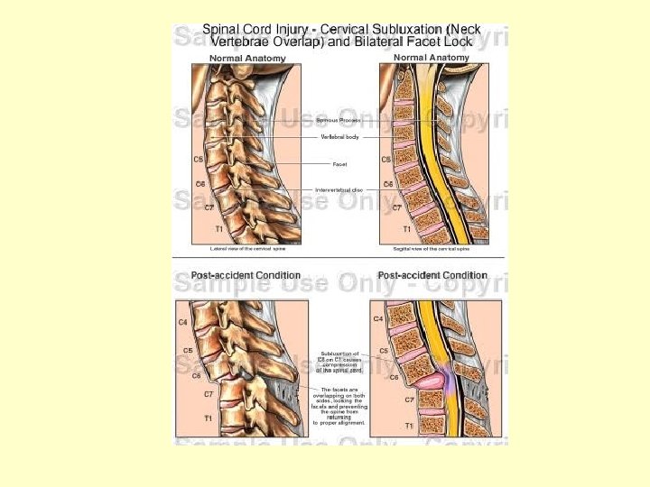
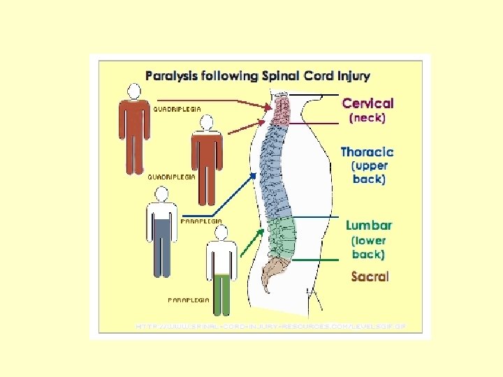
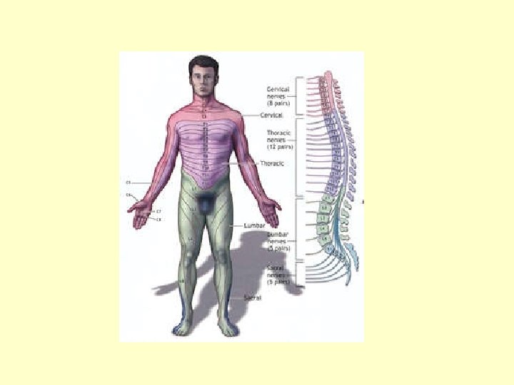
Spinal cord injury • A severe spinal cord injury often causes loss of feeling and paralysis • the loss of movement and voluntary control over the muscles in the body. • Spinal cord damage also causes loss of reflex function below the point of injury interrupting bodily functions such as breathing, bowel control, and bladder control. • In the event of a spinal injury prompt medical attention can help to minimize further spinal cord damage.
Spinal cord injury
Spinal cord injury
Spinal cord injury – first aid