Fig 1 Male reproductive system Fig 3 Structural


.")


 low and (b) high magnification.")


 front view and (b) lateral view.")


- Slides: 29
Fig 1: Male reproductive system
Fig. 3: Structural Organization of the Testis
Fig. 3 a: Section of testis in low magnification.
Fig. 3 b: Section of testis in high magnification.
Fig. 4: Testis (H&E stain, X 10).
Fig. 2: Stratified Seminiferous Epithelium
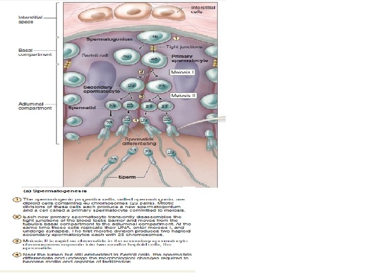
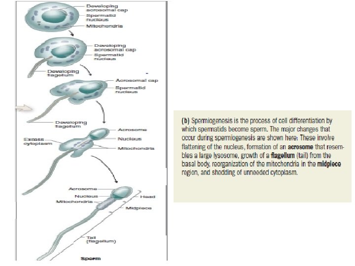
Fig. 5: Spermatogenesis. 2 n denotes diploid and n denotes haploid number of chromosomes.
Fig. 2: Structural Organization of the Testis and Epididymis.
Fig. 6: Section of epididymis in low magnification. Inset shows an enlarged view of a part of epididymis.
Fig. 7: Transverse section of the vas deferens in low magnification
Fig 8: Accessory glands of the male reproductive tract.
Fig 9: Section of prostate in high magnification
Fig 10: Structure of a Prostate
Fig 11: Transverse section of prostate gland showing the arrangement of various glands
Fig 12: Section of seminal vesicle in (a) low and (b) high magnification.
Fig 13: Section of bulbourethral gland in high magnification
Fig 14: Section of penis in low magnification
FEMALE REPRODUCTIVE SYSTEM
Fig 1: The female internal genitalia: (a) front view and (b) lateral view.
Fig 2: Section of ovary in low magnification.
Fig 3: Ovary
Fig 4: Section of ovary in high magnification
Fig 5: Cortex of the ovary showing different types o ovarian follicles
Fig 6: Cortex of the ovary showing primordial and primary follicles
Fig 7: Cortex of the ovary showing atretic follicle
Fig 8: Transverse section of fallopian tube in low magnification
Fig 9: Transverse section of fallopian tube in high magnification