Eczematous skin disorders Dr Ravneet Sekhon PGY 3

















- Slides: 45
Eczematous skin disorders Dr. Ravneet Sekhon, PGY 3 Pediatrics Telehealth Rounds January 27, 2017 Thanks to Dr. Ramien for her feedback and support in preparation of this presentation!
Objectives • • Describe the functions of skin Description of skin lesions Eczema and other dermatides Outpatient management of eczema
Functions of the skin • Protection – – – Protective barrier Radiation shield from UV light Mechanical, chemical, thermal insults Relatively impermeable to water Microbes • Sensation: receptors • Thermoregulation – Insulates from heat loss – Heat loss through evaporation of sweat • Metabolic function – Subcutaneous fat stores energy – Vitamin D synthesized in epidermis in presence of UV light
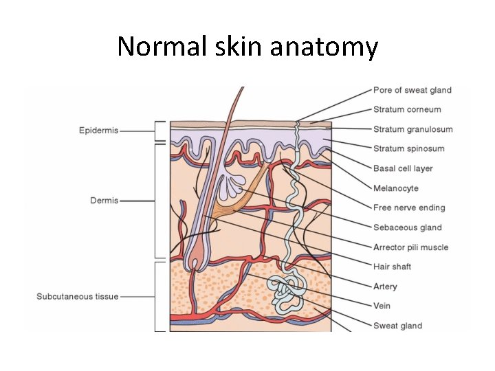
Normal skin anatomy
• Careful and detailed history should be gathered regarding onset, inciting factors, evolution of skin lesions, pruritus, medications/immunizations, family hx
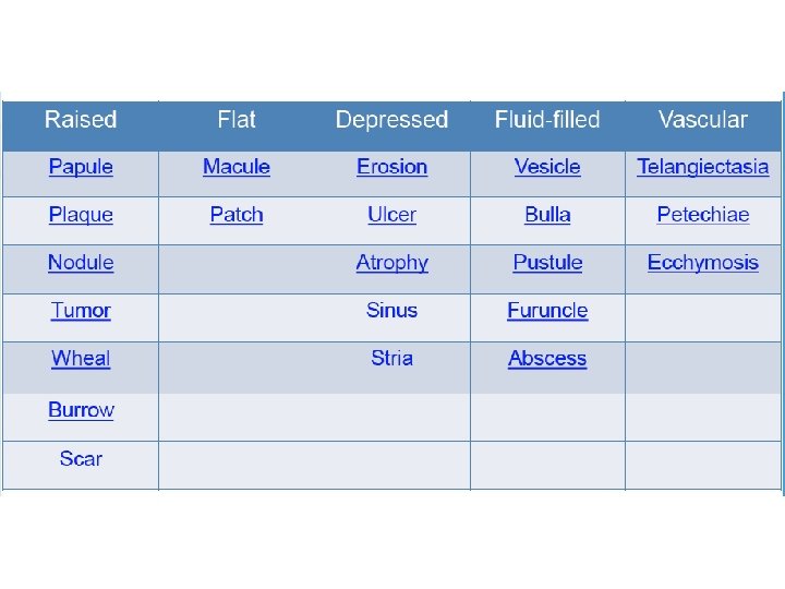
Describing skin lesions • Describe: – Size – Shape – Color – Texture – Configuration (relation to each other) – Distribution (where on the body)
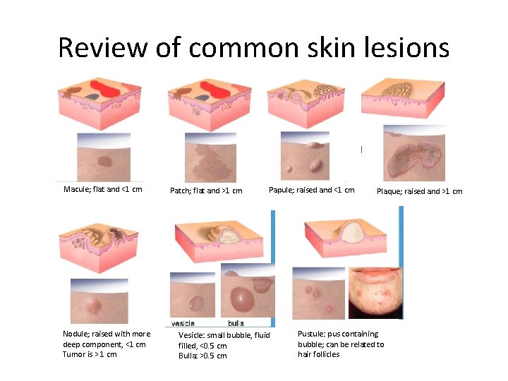
Review of common skin lesions Macule; flat and <1 cm Nodule; raised with more deep component, <1 cm Tumor is > 1 cm Patch; flat and >1 cm Papule; raised and <1 cm Vesicle: small bubble, fluid filled, <0. 5 cm Bulla: >0. 5 cm Plaque; raised and >1 cm Pustule: pus containing bubble; can be related to hair follicles
• Great resource with modules https: //www. aad. org/education/basic-dermcurriculum/suggested-order-ofmodules/morphology (American Academy of Derm)
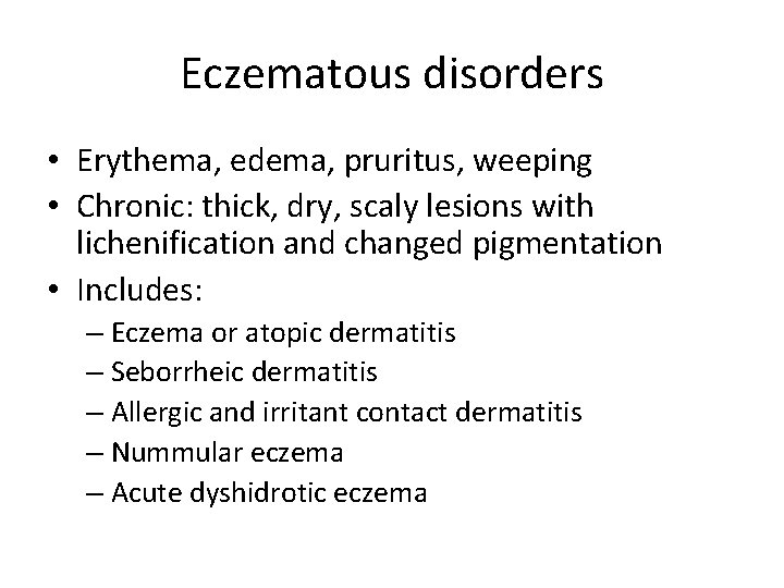
Eczematous disorders • Erythema, edema, pruritus, weeping • Chronic: thick, dry, scaly lesions with lichenification and changed pigmentation • Includes: – Eczema or atopic dermatitis – Seborrheic dermatitis – Allergic and irritant contact dermatitis – Nummular eczema – Acute dyshidrotic eczema
Diagnosis?
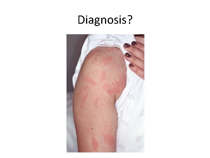
Nickel dermatitis • A type of allergic contact dermatitis • Due to prolonged or repetitive exposure • Improves after removal of stimulus but recurs quickly on re-exposure • Temporary treatment with topical or oral corticosteroids - what steroid, how much, and for how long? • Education of parents about cause
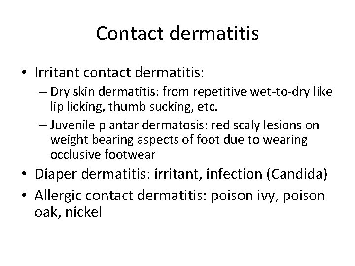
Contact dermatitis • Irritant contact dermatitis: – Dry skin dermatitis: from repetitive wet-to-dry like lip licking, thumb sucking, etc. – Juvenile plantar dermatosis: red scaly lesions on weight bearing aspects of foot due to wearing occlusive footwear • Diaper dermatitis: irritant, infection (Candida) • Allergic contact dermatitis: poison ivy, poison oak, nickel
Diagnosis?
Diagnosis?
Diagnosis?
Diagnosis?
Pityriasis alba • Mainly in kids 3 -16 yo, M=F • Hypopigmented, ill defined, round or oval patches • Can be mildly erythematous and have fine scales • Location: face, neck, upper trunk, prox arms • Frequently misdiagnosed: vitiligo, tinea versicolor, tinea corporis • Treatment: emollient, low potency topical steroid or calicineurin inhibitor
Diagnosis?
Seborrheic dermatitis • Also called cradle cap in infants • Greasy scaling of scalp, axillae, groin folds; in adults face, eyebrows, upper trunk • Salmon pink patches that can flake or peel • Not itchy • Infants<3 mo • Associated with proliferation of various Malassezia species • Treatment: keratolytics, topical antifungals, mild corticosteroids.
Diagnosis?
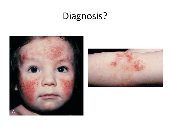
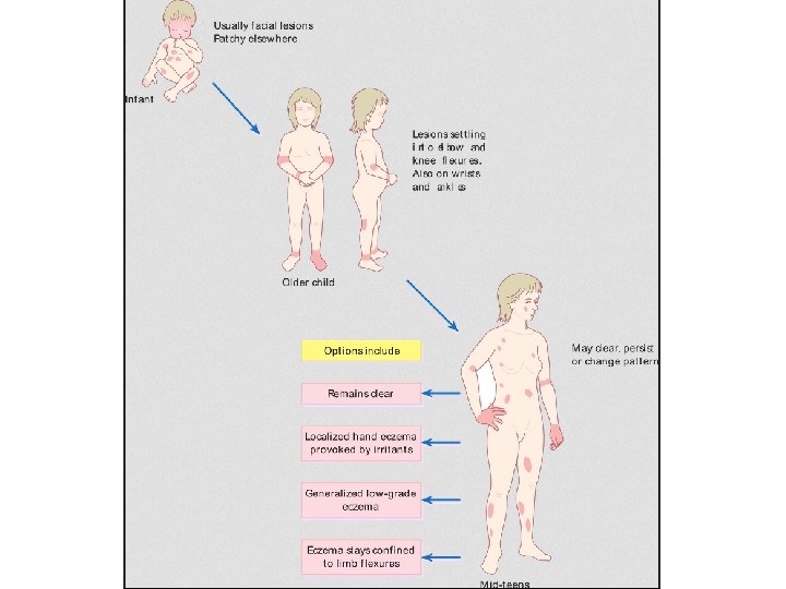
Atopic dermatitis • 10 -30% of kids worldwide • Most common relapsing-remitting skin condition • Occurs with other atopic dz like: asthma, allergic rhinitis, food allergy
Etiology Genetic mutations in filaggrin in up to 50% Defective skin barrier Reduced skin innate immune responses Exaggerated T cell response to environmental allergens and microbes • Chronic skin inflammation • •
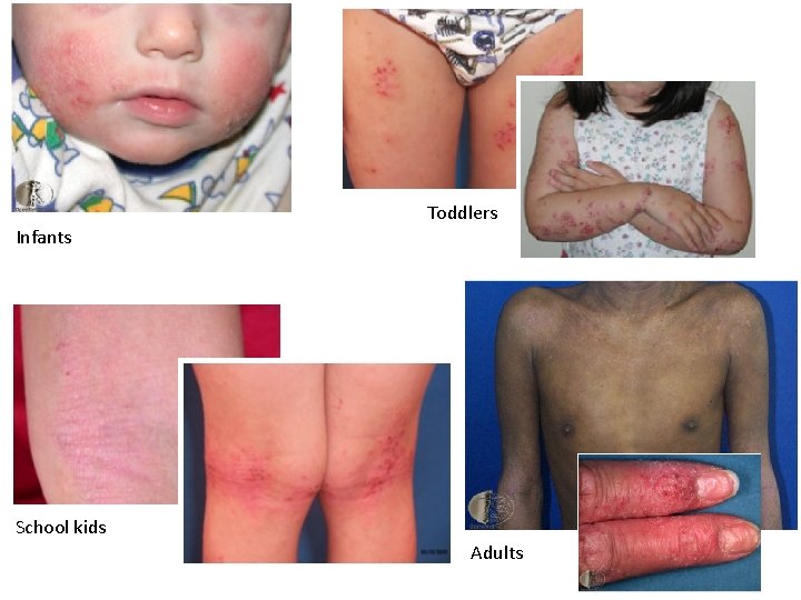
Toddlers Infants School kids Adults
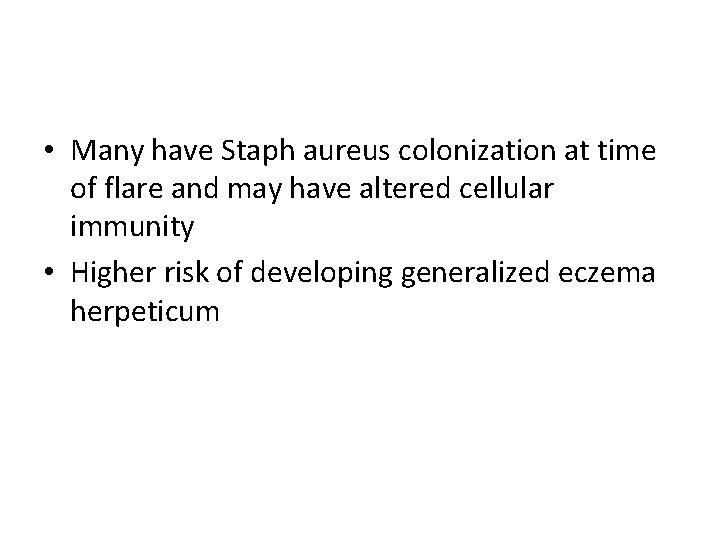
• Many have Staph aureus colonization at time of flare and may have altered cellular immunity • Higher risk of developing generalized eczema herpeticum
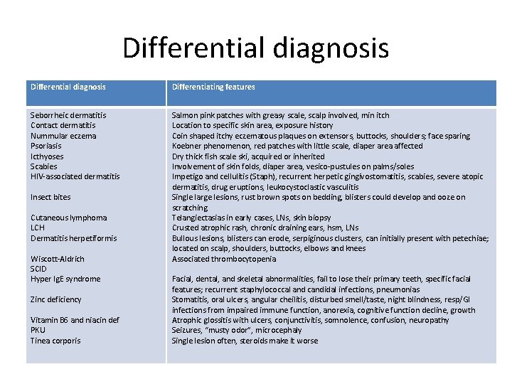
Differential diagnosis Differentiating features Seborrheic dermatitis Contact dermatitis Nummular eczema Psoriasis Icthyoses Scabies HIV-associated dermatitis Salmon pink patches with greasy scale, scalp involved, min itch Location to specific skin area, exposure history Coin shaped itchy eczematous plaques on extensors, buttocks, shoulders; face sparing Koebner phenomenon, red patches with little scale, diaper area affected Dry thick fish scale ski, acquired or inherited Involvement of skin folds, diaper area, vesico-pustules on palms/soles Impetigo and cellulitis (Staph), recurrent herpetic gingivostomatitis, scabies, severe atopic dermatitis, drug eruptions, leukocystoclastic vasculitis Single large lesions, rust brown spots on bedding, blisters could develop and ooze on scratching Telangiectasias in early cases, LNs, skin biopsy Crusted atrophic rash, chronic draining ears, hsm, LNs Bullous lesions, blisters can erode, serpiginous clusters, can initially present with petechiae; located on scalp, shoulders, buttocks, elbows and knees Associated thrombocytopenia Insect bites Cutaneous lymphoma LCH Dermatitis herpetiformis Wiscott-Aldrich SCID Hyper Ig. E syndrome Zinc deficiency Vitamin B 6 and niacin def PKU Tinea corporis Facial, dental, and skeletal abnormalities, fail to lose their primary teeth, specific facial features; recurrent staphylococcal and candidal infections, pneumonias Stomatitis, oral ulcers, angular cheilitis, disturbed smell/taste, night blindness, resp/GI infections from impaired immune function, anorexia, cognitive function decline, growth Atrophic glossitis with ulcers, conjunctivitis, somnolence, confusion, neuropathy Seizures, “musty odor”, microcephaly Single lesion often, steroids make it worse
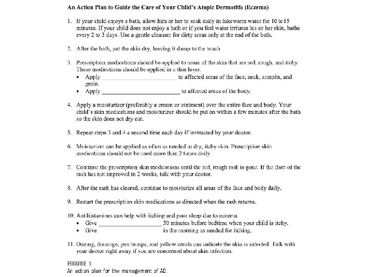
Management • Prevention – Bathing: lukewarm baths or showers; mild skin cleanser (unscented); pat skin dry – Moisturize: traps water in the skin and improves barrier function. Can be applied throughout the day PRN. Aveeno, Glaxal base, Cliniderm, Vaseline, etc. – Clothing: avoid wool, rough clothes; prefer cotton. Loose, light clothes in warm weather. – Bedroom: • Vacuum bedroom carpet daily • Use special mattress and pillow covers under cotton liner or mattress cover • Change furnace filters q 3 -4 months • Wash sheets and mattress cover weekly • Humidifier
– Replace scratching with moisturizing, etc. – Wet wrap overnight, after moisturizing or medicated creams are applied (for flares) WET WRAP VIDEO: https: //www. youtube. com/watch? v=CMX_AEu_Oro
• Treatment – Topical corticosteroids: what can you apply and where? – Treat the itch!: antihistamines orally, also has sedative effect for sleep – Antibiotics for superinfections
What kind of topical preparation to use? • The various preparations: – Ointment: • Less likely to produce burning/stinging (no preservatives) • More effect with the same active ingredient and concentration bc of their occlusive effect • Need only to apply a thin layer to affected areas until they are smooth to touch – Cream: more cosmetically acceptable – Lotion: good for hair-bearing areas – Oil based: easier to spread (esp useful in large areas of skin are involved) and are not too strong (useful for mild-mod eczema); Ex. Dermasmooth – Foam
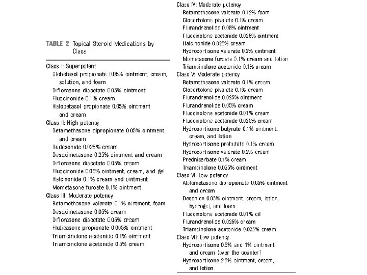
Topical corticosteroids • Mild: hydrocortisone 1% ointment to red raised itchy areas on face or folds BID PRN • Mild-mod: Desonide 0. 05% ointment on face or body BID PRN • Moderate: Betaderm 0. 1% ointment to body BID PRN • Severe: Clobetasol propionate 0. 05% to palms and soles BID PRN; not on body usually
• Risks of steroid use: – Cutaneous atrophy – Striae – Telangiectasias – Systemic absorption -> adrenal suppression – Around mouth: peri-orificial dermatitis – Around eyes: intraocular HTN, cataracts
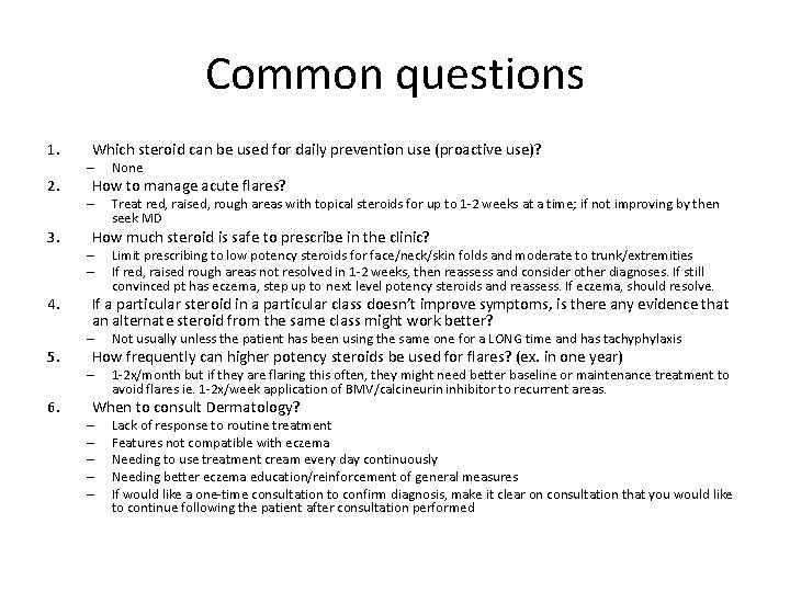
Common questions 1. 2. 3. 4. 5. 6. Which steroid can be used for daily prevention use (proactive use)? – None How to manage acute flares? – Treat red, raised, rough areas with topical steroids for up to 1 -2 weeks at a time; if not improving by then seek MD How much steroid is safe to prescribe in the clinic? – – Limit prescribing to low potency steroids for face/neck/skin folds and moderate to trunk/extremities If red, raised rough areas not resolved in 1 -2 weeks, then reassess and consider other diagnoses. If still convinced pt has eczema, step up to next level potency steroids and reassess. If eczema, should resolve. If a particular steroid in a particular class doesn’t improve symptoms, is there any evidence that an alternate steroid from the same class might work better? – Not usually unless the patient has been using the same one for a LONG time and has tachyphylaxis How frequently can higher potency steroids be used for flares? (ex. in one year) – 1 -2 x/month but if they are flaring this often, they might need better baseline or maintenance treatment to avoid flares ie. 1 -2 x/week application of BMV/calcineurin inhibitor to recurrent areas. When to consult Dermatology? – – – Lack of response to routine treatment Features not compatible with eczema Needing to use treatment cream every day continuously Needing better eczema education/reinforcement of general measures If would like a one-time consultation to confirm diagnosis, make it clear on consultation that you would like to continue following the patient after consultation performed
Topical calcineurin inhibitors • Topical immuno-suppressants • Inhibit T cell activation • 2 forms: – Tacrolimus (Protopic) 0. 03% and 0. 1% ointment; only 0. 03% approved for 2 -15 yo kids – Pimecrolimus 1% cream • • Second line therapy for recurrent areas Adverse effect profile is different from steroids More costly Adverse effects: – Stinging/burning, pruritus – tend to be brief (few days) – Rare cases of skin malignancy and lymphoma reported; continuous long term use not recommended and limit application to affected areas only – Prolonged systemic exposure associated with inc risk of infections – Unknown if interferes with skin response to UV damage; minimize/avoid sun • Health Canada recommends if sx do not improve in 6 weeks of BID treatment, then discontinue and reassess diagnosis
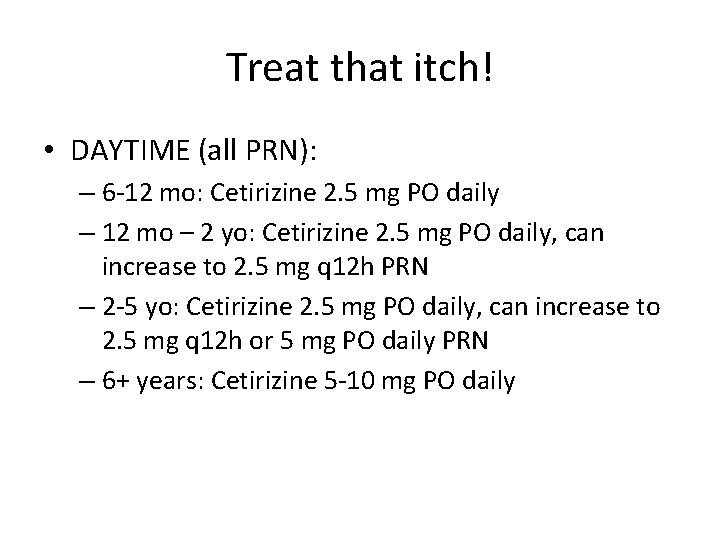
Treat that itch! • DAYTIME (all PRN): – 6 -12 mo: Cetirizine 2. 5 mg PO daily – 12 mo – 2 yo: Cetirizine 2. 5 mg PO daily, can increase to 2. 5 mg q 12 h PRN – 2 -5 yo: Cetirizine 2. 5 mg PO daily, can increase to 2. 5 mg q 12 h or 5 mg PO daily PRN – 6+ years: Cetirizine 5 -10 mg PO daily
• NIGHT TIME (all PRN) – Benadryl 0. 5 mg/kg/dose PO qhs – Hydroxyzine (Atarax) 0. 5 -1 mg/kg/dose PO qhs, max 25 -50 mg single dose
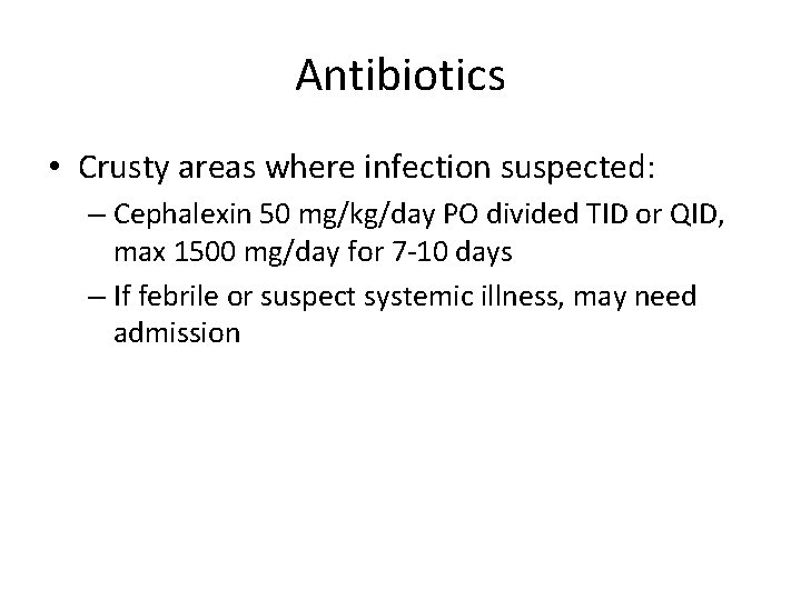
Antibiotics • Crusty areas where infection suspected: – Cephalexin 50 mg/kg/day PO divided TID or QID, max 1500 mg/day for 7 -10 days – If febrile or suspect systemic illness, may need admission
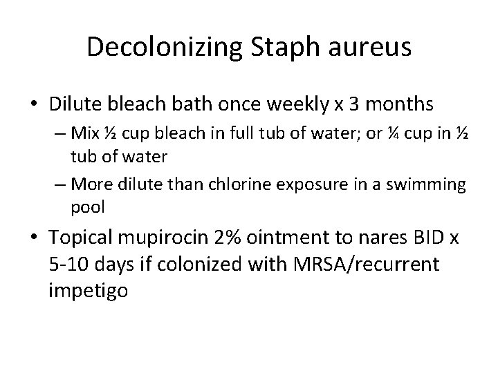
Decolonizing Staph aureus • Dilute bleach bath once weekly x 3 months – Mix ½ cup bleach in full tub of water; or ¼ cup in ½ tub of water – More dilute than chlorine exposure in a swimming pool • Topical mupirocin 2% ointment to nares BID x 5 -10 days if colonized with MRSA/recurrent impetigo
Thank-you!
References 1. Nelson’s textbook of Pediatrics 2. Zitelli’s atlas of pediatric physical diagnoses: 5 th edition 3. New Zealand website on Dermatology http: //www. dermnetnz. org/ 4. AAP Article on Eczema Nov 2014 http: //pediatrics. aappublications. org/conten t/pediatrics/early/2014/11/18/peds. 20142812. full. pdf