SALIVARY GLAND DISEASES omr Introduction Classification of salivary


")


 u Submandibular Ganglion (para)")


 u Myoepithelial cells u")


 per 24 hours. u")


 u. Radiation theraphy u.")


- Slides: 50
SALIVARY GLAND DISEASES omr
Introduction
Classification of salivary glands: Ø Ø Ø According to size. u Major : - Parotid / Submandibular /Sublingual u Minor : - Lingual / buccal / labial / palatine / glossopalatine According to nature of secretion. u Serous : - Parotid u Mucous : - Sublingual / minor u Mixed : - Submandibular / minor According to location of the opening of duct. u Into vestibule : - Parotid / minor u Into oral cavity proper : - Submandibular / Sublingual / minor
Embryology u 6 th-8 th Weeks of Gestation u Parotid u. First to develop u. Last to become encapsulated u Autonomic Nervous System Crucial
Embryology
Anatomy: Parotid Gland u Wedge shaped with 5 processes u 3 Superficial u 2 Deep u Parotid Compartment u Superior – Zygoma u Posterior – EAC u Inferior – Styloid, ICA, Jugular Veins
Anatomy: Parotid Gland u 80% overlies Masseter & Mandible u 20% Retromandibular u Stylomandibular Tunnel, Isthmus of Parotid u Tail of Parotid
Anatomy: Parotid Gland u. Parapharyngeal u. Prestyloid Space Compartment u. Poststyloid Compartment (Paragangliomas)
Anatomy: Parotid Gland u Stensen’s u. Arises u 1. 5 from anterior border cm inferior to Zygomatic arch u. Pierces u 4 -6 u 5 Duct Buccinator at 2 nd Molar cm in length mm in diameter
Anatomy: Parotid Gland u Parotid Capsule u. Superficial layer Deep Cervical Fascia u. Superficial u. Deep layer
Anatomy: Parotid Gland u CN VII u 2 Surgical zones u 3 Motor branches immediately u Pes Anserinus – 1. 3 cm u Temperofacial u Cervicofacial u 5 Division Terminal branches
Anatomy: Parotid Gland
Anatomy: Parotid Gland u Localization of CN VII u Tragal pointer u Tympanomastoid suture u Posterior belly Digastric u Styloid process u Retrograde dissection u Mastoidectomy
Anatomy: Parotid Gland u Great Auricular nerve u Auriculotemporal nerve u Superficial Temporal vessels u Frey’s Syndrome
Anatomy: Parotid Gland u Neural u VII, compartment Great Auricular, Auriculotemporal u Venous compartment u Retromandibular u Arterial vein compartment u Superficial Temporal/Transverse Facial
Anatomy: Parotid Gland u Lymphatics u Paraparotid u Superficial & Intraparotid nodes & Deep Cervical nodes
Anatomy: Submandibular Gland u The ‘Submaxilla’ u Submandibular Triangle u Mylohyoid ‘C’ u Marginal Mandibular branch u Capsule from superficial layer of Deep Cervical fascia
Anatomy: Submandibular Gland u Wharton’s duct u Exits medial surface u Between Mylohyoid & Hyoglossus u 5 cm in length u Lingual nerve & CN XII
Anatomy: Submandibular Gland
Anatomy: Submandibular Gland u Innervation u Superior Cervical Ganglion (symp) u Submandibular Ganglion (para) u Artery: Submental branch of Facial a. u Vein: Anterior Facial vn. u Lymphatics: Deep Cervical and Jugular chains u Facial artery nodes
Anatomy: Sublingual Gland u Between Mandible & Genioglossus u No capsule u Ducts of Rivinus +/- Bartholin’s duct u Sialogram not possible u Innervation: Same as Submandibular u Artery/Vein: Sublingual branch of Lingual & Submental branch of Facial u Lymphatics: Submandibular nodes
Anatomy: Sublingual Gland
Anatomy: Minor Salivary Glands u 600 -1, 000 u Simple ducts u Buccal, Labial, Palatal, Lingual u Tumor sites: Palate, upper lip, cheek u Lingual & Palatine nn.
INVESTIGATIONS: 1. IMAGING u Radiography u Sialography u CT scan u MRI u Ultrasound Contd. .
2. PATHOLOGY u Aspiration cytology & biopsy for definitive diagnosis u Frozen section u Histochemistry u Immunohistochemistry u Electron microscophy Contd. .
3. FUNCTIONAL TESTS u Flow rates & sialochemistry 4. TESTS FOR RELATED / CONTRIBUTORY SYSTEMIC DISEASE u Bacteriology u Haematology u Autoantibody studies Contd. .
Imaging u CT – Inflammatory u MR – Tumor u Children: U/S & MR u NO sialogram during active infection u Parotid is fatty
Microanatomy u The Secretory Unit u Acinus (serous, mucous, mixed) u Myoepithelial cells u Intercalated duct u Striated duct u Excretory duct
Microanatomy u Striated & Intercalated ducts well developed in serous, NOT mucous glands u Striated duct: HCO 3 into, Cl from lumen u Intercalated duct: K into lumen, Na from lumen, producing hypotonic fluid u Excretory ducts do NOT modify saliva
Microanatomy
Microanatomy u The Bicellular Theory u Intercalated duct u Excretory duct u The Multicellular Theory
Microanatomy u Parotid: serous & fatty u Submandibular: mixed serous u Sublingual: mixed mucous u Stroma: Plasma cells
Microanatomy
Microanatomy
Saliva u 600 to 700 ml (upto 1. 5 L) per 24 hours. u PH is 6. 7 (6. 2 – 7. 6) u Contribution of various glands: u. Parotid: - 60 – 65% u. Submandibular: - 20 – 30% u. Sublingual: - 2 – 5% u. Minor glands: - 6 – 7%
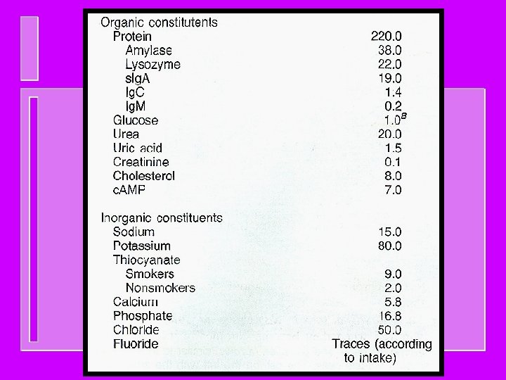
Composition of saliva: u Water : - 94. 0 – 99. 5% u Solids : - 6. 0% (unstimulated), 0. 5% (stimulated) u organic constituents: -urea, uric acid, glucose, aminoacid, lactate, fatty acids, proteins like amylase, peroxidase, lysosyme, Ig. A, Ig. M, Ig. G. u inorganic constituents: - Ca, Mg, F, HCO 3, K, Na, Cl, NH 4. u gases: - CO 2, N 2, O 2. u constituents from oral cavity: desquamative epithelial cells, bacteria.
Function of Saliva u. Inhibition of dental caries u. Water balance u. Lubrication u. Taste action of food u. Buffering u. Hygienic action u Digestion (Amylase, Lipase) u Antibacterial (Lysozyme, Ig. A, Peroxidase, FLOW) u Mineralization u Protective Pellicle
Factors influencing salivary secretion: u. Taste and smell u. Mechanical stimulation of oral mucosa and gingiva u. Mastication of food u. Chemical irritation of oral mucosa & stomach u. Distension / irritation of oesophagus u. Pregnancy
Causes of salivary gland dysfunction: u. Drugs (anticholinergic, antihistamine, antihypertensive) u. Radiation theraphy u. Oncologic chemotheraphy u. Psychological u. Systemic factors diseases u. Malnutrition and neoplasms
Clinical features: u Commonly present as an asymptomatic mass u Pain – infection/ hemorrhage/ infected cyst u Malignant lesions are also symtomless unless involves the nerves u Nasal obstruction – tumor involves paranasal sinuses / nose u Xerostomia
Salivary hypofunction u. Candidiasis u. Lichen Planus u. Burning Mouth u. Aphthous u. Dental ulcers caries u. Xerostomia reliable not
XEROSTOMIA u Causes; u. Depression / chronic anxiety u. Dehydration u. Drugs u. Diseases
XEROSTOMIA u Clinical features; u. Dry mouth u. Salivary u. Rapid froath adhering to mucosa dental decay u. Adherence u. Soreness of food debris to teeth / halitosis
XEROSTOMIA u MANAGEMENT u. Preventive theraphy u. Symptomatic treatment u. Local or topical salivary stimulation u. Systemic salivary stimulation
Effects of Aging u Total salivary flow independent of age u Acinar cells degenerate with age u Submandibular gland more sensitive to metabolic/physiologic change u Unstimulated salivary flow more greatly affected by physiologic changes
CLASSIFICATION OF DISEASES u DEVELOPMENTAL u APLASIA / HYPOPLASIA u ACCESSORY SALIVARY DUCTS u DIVERTICULI u DARIER’S DISEASE u ACQUIRED u REACTIVE LESIONS u INFECTIONS u METABOLIC CONDITIONS u ASSOCIATED IMMUNE DEFECTS u NEOPLASMS
NEOPLASMS BENIGN u. Pleomorphic adenoma u. Monomorphic adenoma u. Ductal papilloma MALIGNANT u Mucoepidermoid carcinoma u Adenoid cystic carcinoma u Acinic cell carcinoma u Salivary duct carcinoma u Squamus cell carcinoma u Basal cell adenocarcinoma u Adenocarcinoma