DYNAMIC AUSCULTATION Dr G Gnanavelu Reader in Cardiology


- Slides: 47
DYNAMIC AUSCULTATION Dr. G. Gnanavelu Reader in Cardiology Madras Medical College, Chennai 3
DEFINITION ‘Technique of altering circulatory dynamics by means of respiration and variety of physiological and pharmacological maneuvers and determining their effects on heart sounds and murmurs’
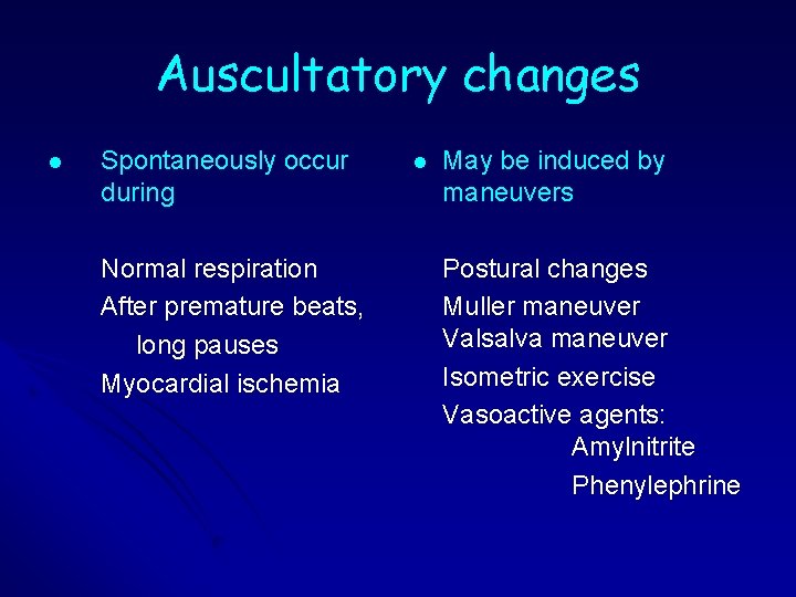
Auscultatory changes l Spontaneously occur during Normal respiration After premature beats, long pauses Myocardial ischemia l May be induced by maneuvers Postural changes Muller maneuver Valsalva maneuver Isometric exercise Vasoactive agents: Amylnitrite Phenylephrine
DYNAMIC AUSCULTATION Proper assessment requires l Good stethoscope l Quiet room l Cooperative patient l Bare chest l Intact autonomic function and normovolemia l Knowledge about the maneuver and the changes expected
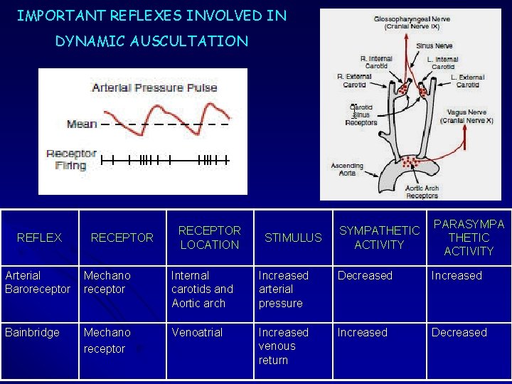
IMPORTANT REFLEXES INVOLVED IN DYNAMIC AUSCULTATION REFLEX RECEPTOR LOCATION STIMULUS SYMPATHETIC ACTIVITY PARASYMPA THETIC ACTIVITY Arterial Baroreceptor Mechano receptor Internal carotids and Aortic arch Increased arterial pressure Decreased Increased Bainbridge Mechano receptor Venoatrial Increased venous return Increased Decreased
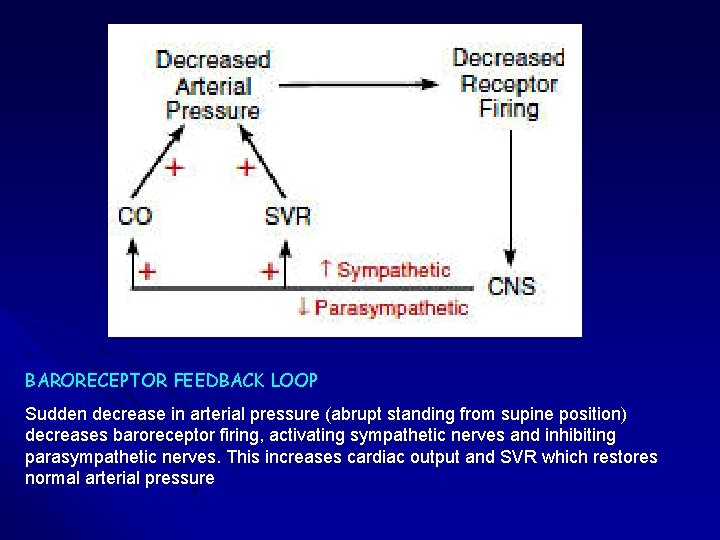
BARORECEPTOR FEEDBACK LOOP Sudden decrease in arterial pressure (abrupt standing from supine position) decreases baroreceptor firing, activating sympathetic nerves and inhibiting parasympathetic nerves. This increases cardiac output and SVR which restores normal arterial pressure
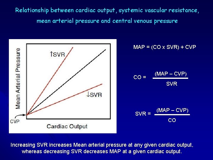
Relationship between cardiac output, systemic vascular resistance, mean arterial pressure and central venous pressure MAP = (CO x SVR) + CVP CO = (MAP – CVP) SVR = (MAP – CVP) CO Increasing SVR increases Mean arterial pressure at any given cardiac output, whereas decreasing SVR decreases MAP at a given cardiac output.
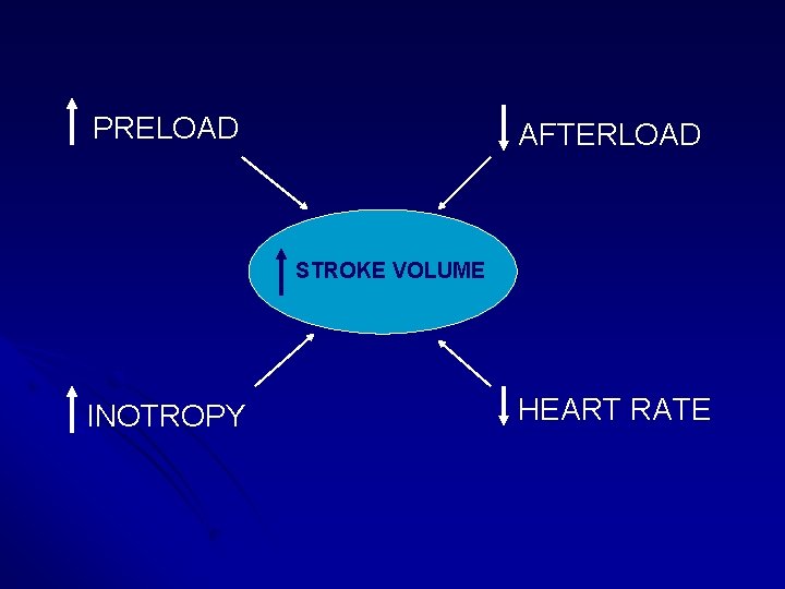
PRELOAD AFTERLOAD STROKE VOLUME INOTROPY HEART RATE
RESPIRATION l l l Assess changes during normal respiration Patient should be in semiupright or sitting posture More pronounced & consistent alterations on murmur originating from right than from left side of heart In RV failure and PHT, no increase in venous return with inspiration, hence no inspiratory augmentation of right sided murmurs and gallops Absence of respiratory influence is of no particular diagnostic value. Effects of inspiration may be accentuated by Muller maneuver.
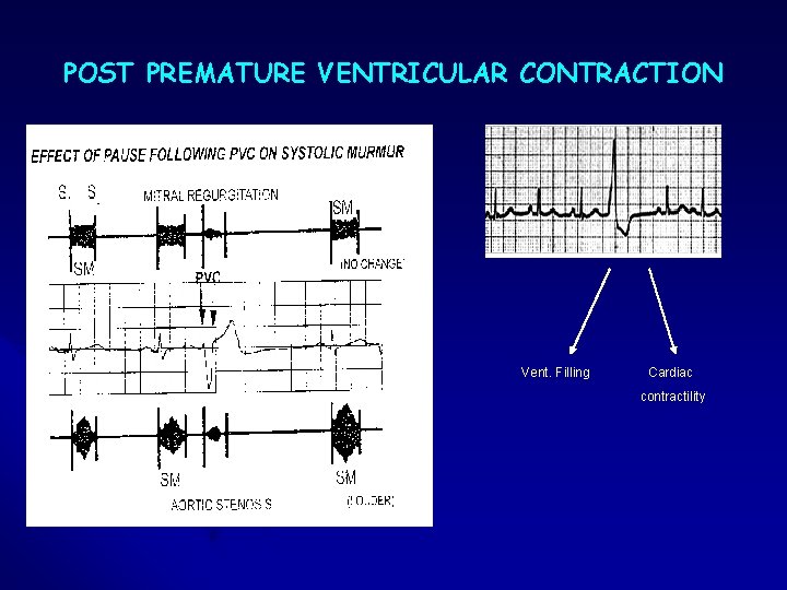
POST PREMATURE VENTRICULAR CONTRACTION Vent. Filling Cardiac contractility
TRANSIENT MYOCARDIAL ISCHEMIA Hemodynamic changes during spontaneous angina or induced by exercise: Increase in LVEDP Papillary muscle dysfunction Reduced stroke volume Increase in arterial pressure Auscultatory changes: Paradoxic splitting of S 2 becomes obvious S 4 may develop during angina Late or midsystolic murmur of Papillary muscle dysfunction may appear or gets augmented
STANDING Rapid standing or sitting up from lying position or rapid standing from squatting posture results in * decreased venous return due to venous pooling in legs and splanchnic vessels leading to decreased stroke volume, decreased mean arterial pressure & decrease in heart size followed by reflex increase in heart rate & systemic resistance
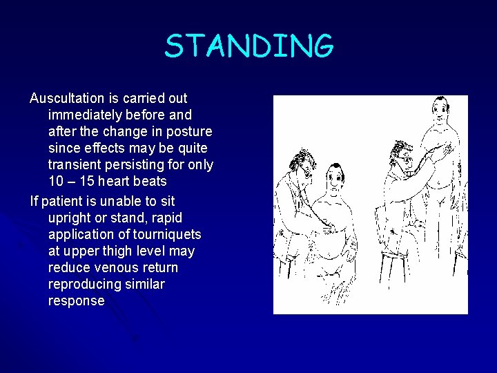
STANDING Auscultation is carried out immediately before and after the change in posture since effects may be quite transient persisting for only 10 – 15 heart beats If patient is unable to sit upright or stand, rapid application of tourniquets at upper thigh level may reduce venous return reproducing similar response
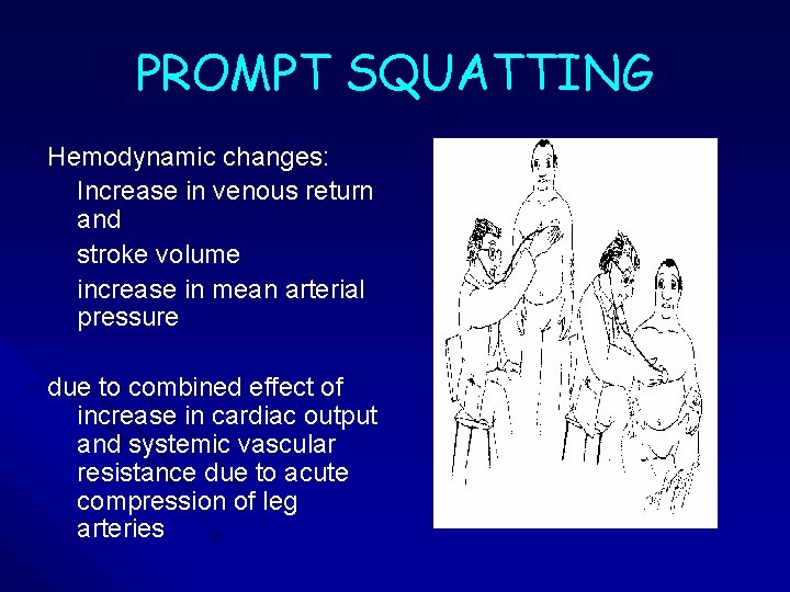
PROMPT SQUATTING Hemodynamic changes: Increase in venous return and stroke volume increase in mean arterial pressure due to combined effect of increase in cardiac output and systemic vascular resistance due to acute compression of leg arteries
PROMPT SQUATTING Examiner sits on a chair with stethoscope in place while patient is still standing to assess changes occurring within first few beats after squatting In patients unable to squat, similar circulatory changes can be produced by bending patient’s knees on his abdomen in supine position
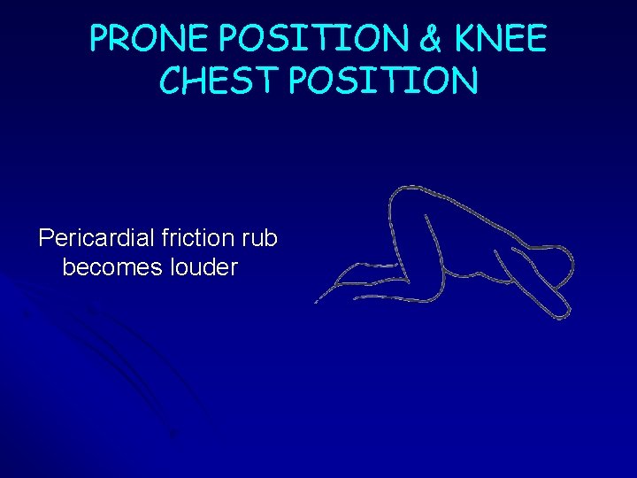
PRONE POSITION & KNEE CHEST POSITION Pericardial friction rub becomes louder
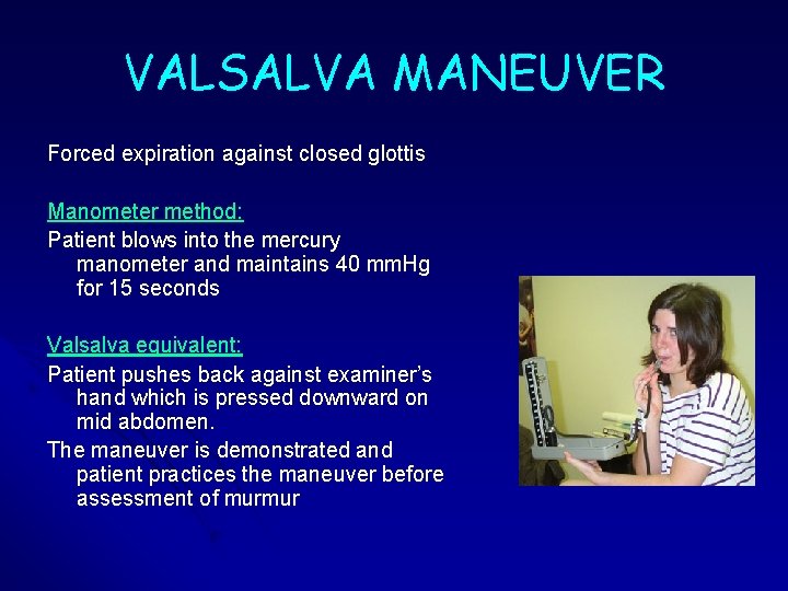
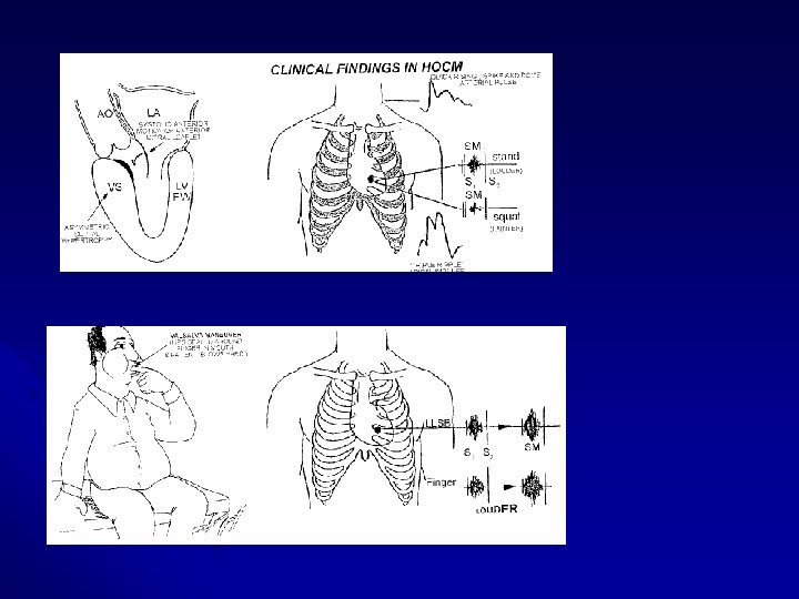
VALSALVA MANEUVER Forced expiration against closed glottis Manometer method: Patient blows into the mercury manometer and maintains 40 mm. Hg for 15 seconds Valsalva equivalent: Patient pushes back against examiner’s hand which is pressed downward on mid abdomen. The maneuver is demonstrated and patient practices the maneuver before assessment of murmur
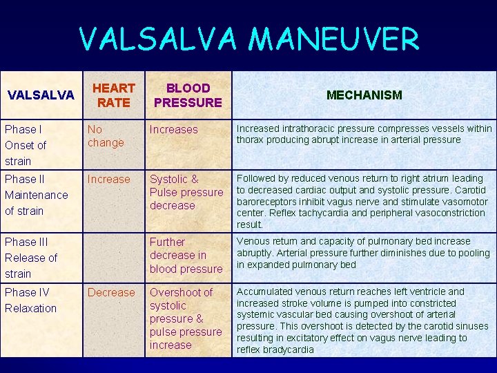
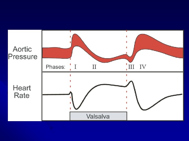
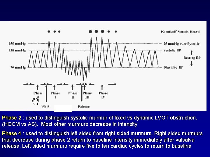
VALSALVA MANEUVER VALSALVA HEART RATE BLOOD PRESSURE MECHANISM Phase I Onset of strain No change Increases Increased intrathoracic pressure compresses vessels within thorax producing abrupt increase in arterial pressure Phase II Maintenance of strain Increase Systolic & Pulse pressure decrease Followed by reduced venous return to right atrium leading to decreased cardiac output and systolic pressure. Carotid baroreceptors inhibit vagus nerve and stimulate vasomotor center. Reflex tachycardia and peripheral vasoconstriction result. Further decrease in blood pressure Venous return and capacity of pulmonary bed increase abruptly. Arterial pressure further diminishes due to pooling in expanded pulmonary bed Overshoot of systolic pressure & pulse pressure increase Accumulated venous return reaches left ventricle and increased stroke volume is pumped into constricted systemic vascular bed causing overshoot of arterial pressure. This overshoot is detected by the carotid sinuses resulting in excitatory effect on vagus nerve leading to reflex bradycardia Phase III Release of strain Phase IV Relaxation Decrease
Phase 2 : used to distinguish systolic murmur of fixed vs dynamic LVOT obstruction. (HOCM vs AS). Most other murmurs decrease in intensity Phase 4 : used to distinguish left sided from right sided murmurs. Right sided murmurs that decrease during phase 2 return to baseline intensity immediately after valsalva release. Left sided murmurs require five to ten cardiac cycles to return to baseline
VALSALVA MANEUVER Mechanical and reflex changes depend on 1. level of cardiac function and effective central blood volume 2. speed and magnitude of baroreceptor responses to change in arterial pressure
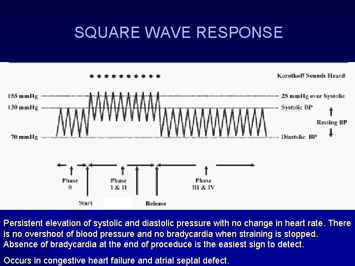
SQUARE WAVE RESPONSE Persistent elevation of systolic and diastolic pressure with no change in heart rate. There is no overshoot of blood pressure and no bradycardia when straining is stopped. Absence of bradycardia at the end of proceduce is the easiest sign to detect. Occurs in congestive heart failure and atrial septal defect.
VALSALVA MANEUVER Apart from induced auscultatory changes Used to assess 1. Autonomic function 2. Left ventricular function Used to treat Supraventricular tachycardias
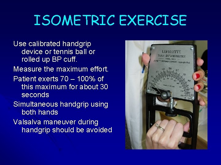
ISOMETRIC EXERCISE Use calibrated handgrip device or tennis ball or rolled up BP cuff. Measure the maximum effort. Patient exerts 70 – 100% of this maximum for about 30 seconds Simultaneous handgrip using both hands Valsalva maneuver during handgrip should be avoided
ISOMETRIC EXERCISE l Avoid in those with ventricular arrhythmias and myocardial ischemia l Contraindicated in recent myocardial infarction, uncontrolled hypertension, cerebrovascular disease, suspected aortic dissection
ISOMETRIC EXERCISE Hemodynamic changes: Significant increase in Arterial pressure Heart rate Cardiac output LV filling pressure Heart size
PHARMACOLOGICAL AGENTS Commonly two agents are used 1. Amylnitrite - Vasodilator 2. Phenylephrine - Vasoconstrictor
AMYL NITRITE Patient in supine position; Perform baseline auscultation 0. 3 ml ampoule of Amyl nitrite placed in gauze is crushed with gloved hand near patient’s nose Warn patient that he will smell some vapors smelling like sweet almond or dirty socks Patient takes three or four deep breaths over 10 to 15 seconds Preferable to have another person to monitor systolic BP and call the level as drug takes effect Patient remains in supine position till the effect wears off
AMYL NITRITE Hemodynamic changes: Initial vasodilatation leading to reduction in systemic arterial pressure Next 1 - 2 mins. - Reflex tachycardia and venoconstriction lead to increase in cardiac output and velocity of blood flow
AMYLNITRITE vs NITROGLYCERIN Both lead to reduction in BP due to systemic vasodilatation l Due to reflex tachycardia and venoconstriction Amylnitrite leads to increased venous return l Venous return falls with Nitroglycerine due to venodilatation l
PHENYLEPHRINE Patient in supine position; Baseline auscultation Dilute 10 mg vial of Phenylephrine in 250 ml of isotonic saline 0. 3 to 0. 5 mg IV over 30 sec. Alternately can be given as infusion 0. 1 mg/min. infusion stopped once 20 mm Hg rise in BP occurs or when intensity of murmur clearly changes It has shorter duration of action and elevates BP for only 3 – 5 mins.
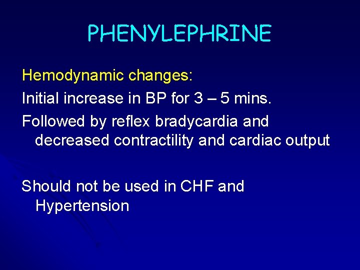
PHENYLEPHRINE Hemodynamic changes: Initial increase in BP for 3 – 5 mins. Followed by reflex bradycardia and decreased contractility and cardiac output Should not be used in CHF and Hypertension
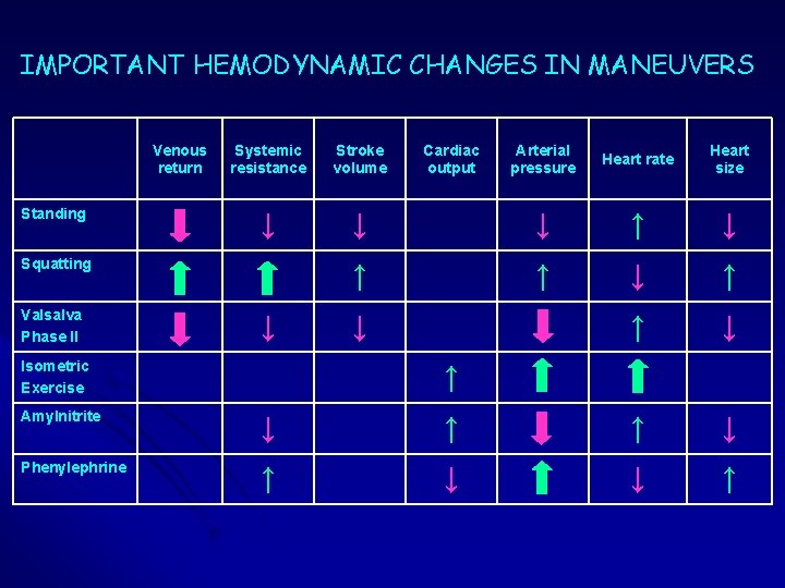
IMPORTANT HEMODYNAMIC CHANGES IN MANEUVERS Venous return Standing Systemic resistance Stroke volume ↓ Squatting Valsalva Phase II ↓ Isometric Exercise Cardiac output Arterial pressure Heart rate Heart size ↓ ↓ ↑ ↑ ↓ ↓ ↑ Amylnitrite ↓ ↑ ↑ ↓ Phenylephrine ↑ ↓ ↓ ↑
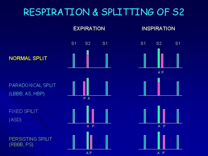
RESPIRATION & SPLITTING OF S 2 EXPIRATION S 1 S 2 S 1 INSPIRATION S 1 S 2 NORMAL SPLIT AP PARADOXICAL SPLIT (LBBB, AS, HBP) P A FIXED SPILIT (ASD) A P AP A P PERSISTING SPILIT (RBBB, PS) S 1
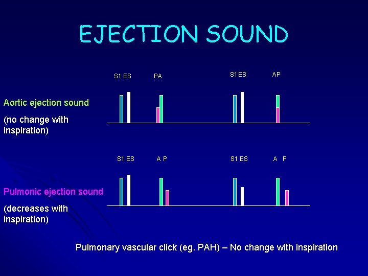
EJECTION SOUND S 1 ES PA S 1 ES AP S 1 ES A P Aortic ejection sound (no change with inspiration) S 1 ES AP Pulmonic ejection sound (decreases with inspiration) Pulmonary vascular click (eg. PAH) – No change with inspiration
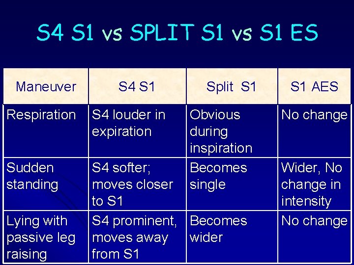
S 4 S 1 vs SPLIT S 1 vs S 1 ES Maneuver S 4 S 1 Split S 1 Respiration S 4 louder in expiration Sudden standing S 4 softer; moves closer to S 1 S 4 prominent, Becomes moves away wider from S 1 Lying with passive leg raising Obvious during inspiration Becomes single S 1 AES No change Wider, No change in intensity No change
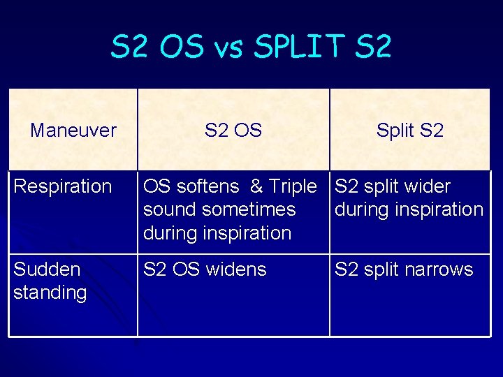
S 2 OS vs SPLIT S 2 Maneuver S 2 OS Split S 2 Respiration OS softens & Triple S 2 split wider sound sometimes during inspiration Sudden standing S 2 OS widens S 2 split narrows
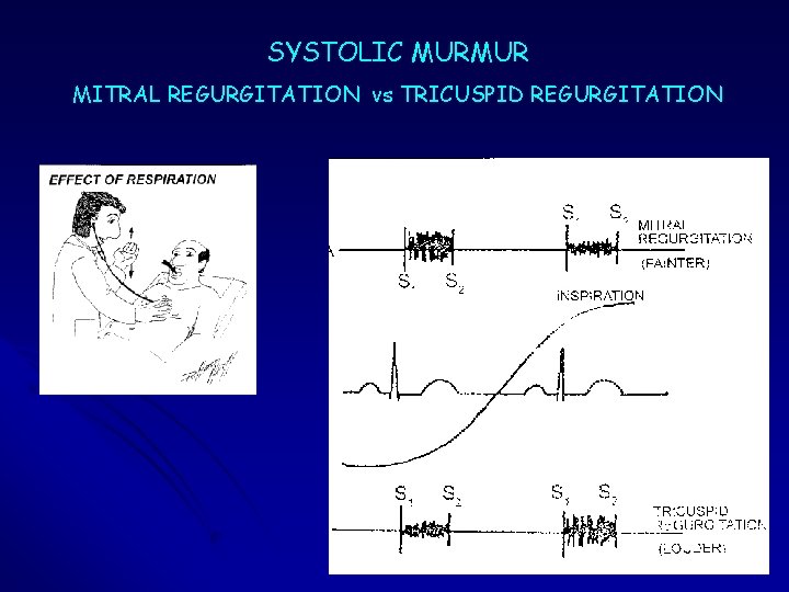
SYSTOLIC MURMUR MITRAL REGURGITATION vs TRICUSPID REGURGITATION
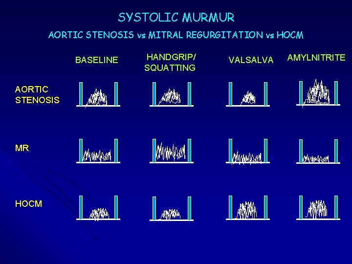
SYSTOLIC MURMUR AORTIC STENOSIS vs MITRAL REGURGITATION vs HOCM BASELINE AORTIC STENOSIS MR HOCM HANDGRIP/ SQUATTING VALSALVA AMYLNITRITE
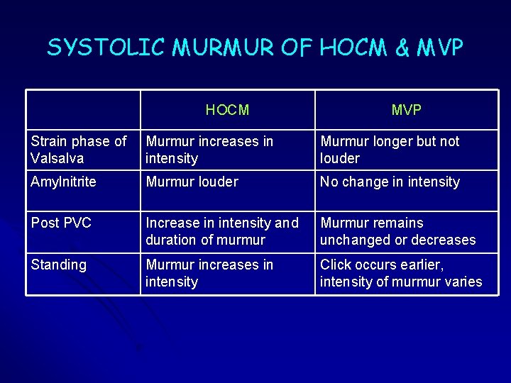
SYSTOLIC MURMUR OF HOCM & MVP HOCM MVP Strain phase of Valsalva Murmur increases in intensity Murmur longer but not louder Amylnitrite Murmur louder No change in intensity Post PVC Increase in intensity and duration of murmur Murmur remains unchanged or decreases Standing Murmur increases in intensity Click occurs earlier, intensity of murmur varies
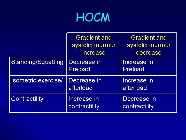
HOCM Gradient and systolic murmur increase Standing/Squatting Decrease in Preload Gradient and systolic murmur decrease Increase in Preload Isometric exercise/ Decrease in afterload Increase in afterload Contractility Decrease in contractility Increase in contractility
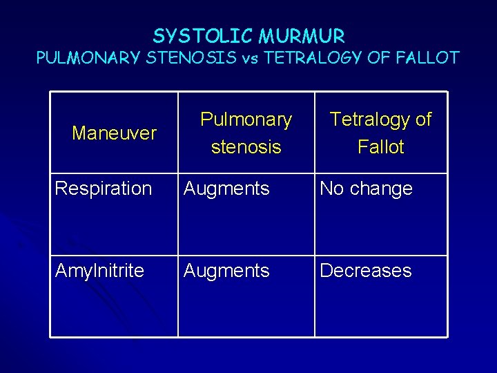
SYSTOLIC MURMUR PULMONARY STENOSIS vs TETRALOGY OF FALLOT Maneuver Pulmonary stenosis Tetralogy of Fallot Respiration Augments No change Amylnitrite Augments Decreases
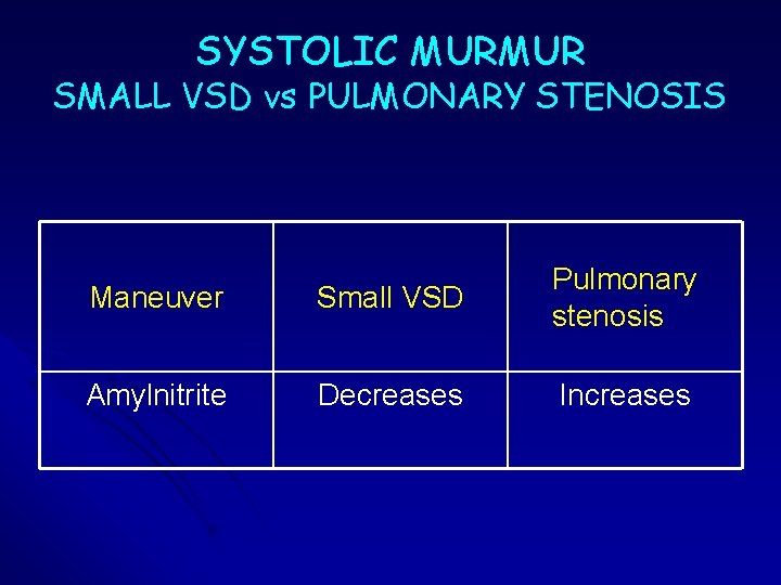
SYSTOLIC MURMUR SMALL VSD vs PULMONARY STENOSIS Maneuver Small VSD Pulmonary stenosis Amylnitrite Decreases Increases
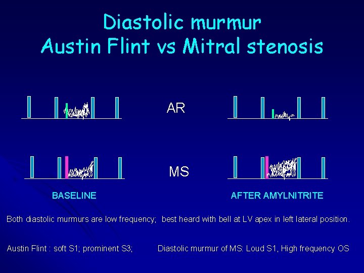
Diastolic murmur Austin Flint vs Mitral stenosis AR MS BASELINE AFTER AMYLNITRITE Both diastolic murmurs are low frequency; best heard with bell at LV apex in left lateral position. Austin Flint : soft S 1; prominent S 3; Diastolic murmur of MS: Loud S 1, High frequency OS
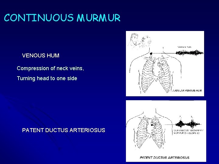
CONTINUOUS MURMUR VENOUS HUM Compression of neck veins, Turning head to one side PATENT DUCTUS ARTERIOSUS