HEALTH PHYSICAL ASSESSMENT IN NURSING Cardiovascular System CARDIOVASCULAR


 VESSELS OF THE HEART. B. POSTERIOR. B")
 node Intra-atrial pathways AV")


 • Paper Recording of Deflections That Represent the Cardiac Cycle • Electrical")


 B. AFTERLOAD IS THE PRESSURE THAT THE VENTRICLES MUST OVERCOME IN ORDER TO")


 AND PULSATIONS • Recall that jugular veins reflect right atrial")


 •")


 POSITIONS FOR AUSCULTATION OF THE HEART. B. LATERAL.")
 POSITIONS FOR AUSCULTATION OF THE HEART. C. SITTING.")


 TETRALOGY OF FALLOT.")


 Electrical representations of the cardiac cycle are documented by deflections")


- Slides: 87
HEALTH & PHYSICAL ASSESSMENT IN NURSING Cardiovascular System
CARDIOVASCULAR SYSTEM • Heart • Vasculature
HEART • • Pericardium Cardiac muscle Chambers Valves Cardiac vessels Conduction system Nerves
FIGURE 17. 1 LAYERS OF THE HEART.
STRUCTURAL COMPONENTS OF THE HEART.
VALVES OF THE HEART.
PERICARDIUM • Thin Sac Composed of Fibroserous Material That Surrounds the Heart • Outer layer • Inner layer • Fluid between the layers
HEART MUSCLE • • • Base Apex Epicardium Myocardium Endocardium
CHAMBERS IN THE HEART • Left and right atria • Left and right ventricles
VALVES • Permit the Flow of Blood Between Chambers and Into Blood Vessels • Atrioventricular (AV) • Tricuspid • Mitral • Semilunar • Pulmonary • Aortic
HEART SOUNDS • S 1 • S 2
HEART SOUNDS IN SYSTOLE AND DIASTOLE.
TABLE 17. 1 CHARACTERIS TICS OF HEART SOUNDS
TABLE 17. 3 DISTINGUISHI NG HEART MURMURS
CONTRACTION AND RELAXATION PHASES OF THE HEART • Systole • Diastole
PULMONARY AND SYSTEMIC CIRCULATION. THE LEFT SIDE OF THE HEART PUMPS OXYGENATED BLOOD (INDICATED IN RED) INTO THE ARTERIES OF THE SYSTEMIC CIRCULATION, WHICH PROVIDES OXYGEN AND NUTRIENTS TO THE CELLS. DEOXYGENATED BLOOD (INDICATED IN BLUE) RETURNS VIA THE VENOUS SYSTEM INTO THE RIGHT SIDE OF THE HEART, WHERE IT IS TRANSPORTED TO THE PULMONARY ARTERIAL SYSTEM TO BE REOXYGENATED.
CIRCULATION OF THE HEART • Coronary arteries • • Left main Right coronary Left anterior descending Circumflex
VESSELS OF THE HEART. A. ANTERIOR. A
(CONTINUED) VESSELS OF THE HEART. B. POSTERIOR. B
CONDUCTION SYSTEM OF THE HEART • • • Sinoatrial (SA) node Intra-atrial pathways AV node Bundle of His Right and left bundle branches Purkinje fibers
CONDUCTION SYSTEM OF THE HEART.
CARDIAC CYCLE • Contraction and Relaxation of the Chambers
THE CARDIAC CYCLE.
CARDIAC CYCLE • Ventricular filling • Ventricular systole • Isovolumetric relaxation
ELECTROCARDIOGRAM (ECG) • Paper Recording of Deflections That Represent the Cardiac Cycle • Electrical deflections • • P wave PR interval QRS interval T wave
ELECTROCARDIOGRAM WAVE.
CARDIAC FUNCTION • Stroke volume • Amount of blood that is ejected with each heartbeat • Cardiac output • Amount of blood ejected from the left ventricle over 1 minute • Cardiac index • Measurement accounting for an individual’s weight when evaluating the pumping action of the heart
PUMPING ACTION OF THE HEART • Preload volume overload • Afterload pressure overload • Cardiac output: stroke volume x heart rate • Blood pressure: cardiac output x systemic vascular resistance
A. PRELOAD IS RELATED TO THE AMOUNT OF BLOOD AND STRETCHING OF THE VENTRICULAR MYOCARDIAL FIBERS. A
(CONTINUED) B. AFTERLOAD IS THE PRESSURE THAT THE VENTRICLES MUST OVERCOME IN ORDER TO OPEN THE AORTIC AND PULMONIC VALVULAR CUSPS. B
LANDMARKS FOR CARDIAC ASSESSMENT • • Sternum Clavicles Ribs Second through fifth intercostal spaces
LANDMARKS FOR CARDIOVASCULAR ASSESSMENT.
FOCUSED INTERVIEW • General questions
FOCUSED INTERVIEW • Specific questions • • Illness Symptoms Behaviors Infants and children Pregnant female Older adult Environment
EQUIPMENT • • Examination gown Stethoscope Metric rulers Doppler
OTHER CONSIDERATIONS • • Age Gender Language Culture
PHYSICAL ASSESSMENT OF THE CARDIOVASCULAR SYSTEM • Techniques • • Inspection Palpation Percussion Auscultation
SPECIFIC AREAS OF THE CARDIOVASCULAR ASSESSMENT • • • Inspection of the face, lips, ears, and scalp Inspection of the jugular veins Inspection of the carotid arteries Inspection of the hands and fingers Inspection of the chest, abdomen, legs, and skeletal structure
JUGULAR VENOUS PRESSURE (JVP) AND PULSATIONS • Recall that jugular veins reflect right atrial pressure • Steps for examination • • • Raise the head of the bed or examining table to 30° Turn the patient’s head gently to the left Identify the topmost point of the flickering venous pulsations Place a centimeter ruler upright on the sternal angle Place a card or tongue blade horizontally from the top of the JVP to the ruler, making a right angle • Measure the distance above the sternal angle in centimeters: a 3 - to 4 -centimeter elevation is normal
ASSESSMENT OF CENTRAL VENOUS PRESSURE.
• Top line – level of the higest visible point of distention • Bottom line – level of the sternal angle • Measure: the vertical distance between the sternal angle and the highest level of jugular distention
SPLINTER HEMORRHAGE.
LANDMARKS IN PRECORDIAL ASSESSMENTS.
SPECIFIC AREAS OF THE CARDIOVASCULAR ASSESSMENT • Palpation of the chest, including the following • • Precordium at the right and left second intercostal spaces Left third intercostal space Left fourth intercostal space Left fifth intercostal space at the midclavicular line
LANDMARKS FOR PALPATION OF THE CHEST.
SPECIFIC AREAS OF THE CARDIOVASCULAR ASSESSMENT • Palpation of the carotid pulses (sequentially) • Percussion of the chest for cardiac border
PALPATING THE CAROTID ARTERY.
ASSESSING THE CAROTID PULSE • Keep the patient’s head elevated to 30° • Place your index and middle fingers on the right then the left carotid arteries, and palpate the carotid upstroke • Never palpate right and left carotid arteries simultaneously • The upstroke may be: • Brisk – normal • Delayed – suggests aortic stenosis • Bounding – suggests aortic insufficiency • Listen with the stethoscope for any bruits
PERCUSSING THE CHEST.
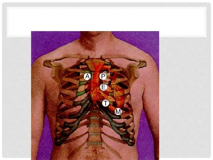
SPECIFIC AREAS OF THE CARDIOVASCULAR ASSESSMENT • Auscultation of the chest using the diaphragm and bell in various positions to include the following locations • Aortic area at the right second intercostal space–S 2 is louder than S 1 • Pulmonic area at the left second intercostal space–S 2 is louder than S 1 • Erb’s point at the left third intercostal space–S 1 and S 2 are heard equally
SPECIFIC AREAS OF THE CARDIOVASCULAR ASSESSMENT • Auscultation of the chest using the diaphragm and bell in various positions to include the following locations • Tricuspid area at the left fourth intercostal space–S 1 is louder than S 2 • Apex at the left fifth intercostal space at the midclavicular line–S 1 is louder than S 2
• Listen in all 5 listening areas for S 1 and S 2 using the diaphragm of the stethoscope • Then listen at the apex with the bell • The diaphragm and the bell. . . • The diaphragm is best for detecting high-pitched sounds like S 1, S 2, and also S 4 and most murmurs • The bell is best for detecting low-pitched sounds like S 3 and the rumble of mitral stenosis
AUSCULTATING THE CHEST OVER FIVE KEY LANDMARKS.
POSITIONS FOR AUSCULTATION OF THE HEART. A. SUPINE.
(CONTINUED) POSITIONS FOR AUSCULTATION OF THE HEART. B. LATERAL.
(CONTINUED) POSITIONS FOR AUSCULTATION OF THE HEART. C. SITTING.
SPECIFIC AREAS OF THE CARDIOVASCULAR ASSESSMENT • Auscultation of the carotid arteries using the diaphragm and bell • Comparison of the apical pulse to a carotid pulse
COMPARING THE CAROTID AND APICAL PULSES.
ABNORMAL FINDINGS IN THE CARDIOVASCULAR SYSTEM • • • Myocardial and pump disorders Valvular disease Septal defects Congenital heart disease Electrical rhythm disturbances
MYOCARDIAL AND PUMP DISORDERS • • Myocardial ischemia Myocardial infarction Congestive heart disease Ventricular hypertrophy
VALVULAR DISEASES • Mitral, aortic, tricuspid, and pulmonic stenosis • Mitral and aortic regurgitation • Mitral valve prolapse
MITRAL STENOSIS.
AORTIC STENOSIS.
MITRAL REGURGITATION.
PULMONIC STENOSIS.
TRICUSPID STENOSIS.
MITRAL VALVE PROLAPSE.
AORTIC REGURGITATION.
SEPTAL DEFECTS • Openings between the right and left atria or right and left ventricles
VENTRICULAR SEPTAL DEFECT.
ATRIAL SEPTAL DEFECT.
CONGENITAL HEART DISEASES • Coarctation of the aorta • Patent ductus arteriosus • Tetralogy of Fallot
COARCTATION OF THE AORTA.
PATENT DUCTUS ARTERIOSUS.
TETRALOGY OF FALLOT.
(CONTINUED) TETRALOGY OF FALLOT.
ELECTRICAL RHYTHM DISTURBANCES • Ventricular tachycardia • Ventricular fibrillation
VENTRICLE TACHYCARDIA.
VENTRICULAR FIBRILLATION.
OBJECTIVES OUTLINED IN HEALTHY PEOPLE • Coronary heart disease • High blood cholesterol
KEY OBJECTIVES FOR CORONARY HEART DISEASE • Screening for risk factors • Individual, community, culturally linguistically appropriate education and counseling • Education about symptoms and emergency care • Weight reduction programs • Programs to increase physical activity
KEY OBJECTIVES FOR HIGH TOTAL BLOOD CHOLESTEROL • Education about risks • Education about diet and exercise • Education about screening
GLOSSARY • Atrioventricular Valves that separate the atria from the ventricles. • Bruit A loud blowing sound, an abnormal finding, most often associated with a narrowing or stricture of the carotid artery usually associated with atherosclerotic plaque. • Bundle Branches Expressways of conducting fibers that spread the electrical current through the ventricular myocardial tissue. • Bundle of His Atrioventricular Nodes that are intricately connected and function to receive the current that has finished spreading throughout the atria. • Cardiac Conduction System The heart's conduction system which can initiate an electrical charge and transmit that charge via cardiac muscle fibers throughout the myocardial tissue. • Cardiac Cycle The events of one complete heartbeat, the contraction and relaxation of the atria and ventricles. • Cardiac output The amount of blood ejected from the left ventricle over 1 minute. • Diastole The phase of ventricular relaxation in which the ventricles relax and are filled as the atria contract.
GLOSSARY • Electrocardiogram (EKG) Electrical representations of the cardiac cycle are documented by deflections on recording paper. • Endocardium The innermost layer of the heart, a smooth layer that provides an inner lining for the chambers of the heart. • Heart An intricately designed pump composed of a meticulous network of synchronized structures. • Infective Endocarditis A condition caused by bacterial infiltration of the lining of the heart’s chambers. • Left Atrium Forms the posterior aspect of the heart. • Left Ventricle Egg shaped, most muscular chamber of the heart, located behind the right ventricle and forms the left border of the heart. • Marfan's Syndrome A degenerative disease of the connective tissue, which over time may cause the ascending aorta to either dilate or dissect, leading to abrupt death. • Mediastinal space The area where the heart sits obliquely within the thoracic cavity between the lungs and above the diaphragm.
GLOSSARY • Myocardium The second, thick, muscular layer of the heart, made up of bundles of cardiac muscle fibers reinforced by a branching network of connective tissue fibers called the fibrous skeleton of the heart. • Pericardium A thin sac composed of a fibroserous material that surrounds the heart. • Purkinje Fibers that fan out and penetrate into the myocardial tissue to spread the current into the tissues themselves. • Right Atrium A thin-walled chamber located above and slightly to the right of the right ventricle that forms the right border of the heart. • Right Ventricle Part of heart formed triangularly and comprises much of the anterior or sternocostal surface of the heart. • S 1 The first heart sound (lub), is heard when the AV valves close. Closure of these valves occurs when ldrslt the ventricles have been filled. • S 2 The second heart sound (dub), occurs when the aortic and pulmonic valves close, they close when the ventricles have emptied their blood into the aorta and pulmonary arteries. • Semilunar Valves that separate the ventricles from the vascular system.
GLOSSARY • Sinoatrial Node The node located at the junction of the superior vena cava and right attrium that initiates the electrical impulse. • Sternum The flat, narrow center bone of the upper anterior chest. • Stroke Volume The amount of blood that is ejected with every heartbeat. • Systole The phase of ventricular contraction in which the ventricles have been filled, then contract to expel blood into the aorta and pulmonary arteries. • Visceral Layer The inner layer, which lines the surface of the heart. • Xanthelasma Yellowish cholesterol deposits seen on the eyelids and are indicative of premature atherosclerosis.