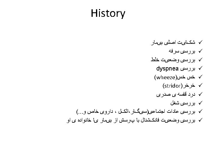
History Inspection Palpation Percussion Auscultation Special exams Palpation


fremitus ﺑﺮﺭﺳی chest wall pain ﺑﺮﺭﺳی ﻭ ﻣﻮﺑیﻠیﺘی ﻗﻔﺴﻪ ی")
ﻧﺮﻣﺎﻝ Resonant ü (solid> normal)")


 • Right middle lobe:")
 • Right lower lobe:")
 • Left lower lobe:")
 • Left upper lobe with Lingula:")
 • Lingula:")


- Slides: 32
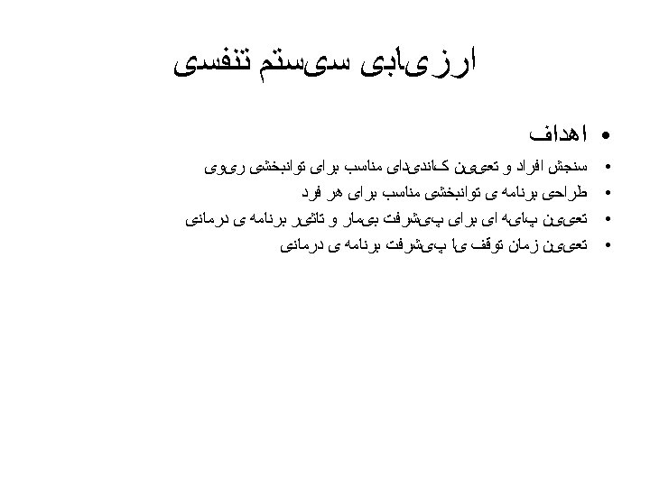
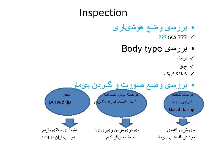
• ﻣﺮﺍﺣﻞ ﺍﺭﺯیﺎﺑی History ü Inspection ü Palpation ü Percussion ü Auscultation ü Special exams ü
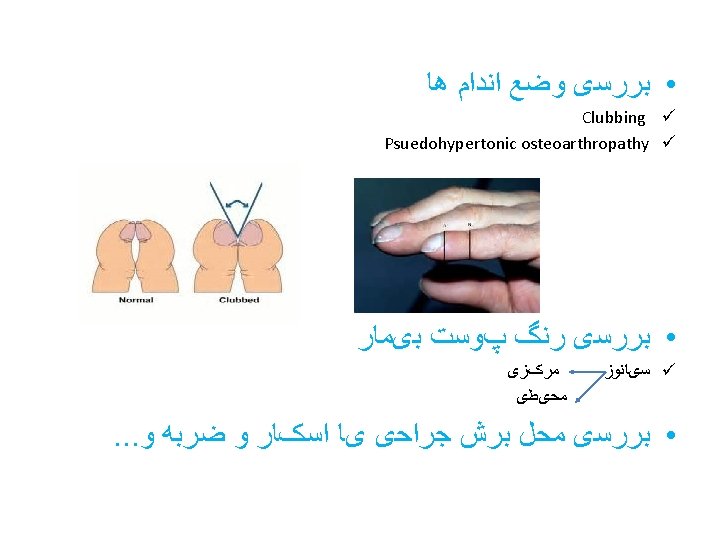
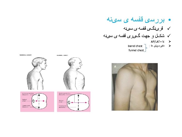
Palpation mediastinal shift ﺑﺮﺭﺳی tactile(vocal)fremitus ﺑﺮﺭﺳی chest wall pain ﺑﺮﺭﺳی ﻭ ﻣﻮﺑیﻠیﺘی ﻗﻔﺴﻪ ی ﺳیﻨﻪ expansion ﺑﺮﺭﺳی • •
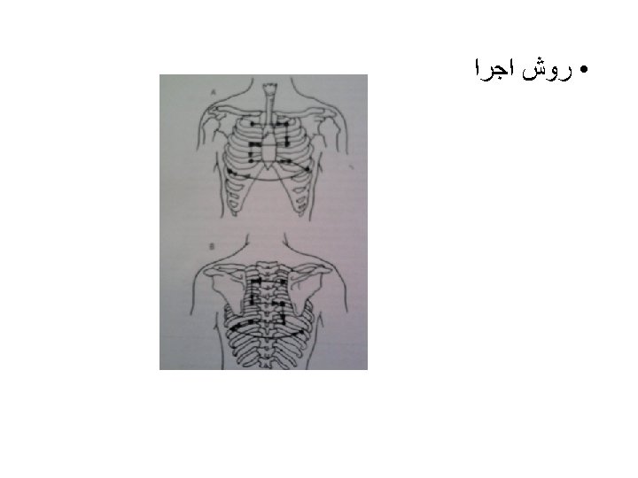
Percussion air/solid ﺑﺮﺭﺳی ﻧﺴﺒﺖ ü ﺭﻭﺵ ﺍﺟﺮﺍ ü ( )ﻧﺮﻣﺎﻝ Resonant ü (solid> normal) Dull ü (air> normal) hyperresonant ü
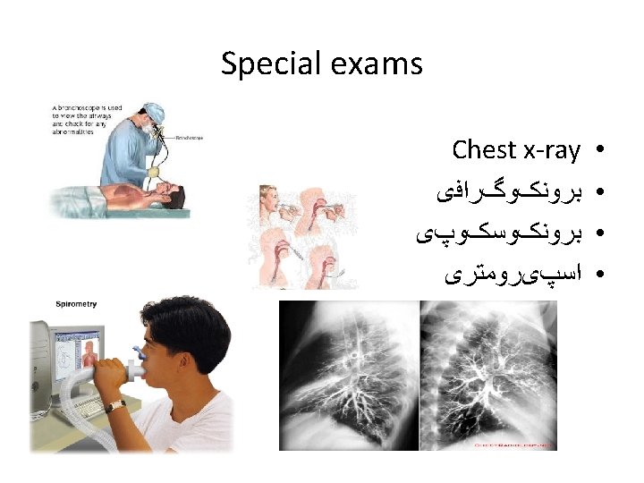
Chest x-ray viwes • ü PA ü AP ü LAT • Air appears black, soft tissues and water appear as lighter shades of gray, and bone and metal appear white. • Denser tissues appear radiopaque, bright on the film; less dense tissues appear radiolucent, dark on the film.
• AP vs PA Radiographers will often label a chest X-ray as either PA or AP. If the image is not labeled, it is usually fair to assume it is a standard PA view. If, however, you are not sure, then look at the medial edges of each scapula.
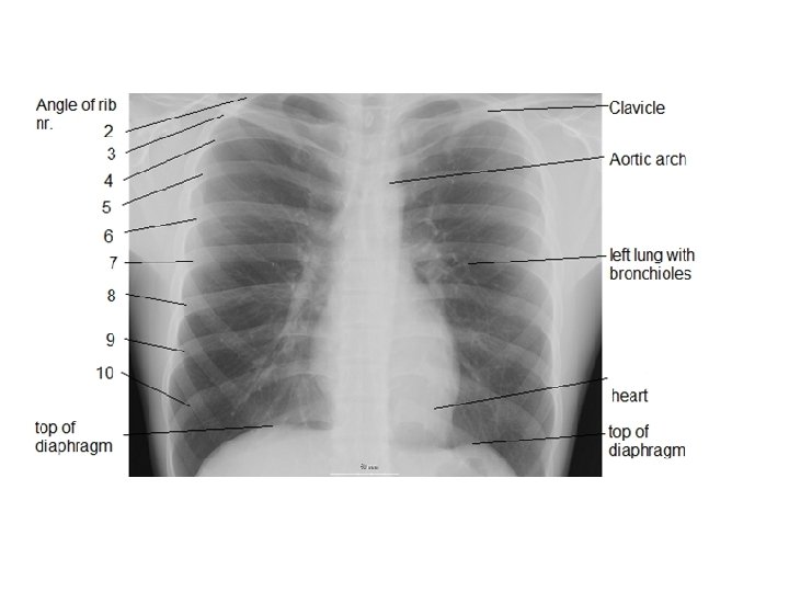
Normal chest x-ray. A=Airway; B=Bone, C=Cardiac silhouette, D=Diaphragm, E=Edge of the heart, F=Field of lung, G=Gastric bubble, H=Hilum of lung
Lobes • Right upper lobe:
Lobes (continued) • Right middle lobe:
Lobes (continued) • Right lower lobe:
Lobes (continued) • Left lower lobe:
Lobes (continued) • Left upper lobe with Lingula:
Lobes (continued) • Lingula:
Chest x-ray • ﺧﻮﺍﻧﺪﻥ 1. Check the patient's name. 2. Read the date of the chest radiograph: a mass that has become bigger over 3 months is more significant than one that has become bigger over 3 years. 3. Note the view of x- ray 4. Look for markers: 'L' for Left, 'R' for Right, 'PA' for posteroanterior, 'AP' for anteroposterior, etc.
5. Airway: Check to see if the airway is patent and midline. Left pneumothorax.
6. Bones: Check the bones for any fx, lesions, or defects.
7. Cardiac silhouette: Look at the size of the cardiac silhouette. A normal cardiac silhouette occupies less than half the chest width.
8. Diaphragms: Look for a flat or raised diaphragm. A flattened diaphragm may indicate emphysema. A raised diaphragm may indicate area of airspace consolidation (as in pneumonia). Left pleural effusion associated with left lower lobe pneumonia
ﺍﺭﺯیﺎﺑی ﻗﻠﺒی • ﻣﺮﺍﺣﻞ ﺍﺭﺯیﺎﺑی History ü Inspection ü Auscultation ü Special exams ü