POSTMORTEM INSPECTION Viscera Inspection 1 Inspection of the


 lymph")


- Slides: 18
POST-MORTEM INSPECTION
Viscera Inspection 1. Inspection of the Esophagus and Trachea • Observe the esophagus for Cysticercus (measles); eosinophilic myositis (EM); and evidence of grub infestation. • Cysticercus and EM conditions require retention. Grub infestation is usually a localized condition requiring affected organs and areas be trimmed or condemned, but the carcass will usually be passed without retention
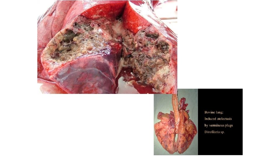
2. Inspection of the Lungs and Heart • Incise and observe lungs lymph nodes - mediastinal [caudal (posterior), middle, cranial (anterior)], and tracheobronchial (bronchial) right and left. • Pneumonia and pleuritis are the most common abnormalities observed. • Acute pneumonia is characterized by enlarged, edematous lymph nodes and/or dark red to purple sections or spots in the lung tissue. Retain this carcass and all parts for disposition
• A chronic pneumonia may be characterized by a localized abscess within the lungs, or many times evidence that the lung has become adhered to the pleura (lining of the thoracic cavity), frequently called pleuritis. • Observe the rest of the viscera and carcass to look for evidence that the condition is generalized. The carcass may appear degenerated. • You should retain the carcass and all parts upon detecting a generalized condition. When the condition is strictly localized, the lungs would be condemned, as well as any contaminated organs, and the carcass retained for removal of the adhesions.
• Tuberculosis may also be detected during incision of the lung's lymph nodes. When TB lesions are detected, the carcass and all parts must be retained.
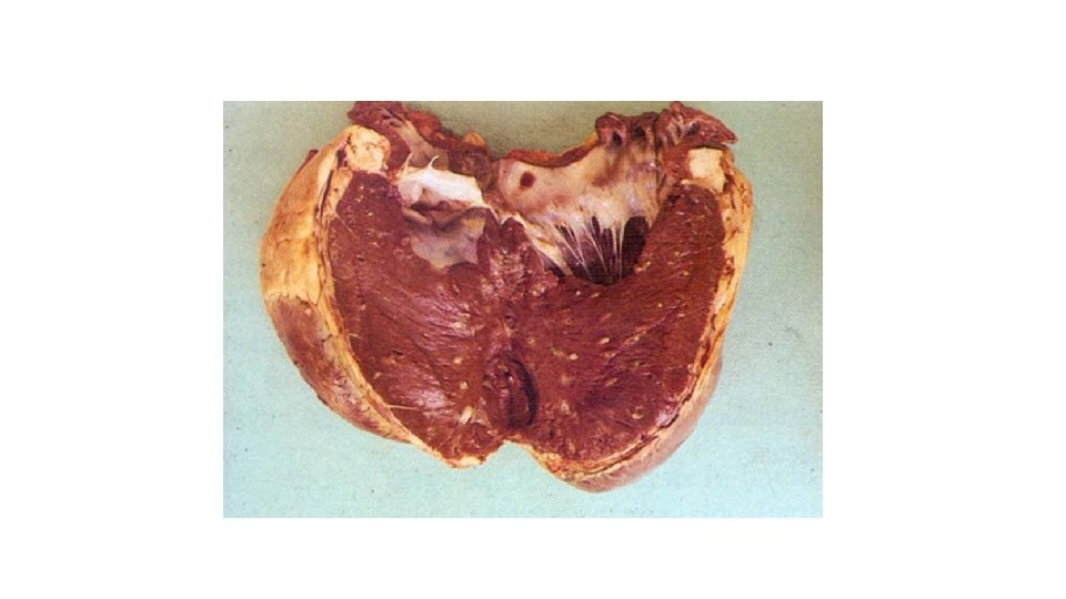
Inspection of the Heart • The inspection of the heart involves opening it by an incision form the base to the apex, or vice-versa. • The usual procedure is to position the heart in a manner that will allow you to safely cut away from your body, and incise the left ventricle about an inch and one-half posterior to the lefts of large vessels leading into the chamber. • Then grasp the opened edge of the ventricle and incise the septum. • By rotating the knife 180 degrees with the cutting edge pointing up, complete opening the ventricles and great vessels with two incisions, causing the heart to lay flat or open.
• Some of the conditions you may detect while inspecting the heart include: Cystircercus (tapeworm cysts, measles, etc. ) Eosinophilic myositis (EM) Neoplasms (tumors) • Pericarditis is an inflammation of the pericardium or heart sac. When an inflammation of the inner lining of the heart occurs, the condition is referred to as endocarditis.
3. Inspection of the Liver In all cases, a liver containing an abscess is condemned as not fit for human consumption • A slight infestation will probably not affect the liver tissue as such. A heavy infestation may cause a cirrhotic effect on the organ, with the surface becoming scarred.
• The primary purpose in opening the bile duct during liver inspection is to detect flukes. • When there is a fluke infestation the bile duct may be thickened and sometimes swollen; frequently you will observe live flukes. • The three liver flukes most often seen in domestic cattle today are: Fascioloides magna; Fasciola hepatica; Dicrocoelium dentricum. • In all cases of liver fluke infestation the liver is condemned and not eligible for human consumption
• The cyst will vary in size but may be as large as two to four inches in diameter. The fluid inside the cyst is usually clear and colorless. You must be careful not to confuse the hydatid cyst with an accessory gall bladder. • Hydatid cysts may occasionally affect livestock. Most domestic food animals are the intermediate host for this tapeworm cyst, which usually is a result of the tapeworm (Enchinococcus granulosus) of dogs. • The organ or part affected with a hydatid cyst is condemned and is not suitable for use in animal food.
4. Inspection of the Spleen • The inspection of the spleen is done by observation. • If tuberculosis is suspected, the carcass and all parts will be retained for veterinary disposition. • You will see physical differences between normal and abnormal. • There may be a definite swelling or size difference, or a color difference. • When an abnormal spleen is detected, retain it as well as the carcass and all parts. • The spleen may be helpful in making a final disposition on any carcass. Ensure that the spleen is included with the viscera whenever a carcass is retained for a disease condition.
5. Inspection of the Abdominal Viscera • Observe cranial and caudal mesenteric (mesenteric) lymph nodes • Observe and palpate rumino-reticular junction. • Abscesses are frequently detected during the palpation and observation of the rumino-reticular junction. These abscesses are usually localized and required only that the viscera be condemned. You should be alert though, to the overall condition of the carcass, and thoracic viscera.
• The mesenteric lymph nodes may show evidence of tuberculosis, neoplasms, and in some cases pigmentary color changes • You must retain the carcass and all parts when you detect tuberculosis and tumors.
• The small intestines may appear dark red to purple; this would indicate a condition called enteritis. The determination whether the condition is acute or chronic must be made. • Evidence of adhesions may be seen. If the condition appears localized, or chronic, and no further carcass or viscera involvement is observed,
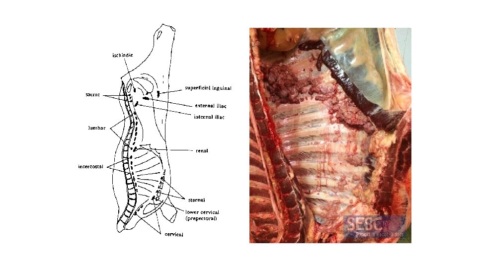
Carcass Inspection • Observe back of skinned carcass while eviscerated • Palpate superficial inguinal, or supramammary, and internal iliac lymph nodes • Observe body cavities. • Observe and palpate kidneys • Observe diaphragm's pillars and peritoneum • Observe and palpate diaphragm. • Observe pleura, cut surfaces of muscles and bones, neck, and carcass exterior