DEVELOPMENT of the DENTAL TISSUE INDUCTION ORAL Epith


- Slides: 32
DEVELOPMENT of the DENTAL TISSUE
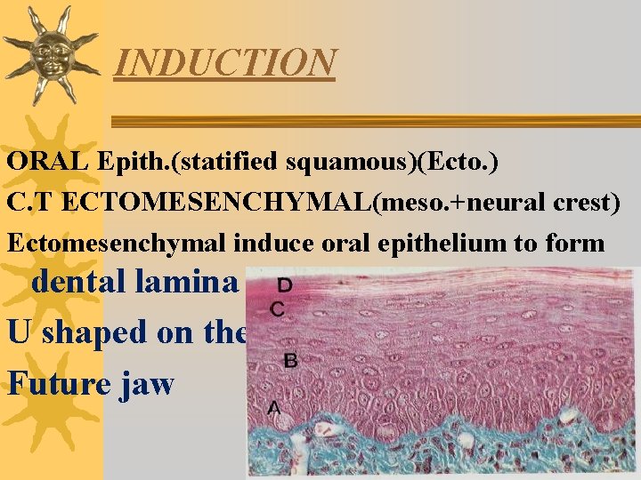
INDUCTION ORAL Epith. (statified squamous)(Ecto. ) C. T ECTOMESENCHYMAL(meso. +neural crest) Ectomesenchymal induce oral epithelium to form dental lamina U shaped on the Future jaw
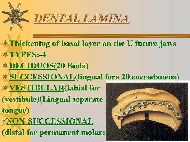
DENTAL LAMINA ¬Thickening of basal layer on the U future jaws ¬TYPES: -4 ¬DECIDUOS(20 Buds) ¬SUCCESSIONAL(lingual fore 20 succedaneus) ¬VESTIBULAR(labial for (vestibule)(Lingual separate tongue) *NON-SUCCESSIONAL (distal for permanent molars)
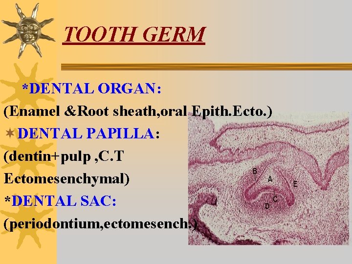
TOOTH GERM *DENTAL ORGAN: (Enamel &Root sheath, oral Epith. Ecto. ) ¬DENTAL PAPILLA: (dentin+pulp , C. T Ectomesenchymal) *DENTAL SAC: (periodontium, ectomesench. )
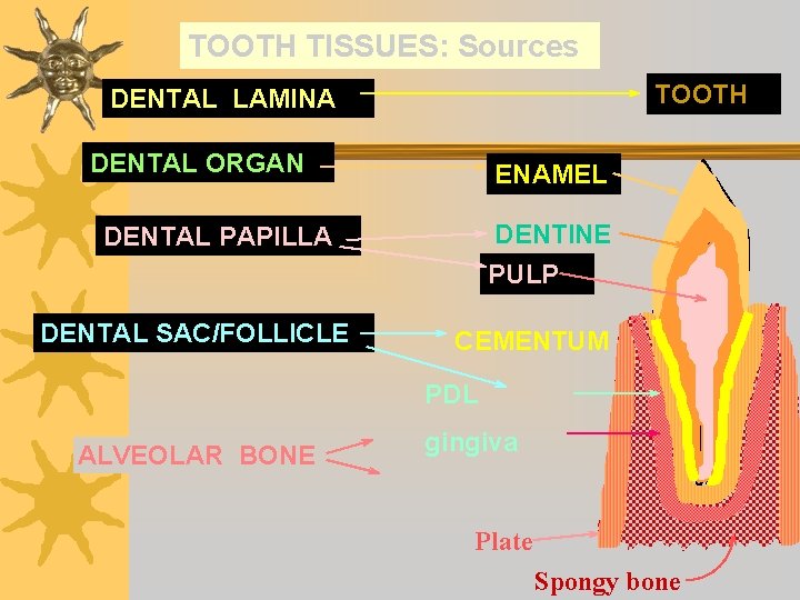
TOOTH TISSUES: Sources TOOTH DENTAL LAMINA DENTAL ORGAN ENAMEL DENTINE DENTAL PAPILLA PULP DENTAL SAC/FOLLICLE CEMENTUM PDL ALVEOLAR BONE gingiva Plate Spongy bone
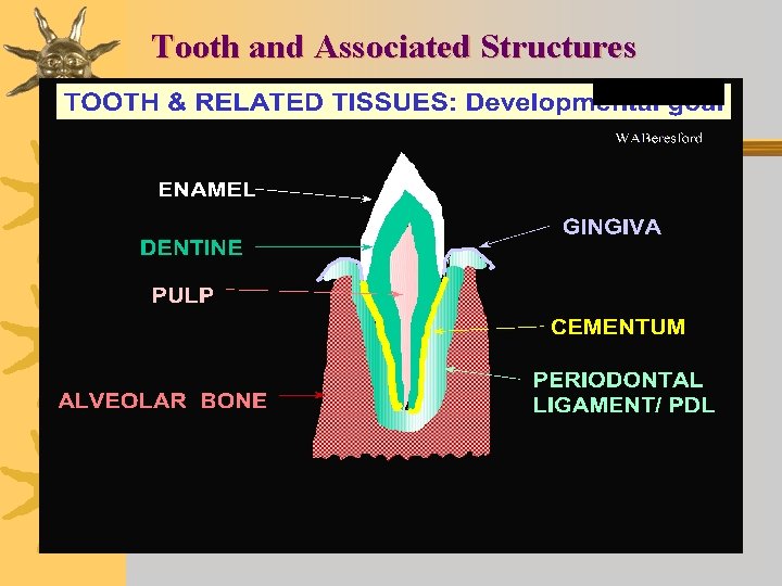
Tooth and Associated Structures
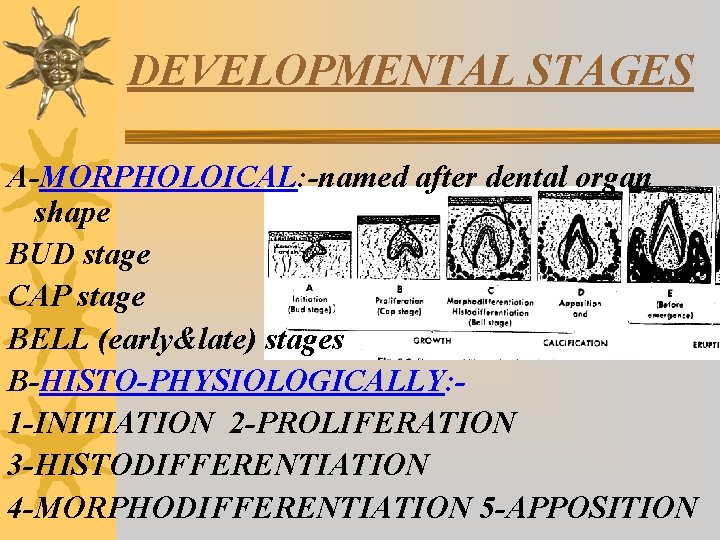
DEVELOPMENTAL STAGES A-MORPHOLOICAL: -named after dental organ shape BUD stage CAP stage BELL (early&late) stages B-HISTO-PHYSIOLOGICALLY: 1 -INITIATION 2 -PROLIFERATION 3 -HISTODIFFERENTIATION 4 -MORPHODIFFERENTIATION 5 -APPOSITION
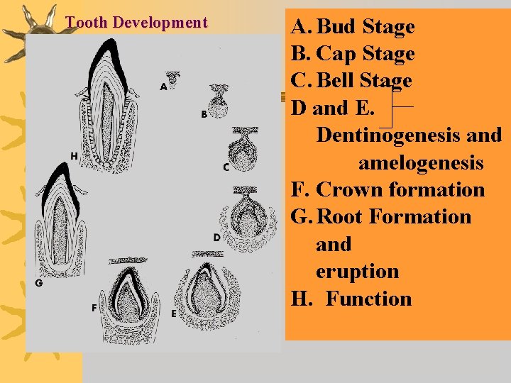
Tooth Development A. Bud Stage B. Cap Stage C. Bell Stage D and E. Dentinogenesis and amelogenesis F. Crown formation G. Root Formation and eruption H. Function
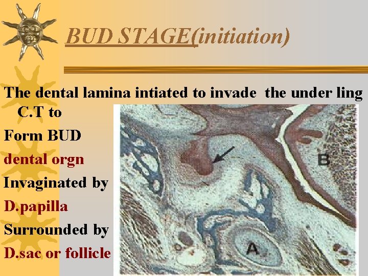
BUD STAGE(initiation) The dental lamina intiated to invade the under ling C. T to Form BUD dental orgn Invaginated by D. papilla Surrounded by D. sac or follicle
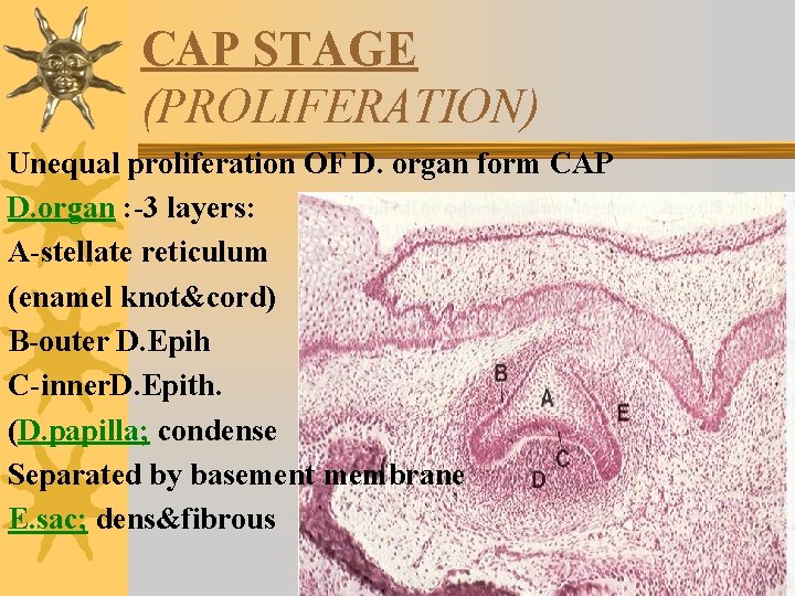
CAP STAGE (PROLIFERATION) Unequal proliferation OF D. organ form CAP D. organ : -3 layers: A-stellate reticulum (enamel knot&cord) B-outer D. Epih C-inner. D. Epith. (D. papilla; condense Separated by basement membrane E. sac; dens&fibrous
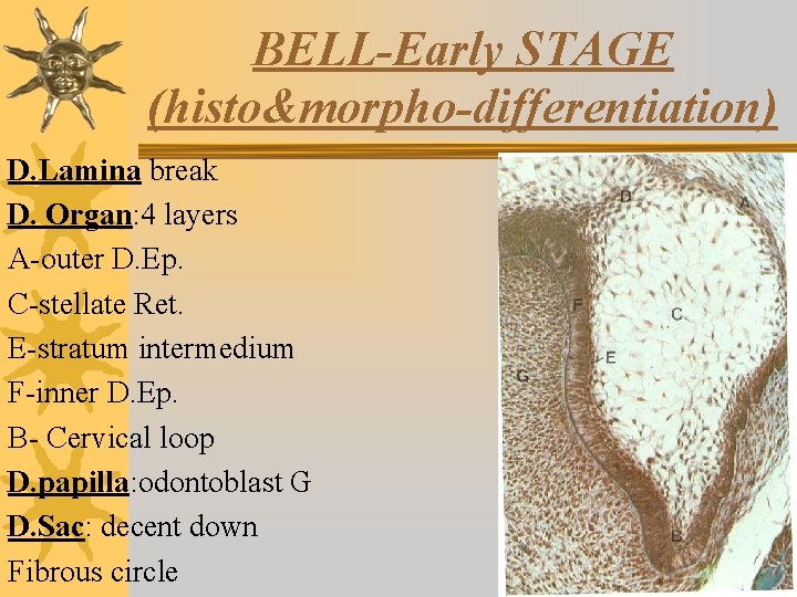
BELL-Early STAGE (histo&morpho-differentiation) D. Lamina break D. Organ: 4 layers A-outer D. Ep. C-stellate Ret. E-stratum intermedium F-inner D. Ep. B- Cervical loop D. papilla: odontoblast G D. Sac: decent down Fibrous circle
Histo-differentiation &mophodifferentiation ¬Histodifferentiaton of inner dental epithelium into tall columnar cells that induce formation of odontoblast from dental papilla ¬The assume its morphology. after formation of dentinenamel junction& cemento-enamel junctions on which the ameloblst arrange according to the tooth form
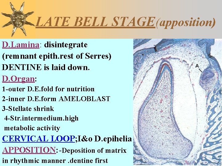
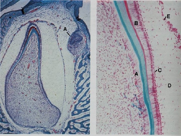
LATE BELL STAGE(apposition) D. Lamina: disintegrate (remnant epith. rest of Serres) DENTINE is laid down. D. Organ: 1 -outer D. E. fold for nutrition 2 -inner D. E. form AMELOBLAST 3 -Stellate shrink 4 -Str. intermedium. high metabolic activity CERVICAL LOOP; I&o D. epihelia APPOSITION: -Deposition of matrix in rhythmic manner. dentine first
Eruption Cyst
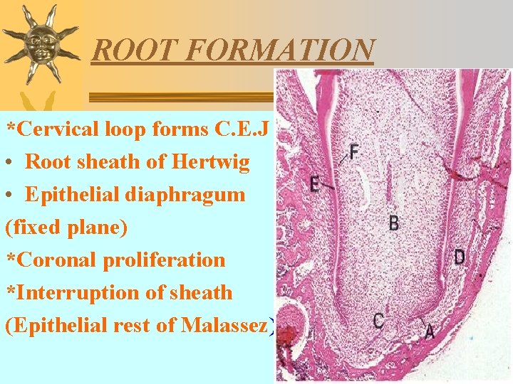
ROOT FORMATION *Cervical loop forms C. E. J • Root sheath of Hertwig • Epithelial diaphragum (fixed plane) *Coronal proliferation *Interruption of sheath (Epithelial rest of Malassez)
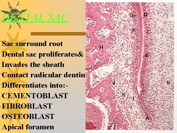
DENTAL SAC Sac surround root Dental sac proliferates& Invades the sheath Contact radicular dentin Differentiates into: CEMENTOBLAST FIBROBLAST OSTEOBLAST Apical foramen
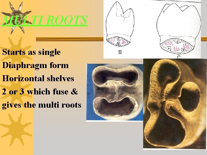
MULTI ROOTS Starts as single Diaphragm form Horizontal shelves 2 or 3 which fuse & gives the multi roots
Rules of “sixes” in dental development 6 weeks old in utero: beginning of dental developmen 6 months old: emergence of the first primary tooth 6 years old: emergence of first permanent tooth Source: http: //www. columbia. edu/itc/hs/dental/d 9903/lectures/lecture 4. pdf
¬Common symptoms of teething include; bulging, red gums which are very tender to touch, the cheeks may be hot and red, the child will also be restless, irritable, and may be suffering from a raised body temperature. Teething pain symptoms are often accompanied by the symptoms of a mild respiratory infection i. e. catarrh and congestion. It is not know whether teething causes this or whether the conditions just frequently occur together
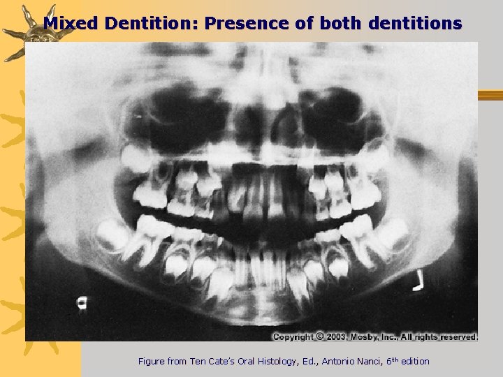
Mixed Dentition: Presence of both dentitions Figure from Ten Cate’s Oral Histology, Ed. , Antonio Nanci, 6 th edition
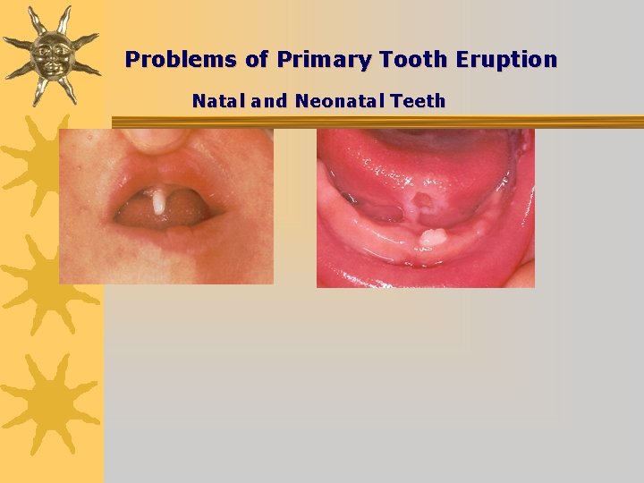
Problems of Primary Tooth Eruption Natal and Neonatal Teeth
Retained Primary teeth
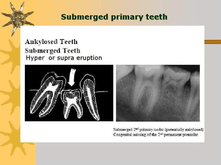
Submerged primary teeth Hyper or supra eruption
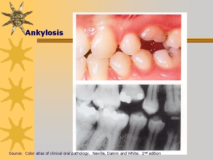
Ankylosis Source: Color atlas of clinical oral pathology. Neville, Damm and White. 2 nd edition
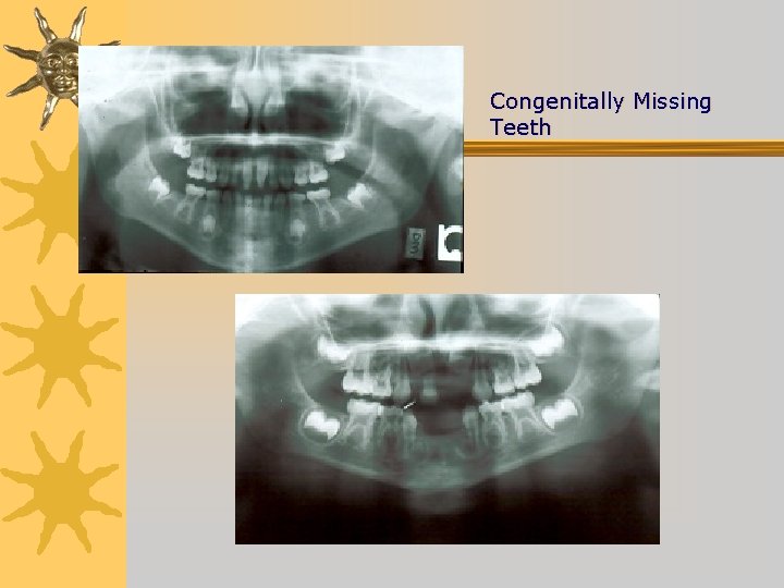
Congenitally Missing Teeth
2/27/2021 29
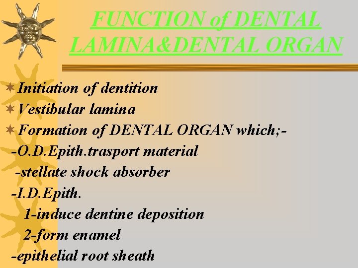
FUNCTION of DENTAL LAMINA&DENTAL ORGAN ¬Initiation of dentition ¬Vestibular lamina ¬Formation of DENTAL ORGAN which; -O. D. Epith. trasport material -stellate shock absorber -I. D. Epith. 1 -induce dentine deposition 2 -form enamel -epithelial root sheath
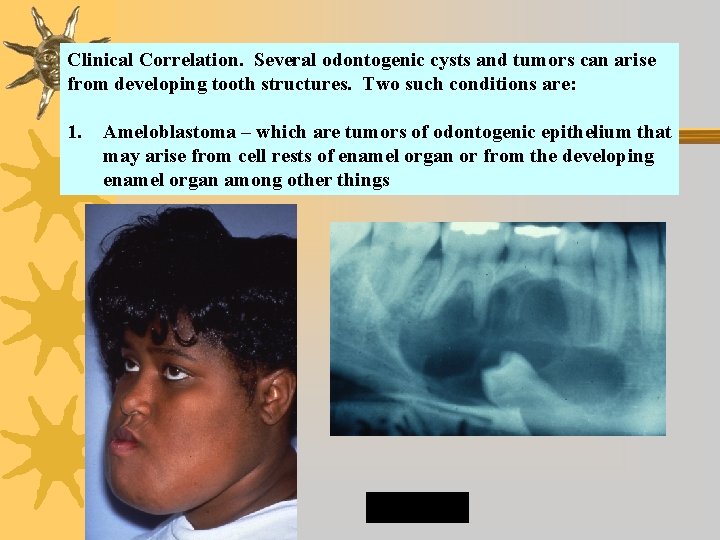
Clinical Correlation. Several odontogenic cysts and tumors can arise from developing tooth structures. Two such conditions are: 1. Ameloblastoma – which are tumors of odontogenic epithelium that may arise from cell rests of enamel organ or from the developing enamel organ among other things