Chapter 23 Heart Sounds Dynamics of Valvular and


- Slides: 32

Chapter 23: Heart Sounds; Dynamics of Valvular and Congenital Heart Defects

The Cardiac Cycle

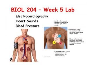
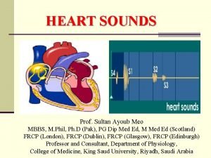
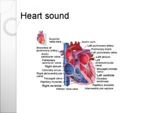
I. 1 st Heart Sound • A. A-V valves close • B. Louder than second sound • C. Low pitch II. 2 nd Heart Sound • Aortic and pulmonary valves close

III. 3 rd Heart Sound • A. Very low pitch • B. Caused by inrushing of blood into ventricles IV. 4 th Heart Sound • A. Atrial contraction late in diastole • B. Hard to hear with stethoscope except in hypertensive patients with a thick left ventricle

Dynamics of Streptococcal Damage to Heart Valves • Streptococcus ¯ • release of M antigen ¯ M Heart valve cell M with M antigens attached M M ¯ • Antibody formed against combination ¯ • Complement damage to heart valves

V. Etiology of murmurs • A. Stenotic damage usually initiated by streptococci • B. Strep. release M antigen • C. Antibody releases complement • D. Mitral valves most common • E. Aortic valve second most common

VI. Results of Heart Valve Damage • • A. Stenosis of valve B. Destruction of valve Þ regurgitation or insufficiency

VII. Mitral stenosis • A. Murmur heard in last 3 rd of diastole • B. Described as a “thrill” over apex of heart • C. Low rumbling murmur

Mitral Stenosis Murmur S 1 S 2 Systole Diastole

Mitral Stenosis Hemodynamics MAP C. O. L. ATRIAL VOL. Teacher draws in responses with powerpoint pen. RT. VENT. PRESS. LAP NORMAL SEVERE

VII. Mitral Stenosis (cont’d) • D. Hemodynamics of mitral stenosis 1. CO and MAP do not ¯ nearly as much as in aortic stenosis 2. Atrial volume leads to atrial fibrillation 3. R. ventricle pressure could lead to r. vent. failure 4. LAP could Þ pulmonary edema 5. L. ventricle normal

VIII. Mitral regurgitation • A. Blowing murmur heard throughout systole - high pitch • B. Best sound heard over l. atrium (too deep); must be heard over l. ventricle.

Mitral Regurgitation Murmur S 1 S 2 Systole Diastole

Mitral Regurgitation Hemodynamics MAP MEAN L. ATRIAL VOL. Teacher draws in responses with powerpoint pen. MEAN L. ATR. PRESS. C. O. NORMAL SEVERE

VIII. Mitral regurgitation (cont’d) • C. Hemodynamics of mitral regurgitation 1. LAP can Þ pul. edema 2. CO falls more if r. heart fails 3. L. atrial volume can Þ atrial fibrillation

IX. Aortic Stenosis • A. 80% of patients are male • B. Diamond shaped - or crescendo and decrescendo - (some are fast ejection murmurs) • C. Pressure in vent. may reach 400 mm Hg Þ vent. hypertrophy • D. Very loud - can be felt with hand if severe • E. Repair with prosthetic valve or porcine valve (as in all murmurs)

Aortic Stenosis Murmur S 1 S 2 Systole Diastole

Aortic Stenosis Hemodynamics L. VENT. SYS. PRESS. MEAN VENT. VOL. AORTIC PRESSURE Teacher draws in responses with powerpoint pen. C. O. LAP NORMAL SEVERE

IX. Aortic Stenosis (cont’d) • F. Hemodynamics of aortic stenosis 1. L. ventricular hypertrophy 2. Repair of valve sometimes leads to regurgitation 3. Angina pain in severe stenosis 4. High mortality in surgery 5. Chronic in blood vol.

X. Aortic Regurgitation • A. Blowing murmur - high pitch • B. Listen over l. ventricle for best sound • C. Short murmur means blood flows back rapidly and is more severe • D. May have stroke vol. of 300 ml with 70 ml going to periphery and 230 leaking back

Aortic Regurgitation Murmur S 1 S 2 Systole Diastole

Aortic Regurgitation Hemodynamics DIAST. VENT. VOL. MAP NET C. O. . Teacher draws in responses with powerpoint pen. LAP NORMAL SEVERE

X. Aortic Regurgitation (cont’d) • E. Hemodynamics of aortic regurgitation 1. Aortic diastolic press. ¯’s rapidly 2. Filling of ventricle can compress inner parts of heart and coronaries 3. L. ventricular hypertrophy

XI. Diagnosis of Murmurs • A. ECG axis deviation showing hypertrophy • B. Echocardiogram • C. X-ray • D. Catheterization • E. Stethoscope or phonocardiogram

Patent Ductus Arteriosus

XII. Patent Ductus Arteriosus • A. Can be treated with prostaglandin blocker, indomethacin • B. Blood recirculates through lungs • C. Net CO ¯ so blood vol. ’s and CO ’s back toward normal • D. ¯ Cardiac reserve • E. Can Þ pul. edema • F. L. vent. hypertrophy • G. Right vent. hypertrophy

Patent Ductus Arteriosus Murmur S 1 S 2 Systole Diastole

Interventricular Septal Defect

XIII. Interventricular Septal Defect • Pan systolic murmur unless hole closes during contraction

Interatrial Septal Defect

XIV. Interatrial Septal Defect • A. Foramen ovale does not close • B. 1/3 of people do not have normal fibrotic closure of f. ovale, but LAP causes it to close

XV. Tetralogy of Fallot • A. Pul. artery stenosis or pul. valve stenosis • B. Aorta displaced over septum • C. Equal systolic pressure in both ventricles • D. Enlarged rt. ventricle • E. Blue baby - blood does not flow through lungs enough • F. Surgery very helpful
 Mitral regurgitation symptoms
 Lvvo heart
 Upper lobe venous diversion
 Causes of valvular heart disease
 Pathophysiology of valvular heart disease
 Right sided heart failure
 Pathophysiology of valvular heart disease
 Causes of valvular heart disease
 Site:slidetodoc.com
 Traslape valvular
 Oral sounds and nasal sounds
 Combination of two vowel sounds
 Heart sounds and murmurs
 Tetralogy of fallot heart sounds
 Pulmonary semilunar valve
 Fixed split s2 causes
 How to take apical pulse
 What are the 5 heart sounds
 膽固醇 正常值
 Splitting heart sounds
 Auscultation of heart sounds
 What are the 5 heart sounds
 Dr mona soliman
 L
L Auscultation of heart sounds
 Apetm heart sounds
 Point of maximal impulse
 Heart sound auscultation areas
 Heart sounds on chest
 Apical pulse auscultation location
 Systolic murmur
 5 cardiac landmarks
 Heart sounds
