Cell Injury and Cell Death Nirush Lertprasertsuke M


 damage cells: O(-), OH(-),")


- Slides: 48
Cell Injury and Cell Death Nirush Lertprasertsuke, M. D. Department of Pathology Faculty of Medicine , Chiang Mai University
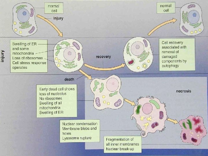
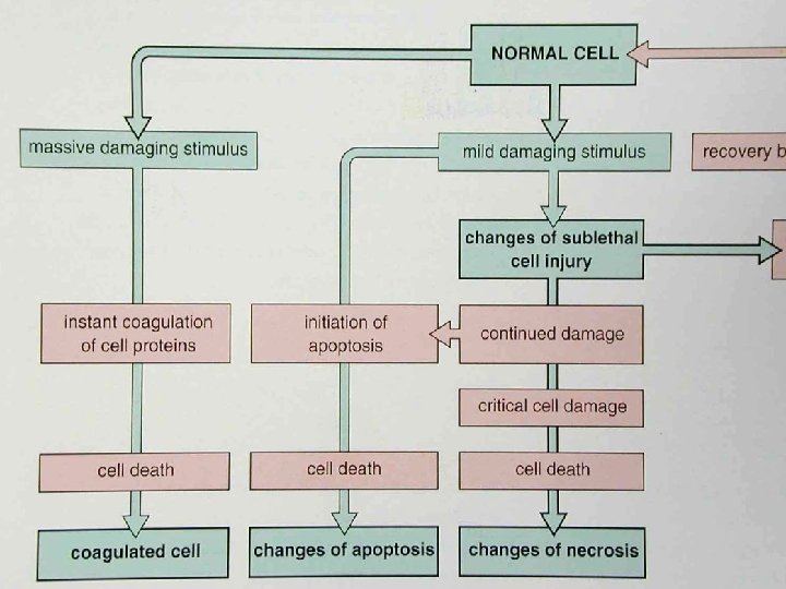
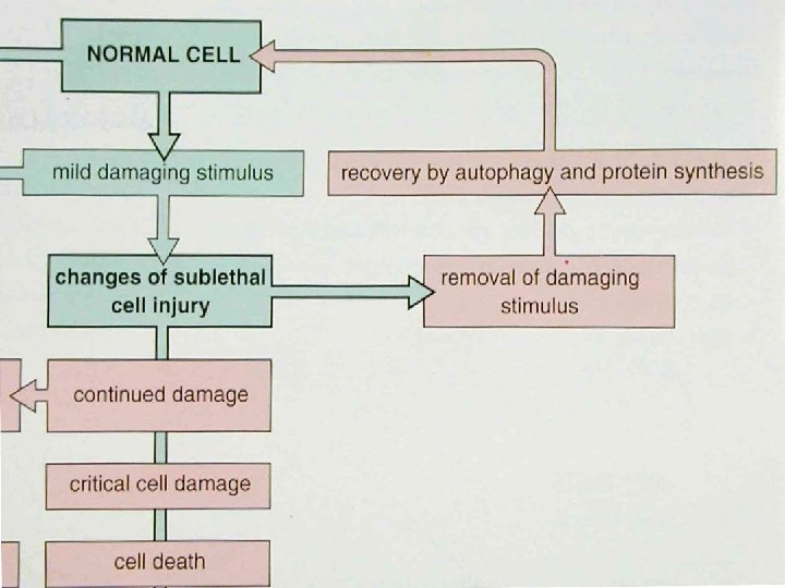
Cell Injury • • Normal cell: homeostasis Sublethal injury: reversible injury Irreversible injury Cell death
Normal homeostasis • Genetic programs – metabolism – differentiation – specialization • Constraints of neighboring cells • Availability of metabotic substrates
Cellular Responses to Injury • Acute cell injury • Reversible cell injury • Cell death • Subcellular alterations in sublethal and chronic injury • Cellular adaptations: ~trophy/~plasia • Intracellular accumulations • Pathologic calcifications • Cell aging
Causes of cell injury • • Oxygen Deprivation: hypoxia/ischemia Physical agents Chemical agents and drugs Infectious agents Immunologic reactions Genetic derangements Nutritional imbalances: self-imposed
Principles of cell injury • Stimulus: type, duration, severity • Cell: type, state, adaptability • Cellular targets – cell membranes: integrity – mitochondria: aerobic respiration – cytoskeleton: protein synthesis – cellular DNA: genetic apparatus • Structural and biochemical elements
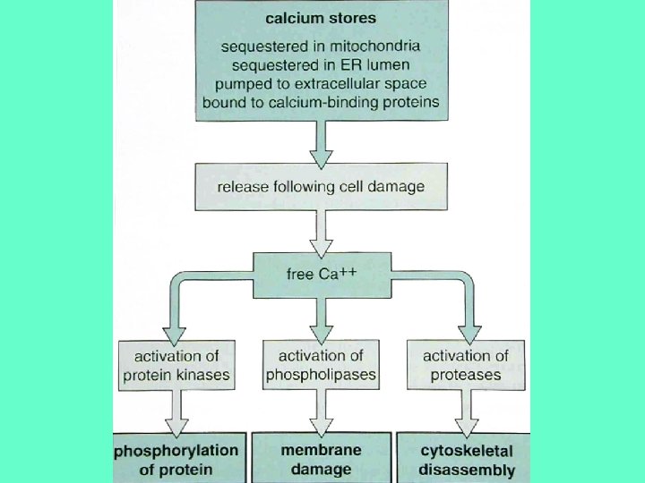
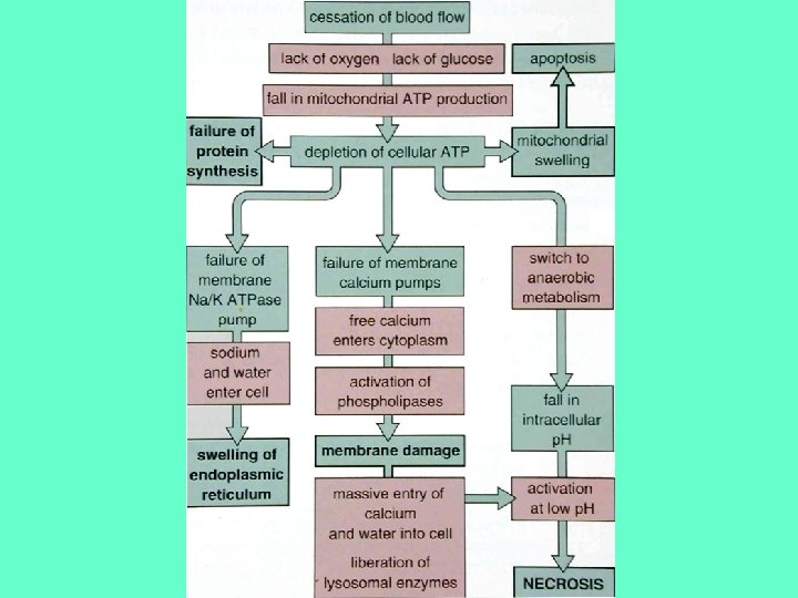
Molecular mechanisms (1 ( • ATP loss causes failure of biosynthesis and ion pumps: ‘cloudy swelling’ • Cytosolic free Ca is a potent destructive agents: activates intracellular enzymes and causes cell death – protein kinases: phosphorylation of protein – phospholipases: membrane damage – proteases: cytoskeletal disassembly
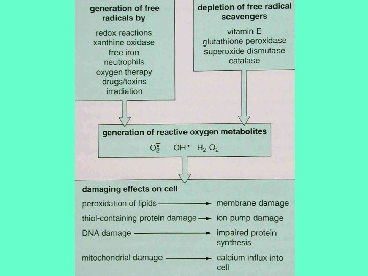
Molecular mechanisms (2 ( • Reactive oxygen metabolites (free radicals) damage cells: O(-), OH(-), H 2 O 2 – peroxidation of lipids (cell memb(. – thiol-containing protein damage (ion pump( – DNA damage (protein synthesis( – mitochondrial damage (Ca influx( • Membrane and cytoskeletal damage – immune-mediated injury
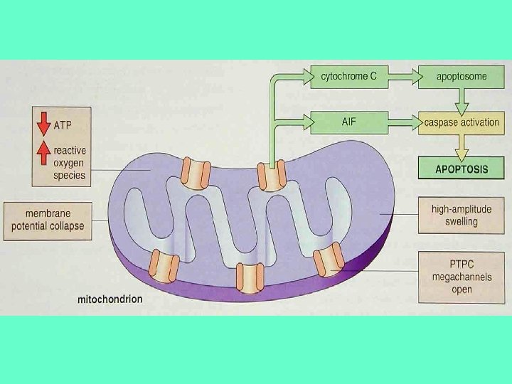
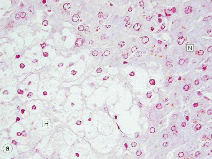
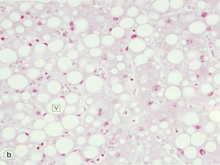
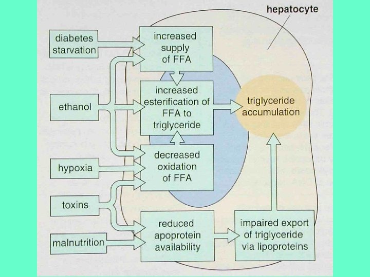
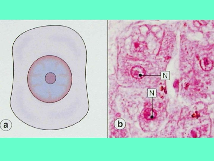
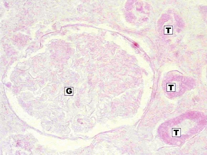
Morphology of Reversible cell injury • Ultrastructural damage to mitochondria – Low-amplitude swelling – )High-amplitude swelling: irreversible( • Swelling of cellular organelles: hydropic degeneration/cloudy swelling • Fatty change: sublethal impairment of metabolism: liver
Morphology of Cell death • Lysis: Disintegration of cellular structure followed by dissolution • Necrosis: spectrum ofmorphologic changes that follow cell death in living tissue • Apoptosis: “programmed cell death”elimination of unwanted host cells
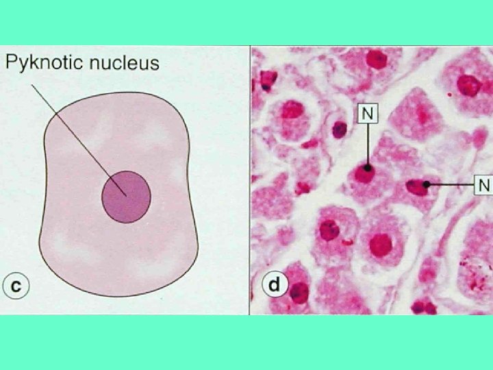
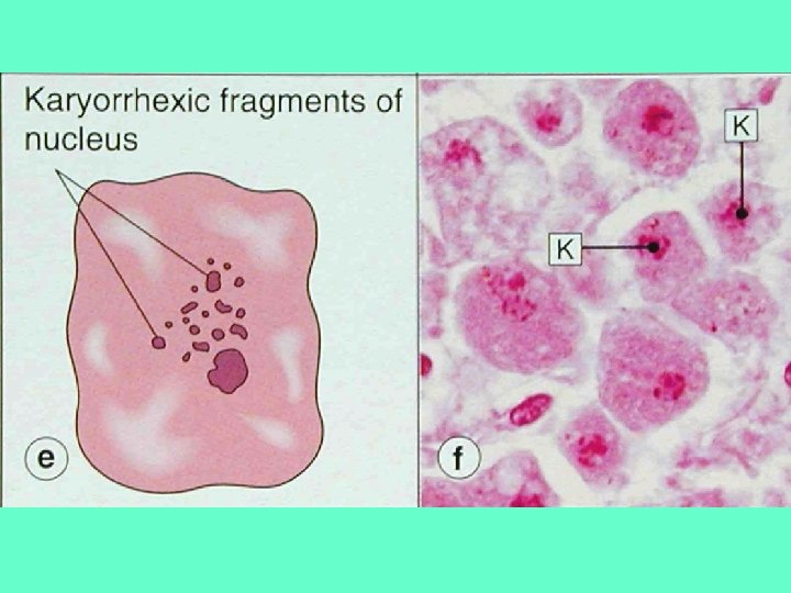
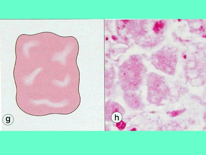
Necrosis • Concurrent processes : – Enzymic digestion: lysis • autolysis: lysosomes of the dead cells • heterolysis: immigrant leukocytes – Denaturation of proteins • Intense eosinophilia • Nonspecific DNA breakdown – Pyknosis – Karyorhexis – Karyolysis
Patterns of Necrosis • • • Coagulative necrosis Liquefactive necrosis Caseous necrosis Fat necrosis Gangrenous necrosis Fibrinoid necrosis
Coagulative necrosis • • • Dead tissue: firm and pale Intact c. outlines and t. architecture Intracellular acidosis denatures enzymes Occlusion of arterial supply Enzymes used in Dx of tissue damage – Myocardium: CK (MB isoform), AST, LDH – Hepatocytes: ALT – Striated muscle: CK (MM isoform ( – Exocrine pancreas: amylase
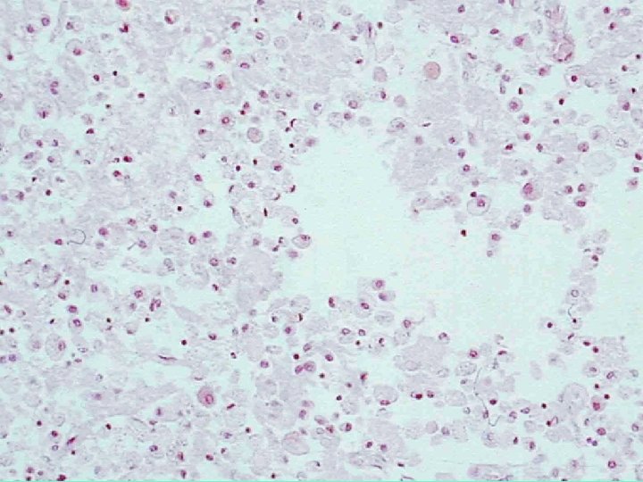
Liquefactive necrosis • Semi-liquid viscous tissue • Potent hydrolytic enzymes • Examples – Hypoxic dead in the CNS: lysosomal enzymes of the neurons and the relative lack of extracellular structural protein – Bacterial infection: pus • neutrophil hydrolases: acute inflammation
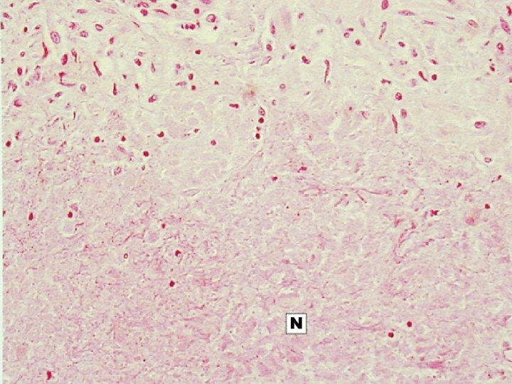
Caseous necrosis • Soft and white: like cream cheese • Amorphous eosinophilic mass, loss of tissue architecture • Associated with granulomatous inflammation(reaction) in Tuberculosis
Fat necrosis • Hard yellow-gray material: fat tissue • Examples: – Retroperitoneal fat necrosis associated with acute of the pancreas – Traumatic fat necosis: breast, buttock
Gangrenous necosis • Mummified darkened and shrinkage • Coagulative necrosis only or modified by liquefactive necrosis • Dry gangrene: limb (lower leg/toe( • Wet gangrene: hollow viscera (GI tract( – hemorrhage within the tissue
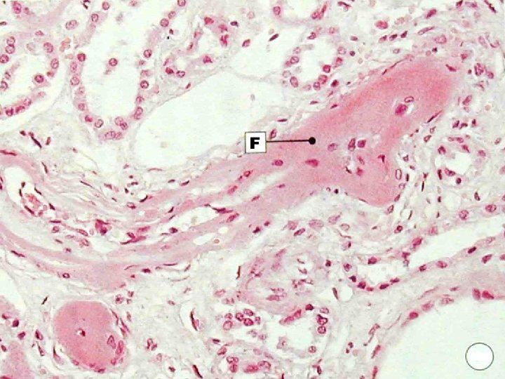
Fibrinoid necrosis • Deposits of fibrin to the wall of necrotic vessels • Causes : – Vasculitis: autoimmune disease – Hypertension
Apoptosis Settings • During development • Homeostatic mechanism to maintain cell populations in tissue: involution • Defense mechanism e. g. immune reaction • Injury – viral infection – low doses of injurious stimuli • Aging
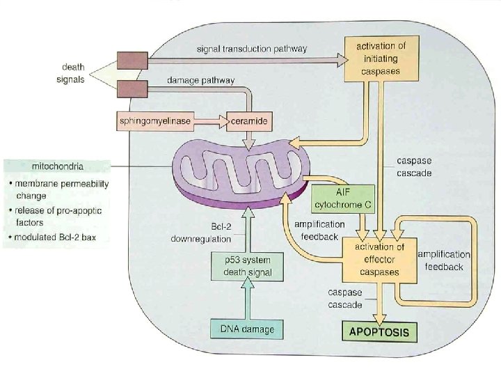
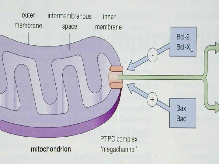
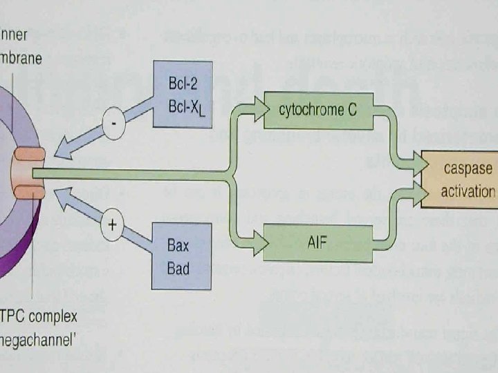
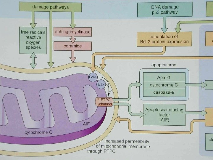
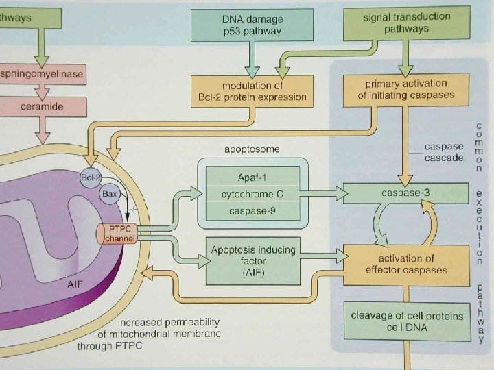
Apoptosis Mechanisms • Signaling pathways – Transmembrane signals: hormone, cytokines – Intracellular signaling: heat, viral infection • Control and integration stage: adaptor proteins, Bcl-2, p 53, granzyme B • Execution phase: endonuclease activation, catabolism of cytoskeleton • Removal of dead cells
Apoptosis Biochemical features • Protein Cleavages: cysteine proteases – caspases: • nuclear scaffold • cytoskeletal proteins • Protein cross-linking: transglutaminase • DNA breakdown: endonucleases – 300~50 kb and then 180~200 bp • Phagocytic recognition – phosphatidylserine
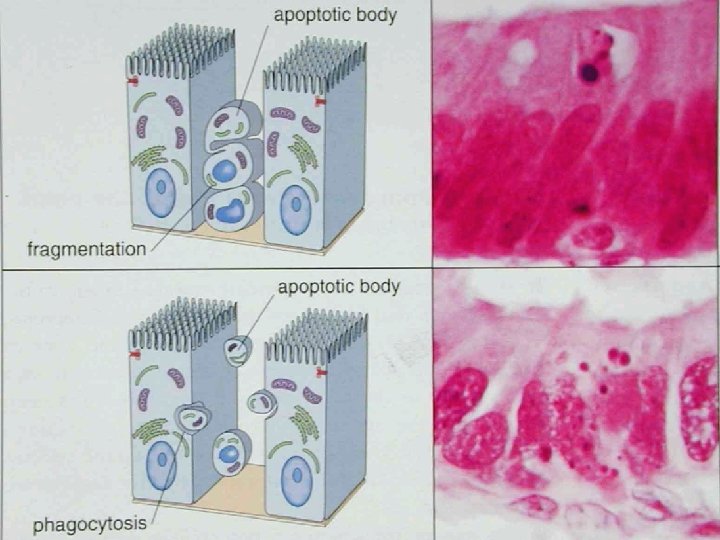
Apoptosis Morphology • Cell shrinkage • Chromatin condensation • Formation of cytoplasmic blebs and apoptotic bodies • Phagocytosis of apoptotic cells/bodies • Single cell or small clusters with intense eosinophilic cytoplasm and dense chromatin fragments