ANATOMY OF URINARY SYSTEM Anatomy of the Urinary

 Lower Urinary Tract Ureters (2) Bladder")


 divide medulla into")


")


 • ~200 ml of urine held • Distension initiates desire")
- Slides: 38
ANATOMY OF URINARY SYSTEM
Anatomy of the Urinary System Kidneys (urine formation) Lower Urinary Tract Ureters (2) Bladder (1) Urethra (1) (urine collection, storage, excretion)
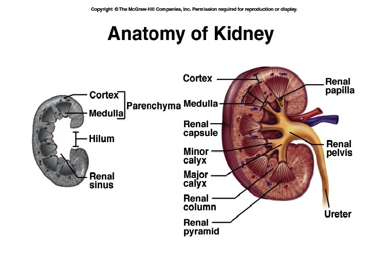
Cortex Medulla Renal hilum Renal artery Renal vein Renal pelvis Ureter Cortex Glomeruli Medulla Renal tubules (with calyces forming the medulary pyramids) Ureter Takes urine to bladder Blood carried to the kidney by the renal artery and taken away by the renal vein.
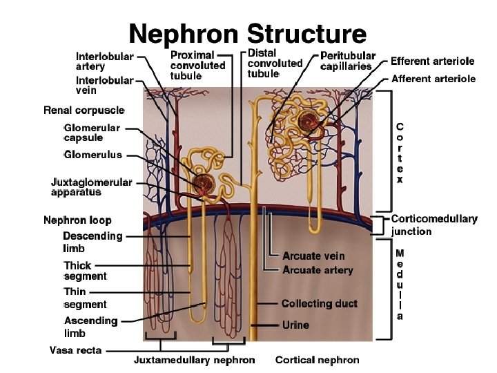
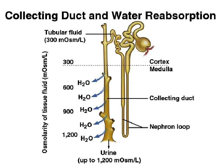
Each KIDNEY consists of 1 million NEPHRONS Each nephron consists of a: GLOMERULUS (found in cortex) forms a protein-free filtrate from blood TUBULE (found in medulla) processes the filtrate to form urine Each TUBULE consists of several segments: Proximal tubule Loop of Henle Distal Tubule Collecting Ducts.
Functional Unit of the Kidney is the NEPHRON Glomerulus Proximal Tubule Loop of Henle Distal Tubule Collecting Duct
Kidney Functions Ø Regulation of water, electrolyte balance, p. H. Ø Removal of waste from blood and excretion of urine. Ø Secretion of hormones Ø Erythropoietin Ø Renin Ø Vitamin D 3
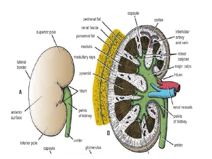
COVERINGS Ø FIBROUS CAPSULE: Outer capsule. Protects from trauma & infections Ø PERIRENAL FAT: Covers fibrous capsule Ø RENAL FASCIA: Encloses kidneys and suprarenal gland Ø PARARENAL FAT: External most forms large quantity of retroperitoneal fat
Gross anatomy • Renal sinus • Renal parenchyma
Renal sinus • Surrounded by renal parenchyma • Contains blood & lymph vessels, nerves, urine-collecting structures
Renal parenchyma • Glandular tissue • Forms urine • Two zones – Outer cortex – Inner medulla
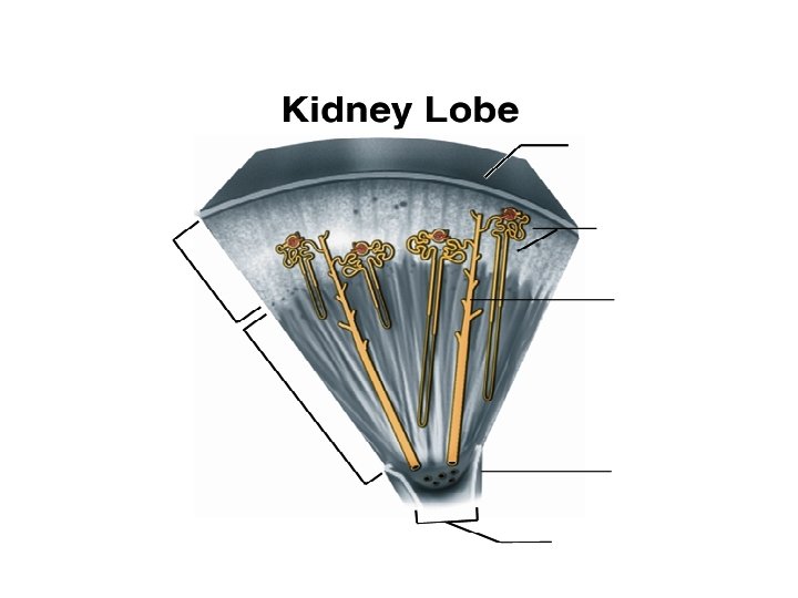
Renal parenchyma • Renal pyramids – Extensions of cortex (renal columns) divide medulla into 6 – 10 renal pyramids – Pyramid + overlying cortex = Lobe – Point of pyramid = Papilla – Papilla nested in cup (minor calyx) – 2 – 3 minor calices Major calyx – 2 – 3 major calices Renal pelvis – Renal pelvis Ureter
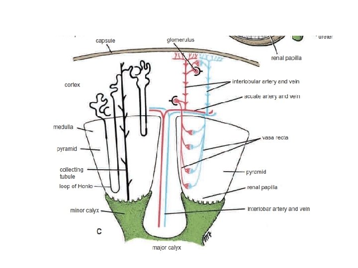
Nephrons • Functional units of kidney • ~1. 2 million per kidney • Three main parts – Blood vessels – Renal corpuscle – Renal tubule
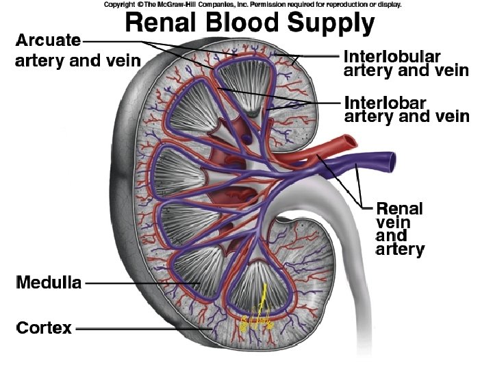
Blood vessels servicing kidney • Supplied by renal artery – ~21% or cardiac output – (Mass in only ~ 0. 4%) – Afferent arterioles – Capillary cluster (glomerulus)
Blood vessels servicing kidney • Glomerulus – Fenestrated capillaries – Capillary filtration in glomerulus initiates urine production – Filtrate lacks cells & proteins – Drained by efferent arteriole – Peritubular capillaries – Renal vein
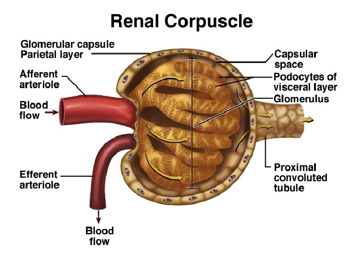
NEPHRON Renal corpuscle • Glomerulus plus capsule • Glomerulus enclosed in two-layered glomerular capsule – “Bowman’s capsule” • Fluid filters from glomerular capillaries – “Glomerular filtrate” • Fluid collects in capsular space • Fluid flows into renal tubule
NEPHRON Renal tubule • Leads from glomerular capsule • Ends at tip of medullary pyramid • ~3 cm long • Four major regions – – Proximal convoluted tubule Nephron loop Distal convoluted tubule Collecting duct
LYMPH SUPPLY: lateral aortic lymph nodes NERVE SUPPLY: Renal sympathetic plexus. Afferent fibers travel through 10 th 11 th 12 th thoracic nerves to spinal cord.
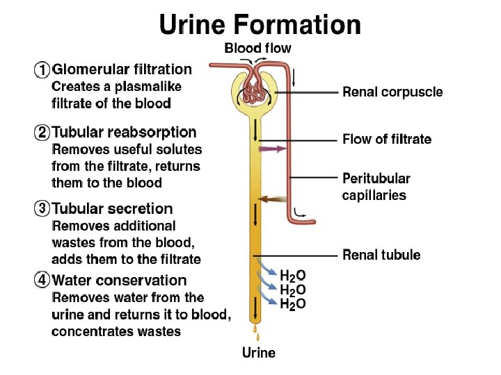
URINE FORMATION Overview • Blood plasma Urine • Four steps – Glomerular filtration – Tubular reabsorption – Tubular secretion – Water conservation
URINE STORAGE Ureters • Carry urine from kidneys to urinary bladder via peristalsis – Rhythmic contraction of smooth muscle • Enter bladder from below • Pressure from full bladder compresses ureters and prevents backflow
Ureters • Small diameter • Easily obstructed or injured by kidney stones (renal calculi) • 3 constrictions: 1) renal pelvis joins the ureter 2) where it crosses the pelvic brim 3) enter into the urinary bladder
• BLOOD SUPPLY • UPPER END: Renal artery • MIDDLE PORTION: testicular artery or Ovarian • LOWER END: In pelvis, sup. Vesical artery • VENOUS Blood drains into the veins corresponding arteries
• Lymph : lateral aortic lymph nodes and iliac nodes • NERVE SUPPLY: Renal, testicular or ovarian and hypogastric plexuses. • Afferent fibers travel with the sympathetic fibers and enter the 1 st and 2 nd lumber segments in the spinal cord.
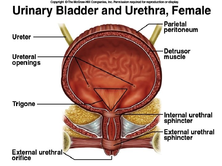
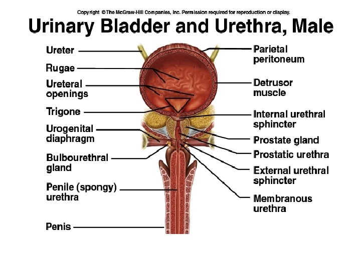
Urinary bladder • Muscular sac • Wrinkles termed rugae • Openings of ureters common site for bladder infection
URINE ELIMINATION Urethra • Conveys urine from body • Internal urethral sphincter – Retains urine in bladder – Smooth muscle, involuntary • External urethral sphincter – Provides voluntary control over voiding of urine
Urethra • 3 – 4 cm long in females – Bound by connective tissue to anterior wall of vagina – Urethral orifice exits body between vaginal orifice and clitoris
Urethra • ~18 cm long in males – Prostatic urethra • ~2. 5 cm long, urinary bladder prostate – Membranous urethra • ~0. 5 cm, passes through floor of pelvic cavity – Penile urethra • ~15 cm long, passes through penis
URINE ELIMINATION Urination (micturition) • ~200 ml of urine held • Distension initiates desire to void • Internal sphincter relaxes involuntarily – Smooth muscle • External sphincter voluntarily relaxes – Skeletal muscle – Poor control in infants • Bladder muscle contracts • Urine forces through urethra