Renal System Urinary System the urinary system is
 • - the urinary system is composed")

 • Medulla: -")


 it contains cuboidal cells that are")


: there")


- Slides: 28
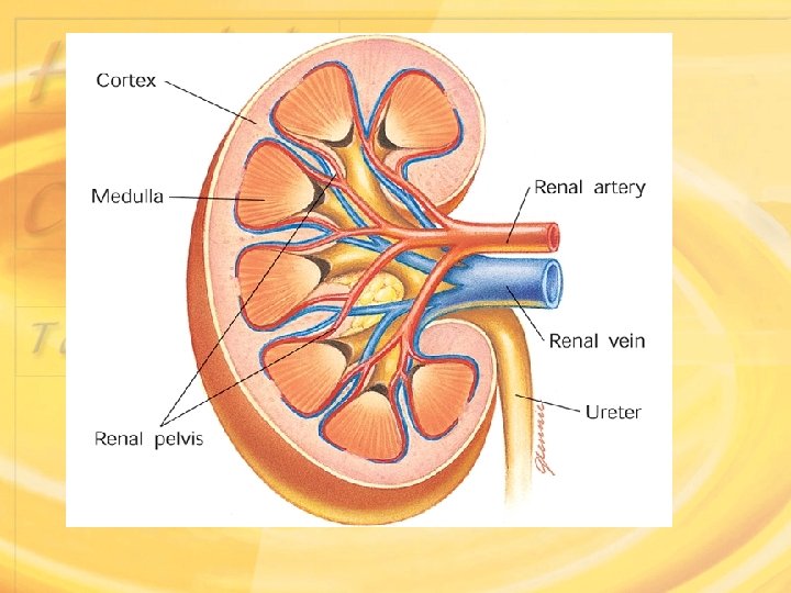
• Renal System: - (Urinary System) • - the urinary system is composed of: 1. two Kidneys. 2. two Ureters. • 3. Urinary Bladder. 4. Urethra. • - the kidney plays a major role in controlling the volume of blood (water) and also controlling ion concentration and PH. • - the kidney is a major excretory organ, responsible for the removal of metabolic waste products from the blood.
• - the kidney act on the plasma first converting it to ultra filtrate to which it later add and remove substances so that the final product is urine. • Renal Functions: • regulation of body water and electrolytes. • excretion of waste products. • regulation of blood pressure (long and short term). • regulation of acid base balance. • regulation of vitamin D 3 production.
• Physiologic anatomy of the kidney: - (Cortex & Medulla) • Medulla: - this tissue appears as pyramids their apex are towards the renal pelvis it appears striated because of the collecting ducts and loops of Henle. • Cortex: - this tissue appears granulated because of the large number of glomeruli, the cortex is surrounded by a connective tissue called Renal Capsule. • *Outside the renal capsule there is a thick layer of fat called Renal Fat Pad which protects the kidney from mechanical shocks
• Functional Anatomy: • The kidney is a complex structure consisting of one million or more, structural and functional unites called Nephrons, separated by connective tissue(interstitial tissue). • The Nephron is the functional unit because it accomplishes the entire complex of processes that result in the formation of urine. • Each individual renal tubule and its glomerulus is a nephron. And the size of the kidney in various species is determined largely by the number of nephrons they contain.
• Each human kidney has approximately 1. 3 million nephrons. (the human needs 1/3 of the 1. 3 million nephrons in order to survive. • ¤ the glomerulus 200µm in diameter is formed by the imagination of a tuft of capillaries into the dilated blind end of the nephron(Bowman's Capsule). • The capillaries are supplied by an afferent arteriole and drained by an efferent arteriole.
Anatomy of the Kidney Copyright © 2008 Pearson Education, Figure 18. 2
• ¤ the tubule starts with: • 1 - Bowman's capsule: the invaginated end of the tubule surrounding the glomerular tuft. {two cellular layers separate the blood from the glomerular filtrate in Bowman's capsule, the capillary endothelium and specialized epithelium of the capsule made up of podocytes. The layers are separated by a basal lamina (basement membrane)}, in addition to the Mesangial cells located between the basal lamina and endothelium (they are contractile and play a role in regulation of glomerular filtration).
Anatomy of the Nephron Figure 18. 3
Renal Corpuscles
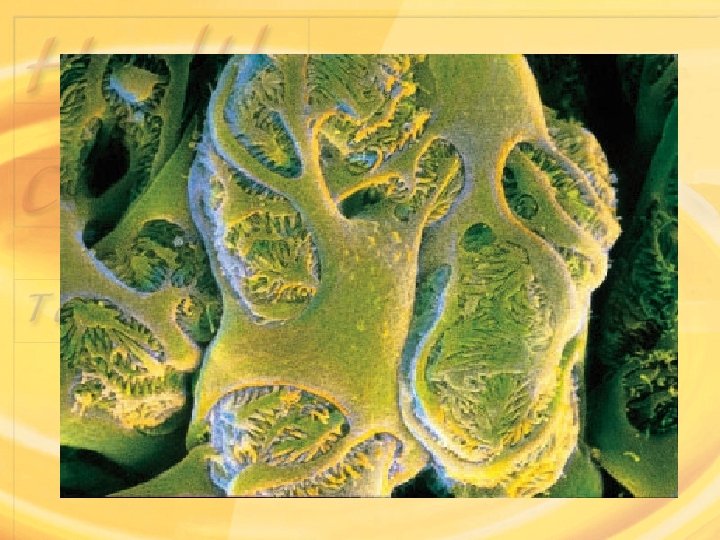
• * the endothelium of glomerular capillaries is fenestrated with pores that are 50 -100 nm in diameter. • * the epithelium(podocytes) form filtration slits approximately 5 -9 nm in diameter.
• 2 - Proximal Convoluted Tubule: (PCT) it contains cuboidal cells that are linked tightly at the apex but contain large lateral intracellular space. Its rich with mitochondria & the surface is covered with micro-villi.
• 3 - Loops of Henle: thin descending loop with high permeability to water & thick ascending loop which is impermeable to water but permit Cl active reabsorption followed by Na movement. • * Thick ascending loop passes between the afferent and efferent arterioles of the same nephron forming juxta glomerular apparatus. • 4 - Distal Convoluted Tubule: (DCT) the first part(diluting segment) continues from the ascending thick loop, then the distal segment which are both under the effect of Aldosteron for Na+-K+ exchange reabsorption. •
• - Collecting Duct: cortical and medullary parts, both under effect of Anti Diuretic Hormone ADH. Contain Principle cells(Pcells) for active Na+ reabsorption and Intercalated cells(I-cells) for H+ secretion. •
• the nephrons in the inner portion of the cortex have long loops of Henle extending down into medullary pyramids and are called Juxta Medullary Nephrons, they form 15%. • While the nephron in the outer portion of the cortex have short loops of Henle and are called Cortical Nephrons which are 85%.
Cortical / Juxtamedullary Nephrons
• Juxta glomerular apparatus: • The thick ascending limb of the loop of Henle reaches the glomerulus of the same nephron which the tubule arose & passes close to its afferent arteriole, the wall of the afferent arteriole contain Renin – secreting Juxta Glomerular cells. At this point, the epithelium of the tubule is modified histologically to form Macula Densa. • The juxta glomerular cells & the macula densa form the Juxta Glomerular Apparatus (JGA) which is a secretary structure secreting renin & erythropoietin.
• * the JGA achieves a regulatory process for Na ions by secreting renin enzyme, the enzyme secretion is stimulated by the sympathetic nerves when sodium is decreased in the blood. • The renin enzyme converts the inactive form of Angiotensinogen (a protein synthesized in liver) to Angiotensin I hormone in which is converted to Angiotensin II by convertaze enzyme in the blood.
• Functions of Angiotensin II are: • A. Short term: • 1. its a potent vasoconstrictive agent due to increasing Blood Pressure(BP). • 2. it also activate the heart which results in increasing the Cardiac Output(CO). • B. Long term: 1. stimulate secretion of Antidiuretic Hormone(ADH) from the Pituitary gland.
• . stimulate secretion of Aldosteron from the Adrenal cortex which regulate the sodium level in the blood. (Aldosteron stimulate reabsorption of Na+ ions & water excretion in urine). • Erythropoietin hormone is secreted in the kidney if either decreased BP or reduced O 2 carried in blood. This hormone stimulates erythropoiesis which increases Blood volume.
• Other endocrine functions of the kidney: • Secretion of prostaglandins (PGs): there are secretary cells called Type I medullary interstitial cells present in the interstitial tissue e. g. PGI 2 & PGE 2. • Converting inactive vitamin D from the inactive form essential for regulation of Ca & Phosphorus in the body
• The blood supply to the kidneys: • Your kidneys receive 20 -25 % of your total CO, in normal individuals about 1200 ml of blood flows through the kidneys each minute. • - each kidney receives blood from a Renal Artery. As it enters the renal sinus the renal artery provides blood to the Segmental Arteries. Segmental arteries further divides into a series of Interlobar Arteries that radiate outward between the renal pyramids. The Interlobar arteries supply blood to the Arcuate Arteries, which arch along the boundary between the cortex & medulla of the kidney. Each arcuate artery gives rise to a number of Interlobular Arteries, and branching from each interlobular artery are Afferent Arterioles (blood reaches the vascular pole of each Glomerulus through an afferent arteriole & leaves in an Efferent Arteriole). •
• Blood travels from the efferent arteriole to form a capillary plexus, a net-work of Peritubular Capillaries (that supplies the PCT & DCT), The Peritubular capillaries provide a route for the pick-up or delivery of substances that are reabsorbed or secreted by these portions of nephrons
• In juxta medullary nephrons, the efferent arteriole & peritubular capillaries are connected to a series of long slender capillary that accompany the loops of Henle into the medulla, these capillaries are known as the Vasa Recta, which absorb & transport solutes and water reabsorbed into the medulla from tubular fluid in the loops of Henle & collecting ducts. (in the normal conditions the removal of solutes and water by the vasa recta balances the rate of solutes and water reabsorbed in the medulla). • - from the peritubular capillaries & vasa recta, blood enters a network of venules and small veins that converge on the Interlobular Veins (in mirror image of the arterial distribution).
• • • • Renal Artery Segmental A. Vein Segmental V. Interlobar V. Interlobar A. Renal Arcuate V. Arcuate A. Interlobular V. Interlobular A. Venules Afferent arteriole Glomerulus Peritubular capillaries & vasa recta Efferent arteriole
• *kidneys receive around 20% of CO, 98% which goes to the cortex & only 2% to medulla. • • Innervation of the Kidneys: • The kidney and ureters are innervated by the Renal Nerves. Most of the nerve fibers involved are sympathetic postganglionic fibers, the sympathetic innervation targets: • Juxta glomerular apparatus. • The smooth muscles in the walls of the afferent & efferent arterioles. • Mesangial cells.
• *Functions of the sympathetic innervation: • → Regulation of glomerular blood flow & pressure. through control of the diameters of the afferent & efferent arterioles and glomerular capillaries. • → Stimulation of renin release from JGA. • → Direct stimulation of water and sodium reabsorption. •