ADVERSE CUTANEOUS DRUG REACTIONS Course 393 Prof Marwan

- Slides: 85
ADVERSE CUTANEOUS DRUG REACTIONS Course 393 (Prof. Marwan Al-Khawajah) College of Medicine King Saud University
ADVERSE CUTANEOUS DRUG REACTIONS - Are common (2 -3% of patients) - Most reactions are mild, accompanied by pruritus and resolve promptly after drug withdrawal - Severe, life threatening ACDRs are rare and unpredictable.
- They can mimic all the morphologic expressions in dermatology. - Must be the first consideration in the differential diagnosis of a suddenly appearing eruption
- Majority are caused by immunologic mechanisms (Gel and coombs types I, III and IV) and in most reactions both cellular and humeral immunity are involved - Provoked by systemic or topical administration including eye/ear drops, suppositories/ pessaries
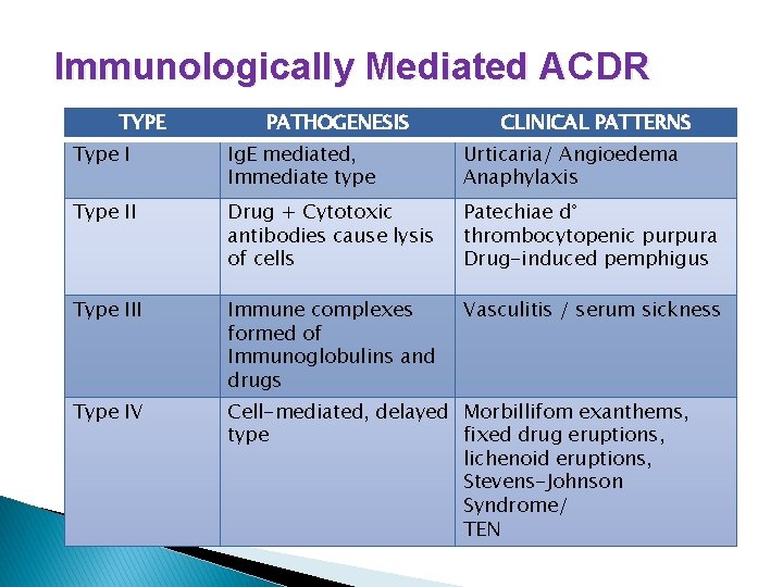
Immunologically Mediated ACDR TYPE PATHOGENESIS CLINICAL PATTERNS Type I Ig. E mediated, Immediate type Urticaria/ Angioedema Anaphylaxis Type II Drug + Cytotoxic antibodies cause lysis of cells Patechiae d° thrombocytopenic purpura Drug-induced pemphigus Type III Immune complexes formed of Immunoglobulins and drugs Vasculitis / serum sickness Type IV Cell-mediated, delayed Morbillifom exanthems, type fixed drug eruptions, lichenoid eruptions, Stevens-Johnson Syndrome/ TEN
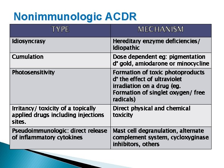
Nonimmunologic ACDR TYPE MECHANISM Idiosyncrasy Hereditary enzyme deficiencies/ Idiopathic Cumulation Dose dependent eg: pigmentation d° gold, amiodarone or minocycline Photosensitivity Formation of toxic photoproducts d° the effect of ultraviolet irradiation on a drug (eg. Formation of singlet oxygen/ free radicals) Irritancy/ toxicity of a topically applied drugs including injections sites. Direct physical and chemical toxicity Pseudoimmunologic: direct release of inflammatory cytokines Mast cell degranulation, alternate complement system, cycloxyginase inhibitors, others
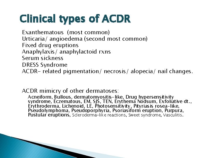
Clinical types of ACDR Exanthematous (most common) Urticaria/ angioedema (second most common) Fixed drug eruptions Anaphylaxis/ anaphylactoid rxns Serum sickness DRESS Syndrome ACDR- related pigmentation/ necrosis/ alopecia/ nail changes. ACDR mimicry of other dermatoses: Acneiform, Bullous, dermatomyositis-like, Drug hypersensitivity syndrome, Eczematous, EM, SJS, TEN, Erythema Nodsum, Exfoliative dt. , Erythroderma, Lichenoid, LE, Photosensitivity, Pityriasis rosea-like, Pseudolymphoma, Pseudoporphyria, Psoriasiform eruption, Purpura, Pustular eruptions, Scleroderma-like reactions, Sweet syndrome, Vasculitis.
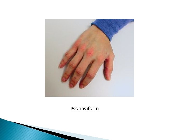
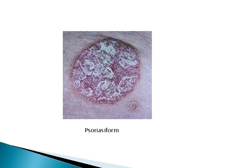
Psoriasiform
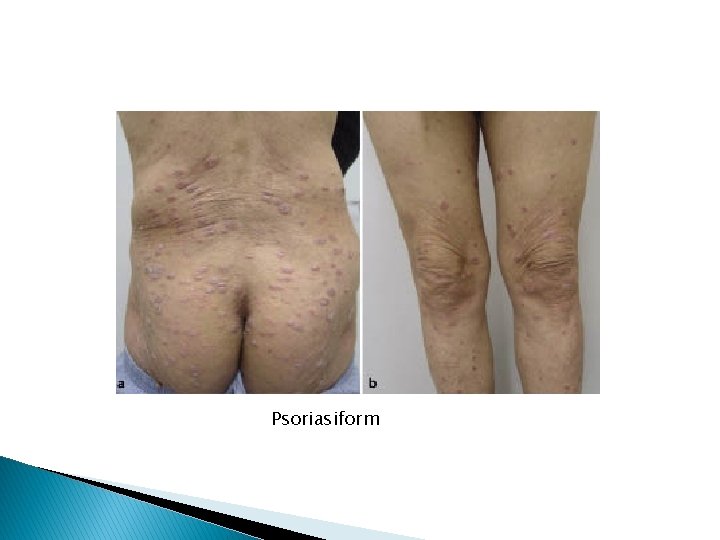
Psoriasiform
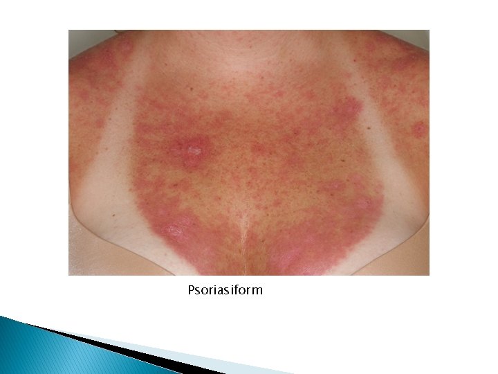
Psoriasiform
Psoriasiform
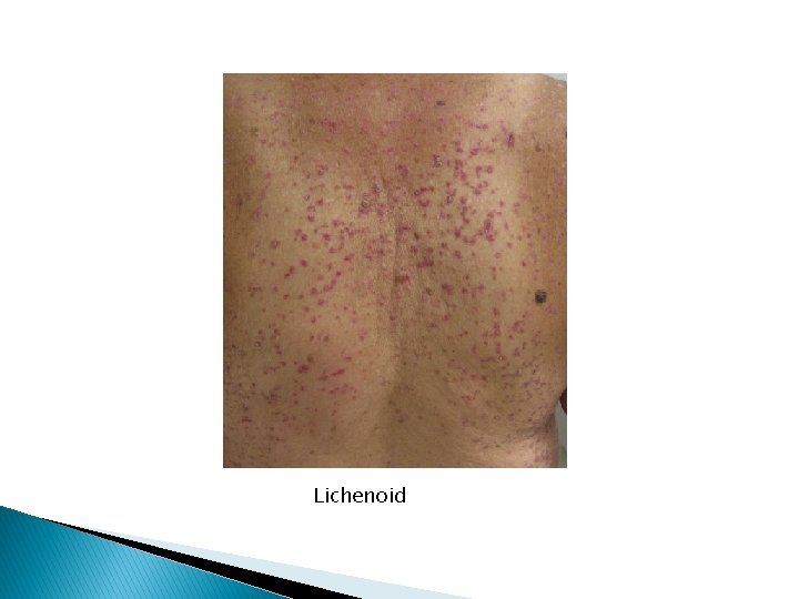
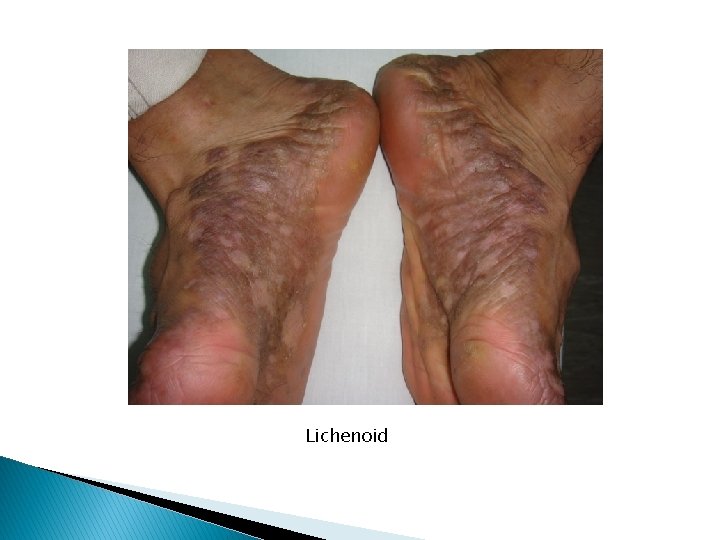
Lichenoid
Lichenoid
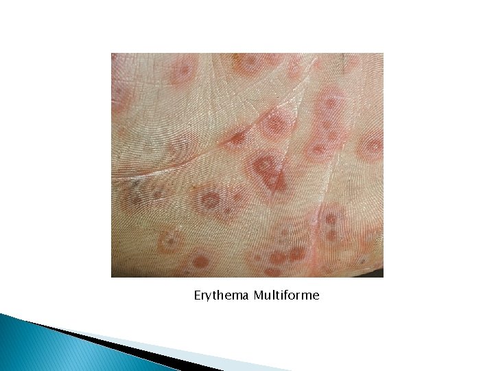
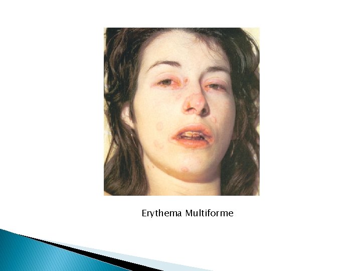
Erythema Multiforme
Erythema Multiforme
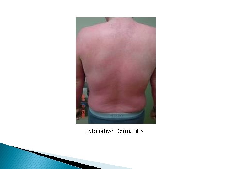
Exfoliative Dermatitis
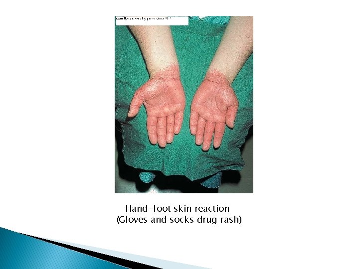
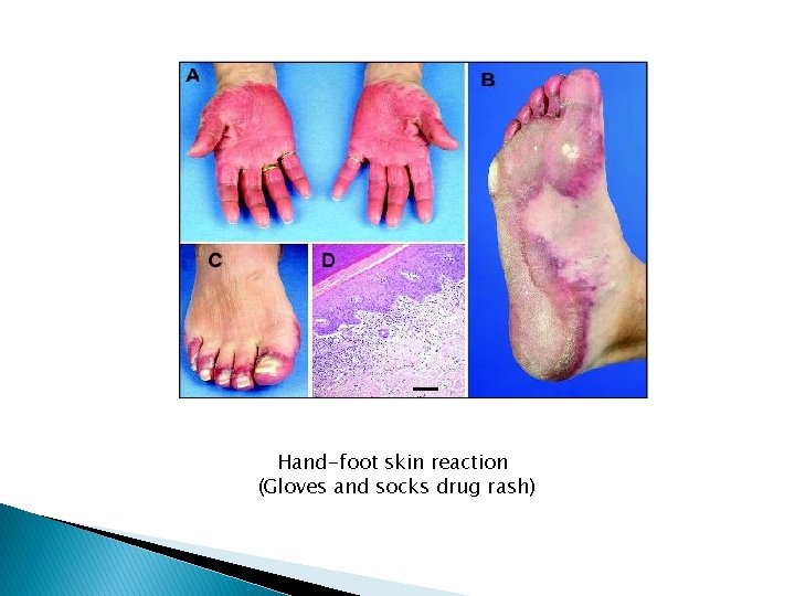
Hand-foot skin reaction (Gloves and socks drug rash)
Hand-foot skin reaction (Gloves and socks drug rash)
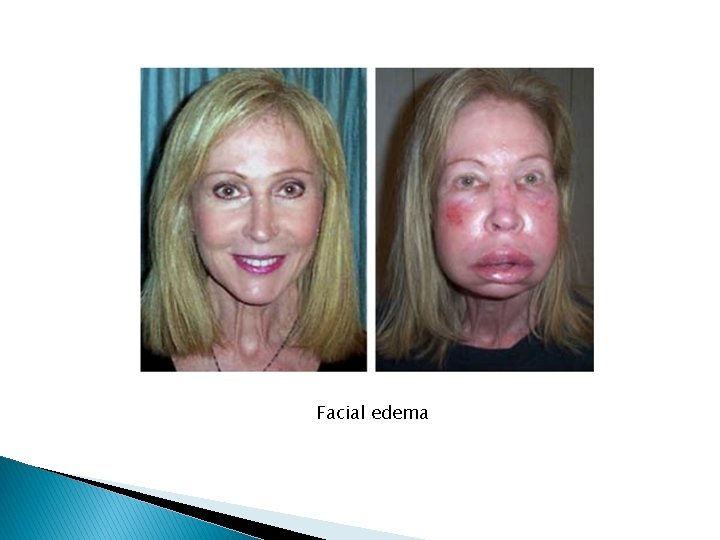
Facial edema
Facial edema
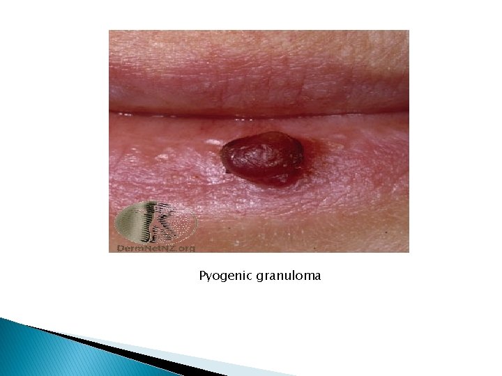
Pyogenic granuloma
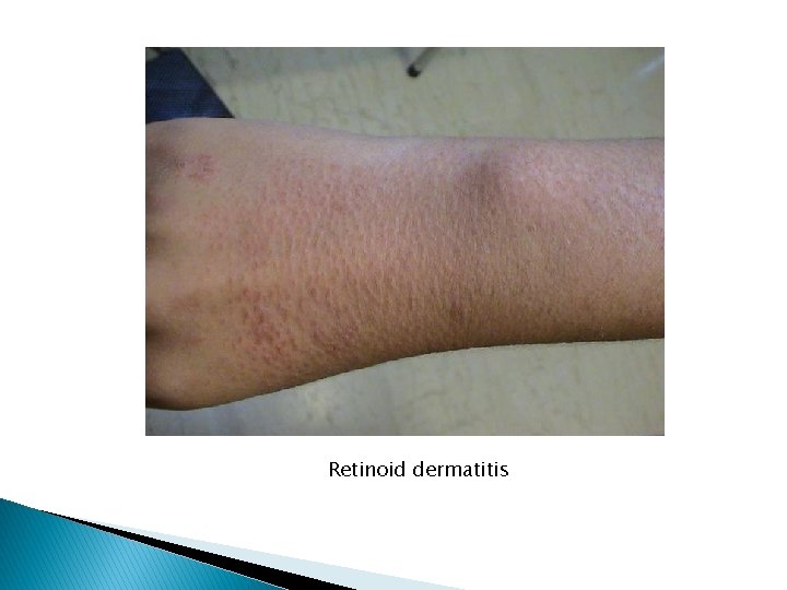
Retinoid dermatitis
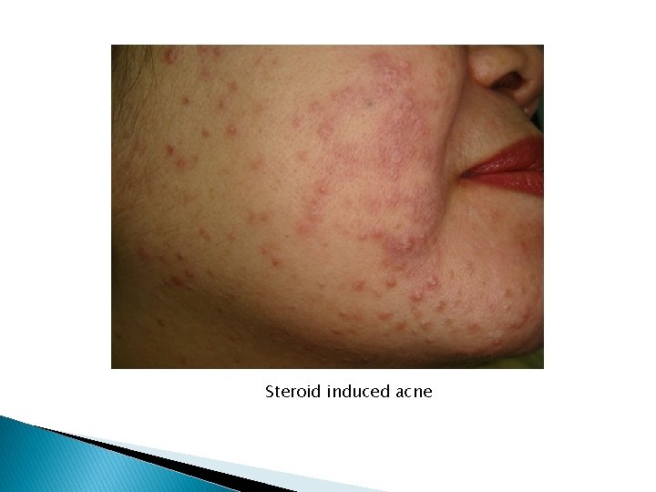
Steroid induced acne
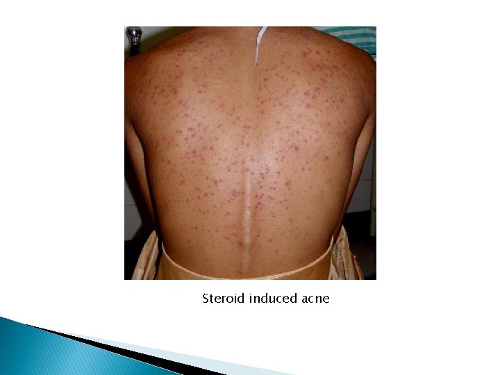
Steroid induced acne
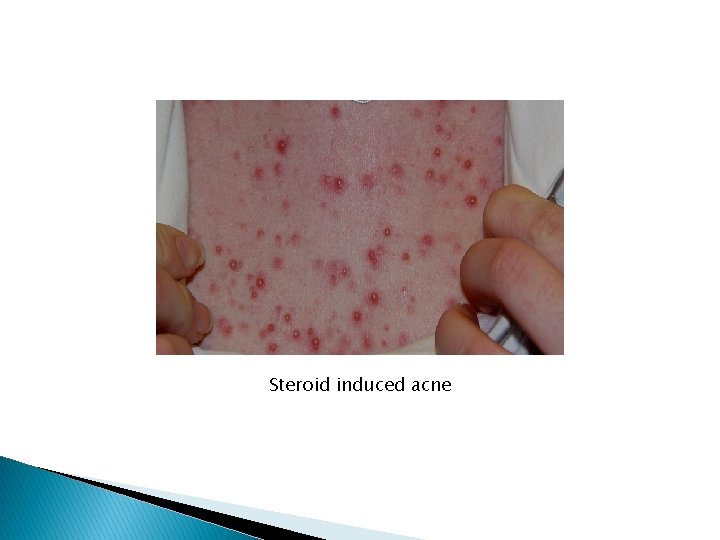
Steroid induced acne
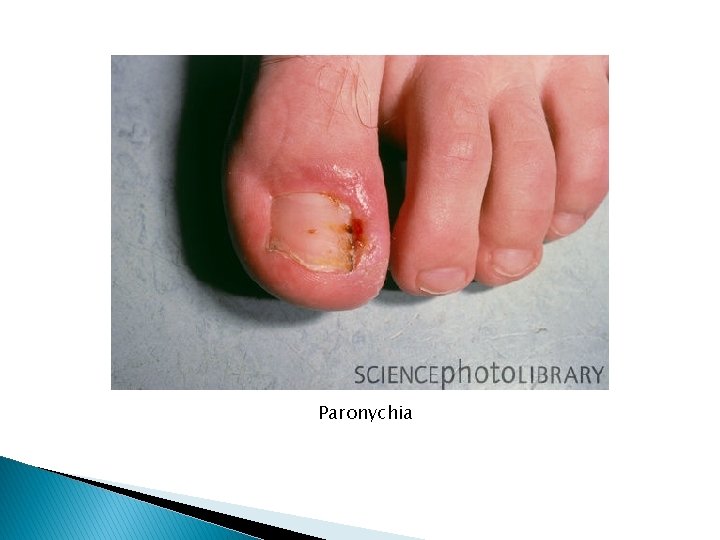
Paronychia
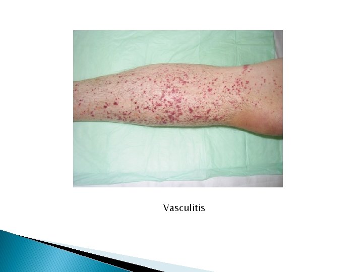
Vasculitis
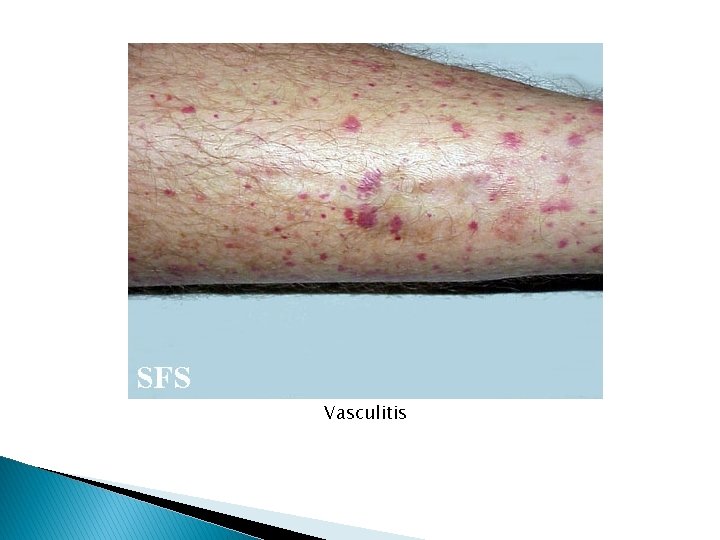
Vasculitis
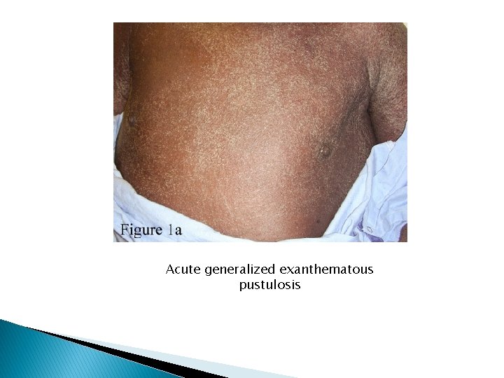
Acute generalized exanthematous pustulosis
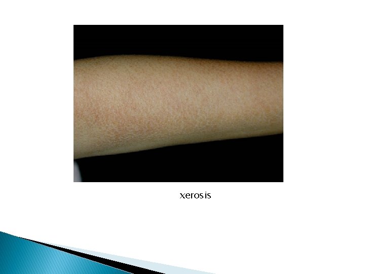
xerosis
Guidelines for assessing possible ACDRs - Exclude other causes esp. infections Examine interval between introduction and induction Determine if similar reactions occurred with the same or similar compounds Note any improvement after withdrawal Note any reaction after readministration
Findings indicating possible lifethreatening ACDR - Arthralgia Blisters/epidermal detachment/ positive Nikolsky sign Confluent erythema Enlarged lymphnodes Facial edema/central facial involvement High fever (>40°c) Mucous membranes erosions Palpable purpura Skin necrosis Skin pain Shortness of breath, wheezing, hypotension Swelling of the tongue/ oral mucosa Urticaria/ Angioedema
Diagnosis is usually made on clinical findings Biopsy is helpful in defining the type of reaction pattern but not in identifying the offending drug. CBC : eosinophil count >1000/micro. L lymphocytosis with atypical lymphocytes Chemistry : abnormal LFT Skin Test/RAST helpful in Ig. E-mediated reaction (penicillin)
Management : - DC the culprit drug/drugs (cf. morbilliform vs. angioedema, SJS and TEN) Symptomatic treatment Prevention: awareness; premedication
Exanthematous Drug Reactions Definition A cutaneous eruption that mimics a measleslike viral exanthem. (synonyms: Morbilliform drug rash, maculopapular drug reaction) Most common type of cutaneous drug reaction but less common in the very young.
Pathogenesis Exact mechanism unknown. Probably delayed hypersensitivity. Most commonly incited drugs (10 -20%): penicillins, carbamazepine, allopurinol, gold salts Less common (3 -5%) : sulfonamides (bacteriostatic, diuretic, antidiabetic), NSAIDs, hydantoin derivatives, isoniazid, chloramphenicol, erythromycin + others (<1%). Special situations: Mononucleosis, HIV, Allopurinol, cross-drug hypersensitivity.
Clinical Manifestations Onset: peak incidence at ninth day after administration, 2 -3 days after readministration. Symptoms: severe pruritis (if painful think TEN) + fever, chills
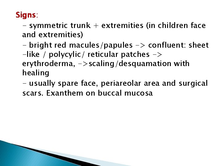
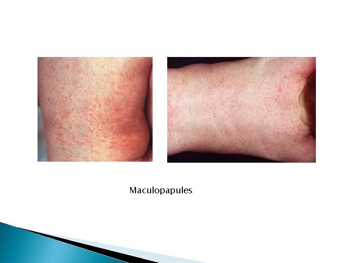
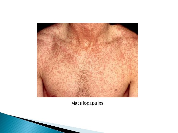
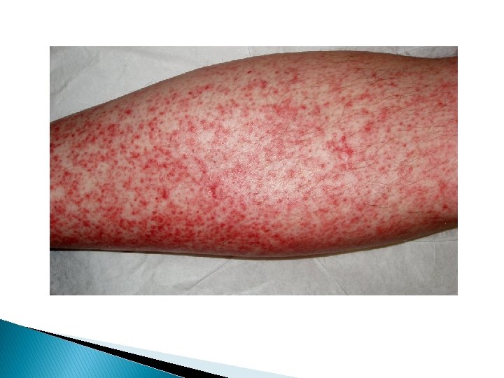
Signs: - symmetric trunk + extremities (in children face and extremities) - bright red macules/papules -> confluent: sheet -like / polycylic/ reticular patches -> erythroderma, ->scaling/desquamation with healing - usually spare face, periareolar area and surgical scars. Exanthem on buccal mucosa
Maculopapules
Maculopapules
Exanthematous
Diagnosis Clinical Diagnosis - Histopathology: perivascular lymphocytes and eosinophils - Blood: eosinophilia Differential Diagnosis - Viral exanthems - Secondary syphilis - Atypical pityriasis rosea - Early widespread allergic contact dermatitis
Prognosis: Good but maybe the initial presentation of a more serious eruption, i. e. SJS, TEN, DRESS, or serum sickness. Treatment: Definitive (cf. indications for discontinuation of a drug) Symptomatic Oral antihistamines, topical and systemic corticosteroids Prevention - Awareness of specific drug and cross-reactants - wearing a bracelet
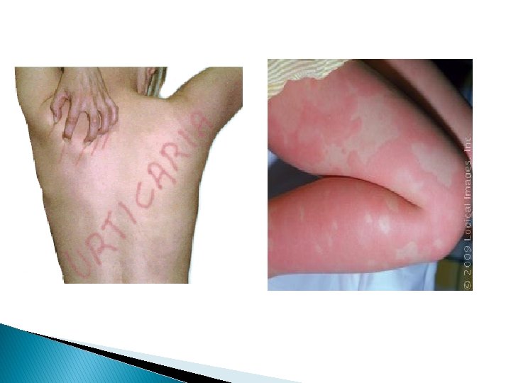
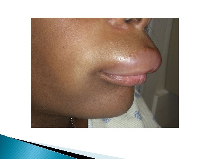
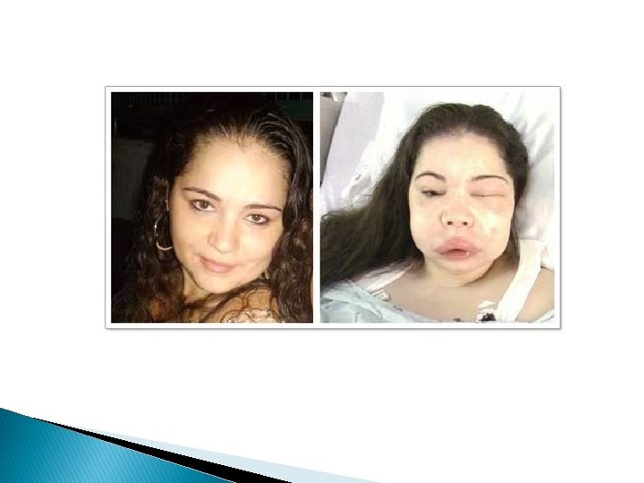
Drug-Induced Acute Urticaria/Angioedema, Edema and Anaphylaxis Definition: transient wheals and edema Pathogenesis Immune-mediated (Ig. E or complement and immune complex) Non allergic: cyclooxygenase inhibitors, direct degranulation of mast cells, direct complement trigger, kinin metabolism inhibitors.
Clinical manifestation Onset: 1 -2 weeks after administration; minutes to hours after readministration Symptoms: - pruritus - burning palms/ soles/ auditory canal, dizziness, tongue numbness, palpitation, sudden fatigue, difficulty breathing, headache substernal pressure, crampy abdominal pain.
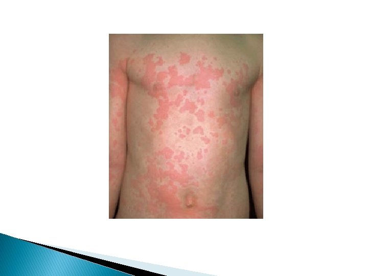
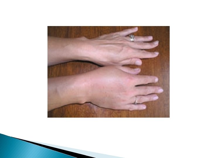
Signs: - Wheals and/or large and deep skin colored swellings - flushing, yawning, airway edema, sneezing, bronchopasm, laryngeal edema, hypotension, vomiting, diarrhea, arthralgia
Diagnosis Clinical Diagnosis - Do biopsy if vasculitis suspected - Measure complement if vasculitis suspected - Ultrasonography if edema of bowel suspected Differential Diagnosis - Acute allergic contact dermatitis - Insect bites - Cellulitis
Prognosis resolves within hours to weeks after drug withdrawal Treatment - Definitive - Symptomatic: subcutaneous epinephrine (0. 30. 5 ml of 1/1000) + airway/ IV access, - H 1/H 2 blockers, sys. glucocorticoids - Prevention: awareness/ wallet card/ bracelet/ pretreatment
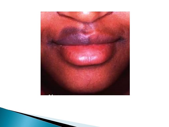
Fixed Drug Eruption Definition Identical skin lesion(s) that recur at the same location. Pathogenesis Unknown Most common drugs: tetracyclines, antimicrobials phenolphthalein, oral contraceptives, NSAIDs, Salicylates, sulfonamides, metronidazole, barbiturates, food coloring (yellow), quinine
Clinical manifestation Onset: Within 30 minutes to 8 hours after ingestion of drug in previously sensitized individual Symptoms: Usually asymptomatic (painful if eroded) May be associated with headache (barbiturate analgesic), constipation (phenolphthalein laxative), Cold (OTC yellow dye) Food (yellow dye, quinine, salicylates)
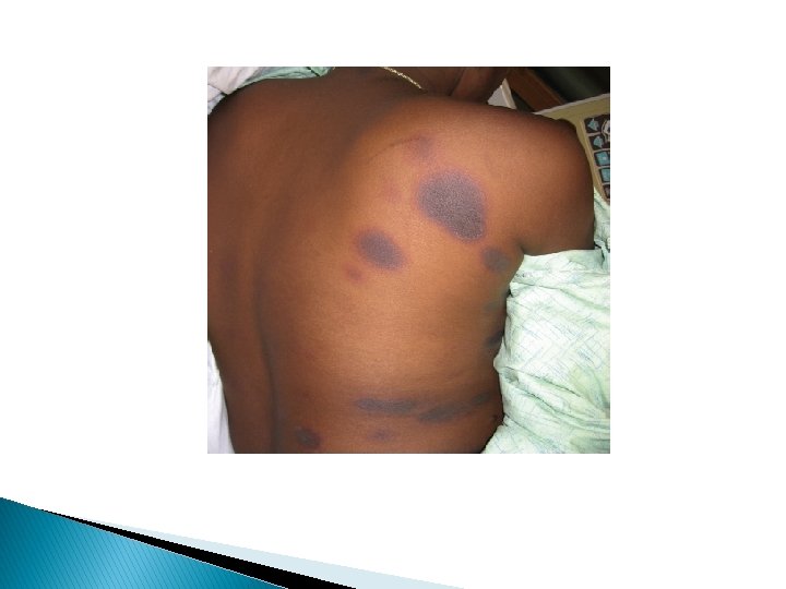
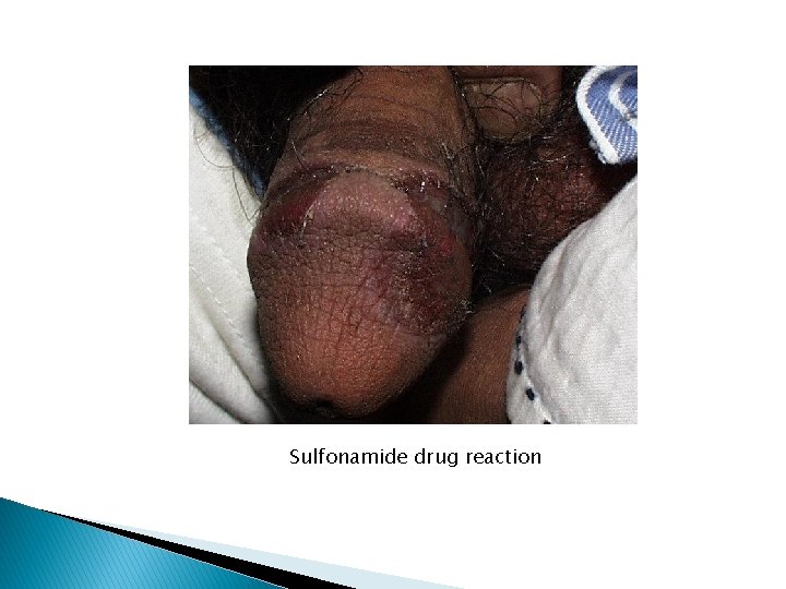
Signs: Round/oval usually solitary, sharply demarcated, erythematous macule -> dusky red/violaceous edematous plaque -> bulla/erosion -> dark brown violaceous post inflammatory hyperpigmentation. Common on genitals and oral mucosa but any site including periorbital, conjunctivae and oropharynx
Sulfonamide drug reaction
Diagnosis Clinical diagnosis: Histopathology similar to EM/TEN Patch test (at the same site) Differential diagnosis: EM; Herpes simplex; Aphthae if extensive: SJS/TEN
Prognosis Resolves within weeks of withdrawal Recurs within hours after a single dose Treatment Non-eroded: potent topical glucocorticoid Eroded: antimicrobial ointment Widespread/ painful mucosal lesions: oral prednisolone 1 mg/kg tapered over few weeks.
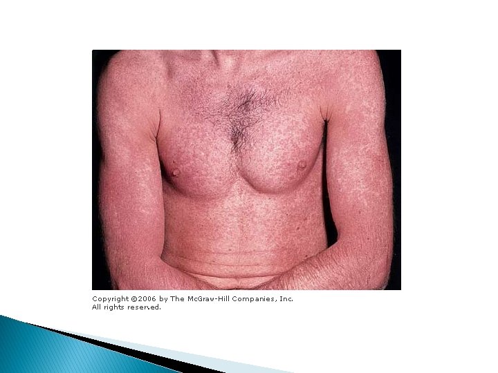
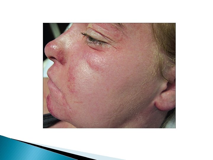
Drug Hypersensitivity Syndrome (DRESS) Definition An idiosyncratic serious adverse drug reaction that involves skin and other organs. Pathogenesis - Hereditary (toxic arene oxide metabolites; slow Nacetylation of sulfonamides) - Idiopathic Most common drugs: - Antiepileptics (phenytoin, carbamazepine, phenobarbital) - Sulfonamides (antimicrobials, dapsone, sulfasalazine).
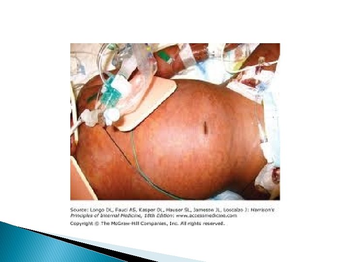
Clinical manifestation Onset: 2 -8 weeks after first drug administration Symptoms: Fever, malaise, ± pruritus Signs: Morbilliform eruption on face, upper trunk and extremities with periorbital edema and mucosal involvement -> generalized exfoliative (erythroderma) ± pustular ± bullous ± purpura on legs -> scaling/desquamation with healing Other: lymphadenopathy, hepatitis, carditis, nephritis, pneumonititis, hematologic, joints, muscles, thyroid, brain.
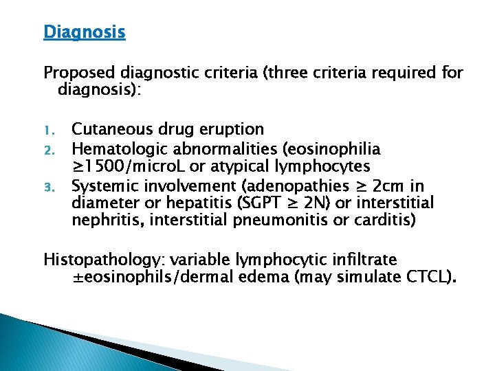
Diagnosis Proposed diagnostic criteria (three criteria required for diagnosis): 1. 2. 3. Cutaneous drug eruption Hematologic abnormalities (eosinophilia ≥ 1500/micro. L or atypical lymphocytes Systemic involvement (adenopathies ≥ 2 cm in diameter or hepatitis (SGPT ≥ 2 N) or interstitial nephritis, interstitial pneumonitis or carditis) Histopathology: variable lymphocytic infiltrate ±eosinophils/dermal edema (may simulate CTCL).
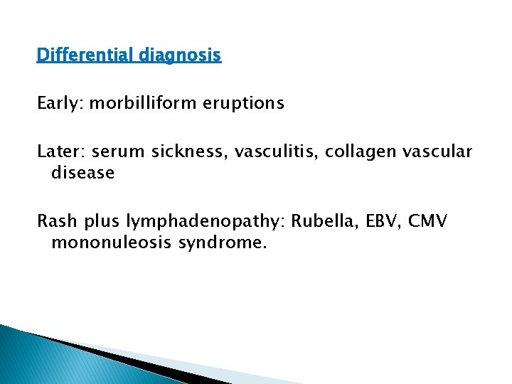
Differential diagnosis Early: morbilliform eruptions Later: serum sickness, vasculitis, collagen vascular disease Rash plus lymphadenopathy: Rubella, EBV, CMV mononuleosis syndrome.
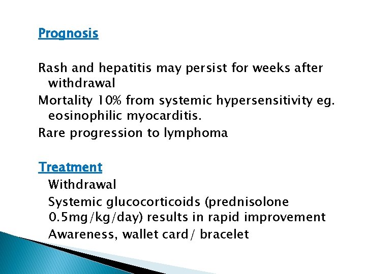
Prognosis Rash and hepatitis may persist for weeks after withdrawal Mortality 10% from systemic hypersensitivity eg. eosinophilic myocarditis. Rare progression to lymphoma Treatment Withdrawal Systemic glucocorticoids (prednisolone 0. 5 mg/kg/day) results in rapid improvement Awareness, wallet card/ bracelet
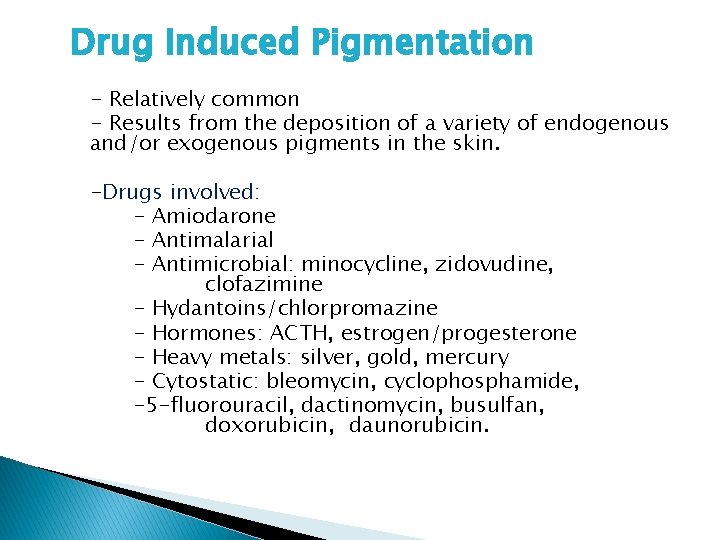
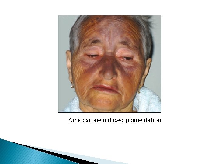
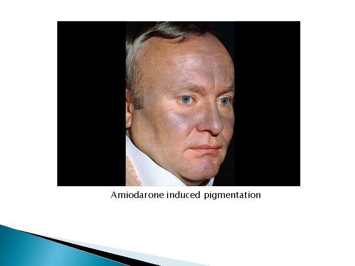
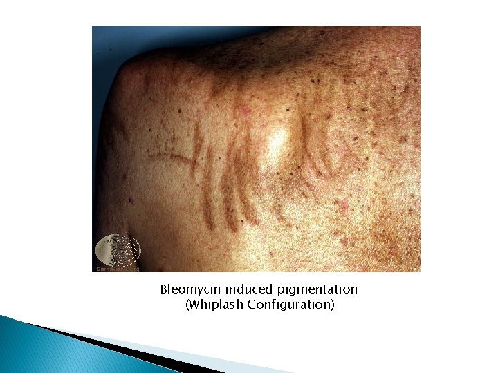
Drug Induced Pigmentation - Relatively common - Results from the deposition of a variety of endogenous and/or exogenous pigments in the skin. -Drugs involved: - Amiodarone - Antimalarial - Antimicrobial: minocycline, zidovudine, clofazimine - Hydantoins/chlorpromazine - Hormones: ACTH, estrogen/progesterone - Heavy metals: silver, gold, mercury - Cytostatic: bleomycin, cyclophosphamide, -5 -fluorouracil, dactinomycin, busulfan, doxorubicin, daunorubicin.
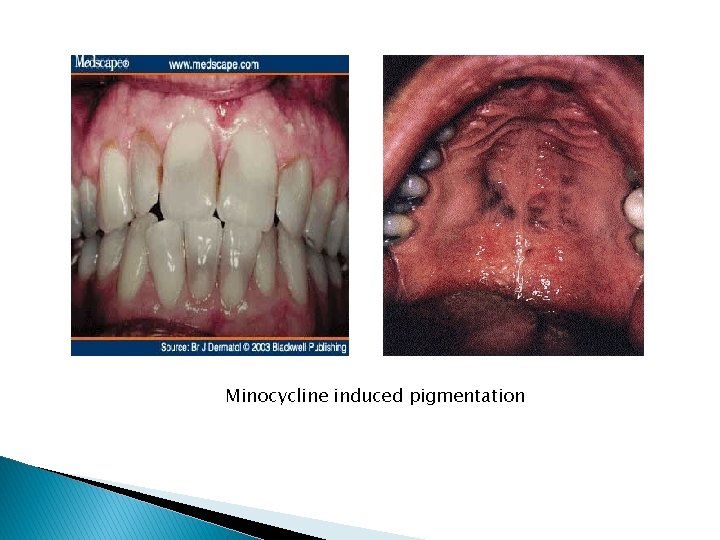
Minocycline induced pigmentation
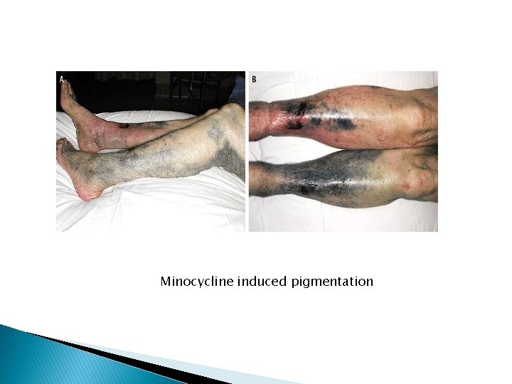
Minocycline induced pigmentation
Amiodarone induced pigmentation
Amiodarone induced pigmentation
Bleomycin induced pigmentation (Whiplash Configuration)
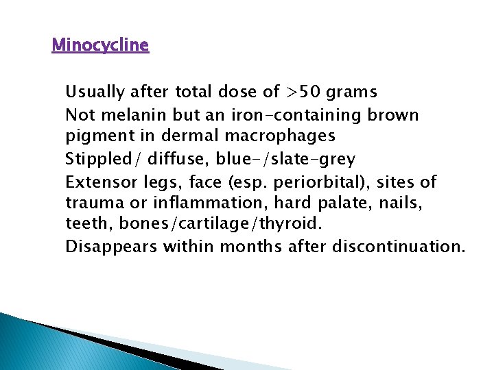
Minocycline Usually after total dose of >50 grams Not melanin but an iron-containing brown pigment in dermal macrophages Stippled/ diffuse, blue-/slate-grey Extensor legs, face (esp. periorbital), sites of trauma or inflammation, hard palate, nails, teeth, bones/cartilage/thyroid. Disappears within months after discontinuation.
Antimalarials Occur in 25% who take the drug for >4 months. Due to melanin/hemosiderin Brownish, grey brown and/or blue black. (quinacrine: yellow-green) Over shins, face, nape of neck, hard palate, under finger- and toenails, cornea, retina, (quinacrine: yellow sclerae) Disappears within few months.
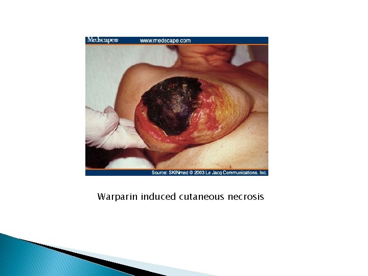
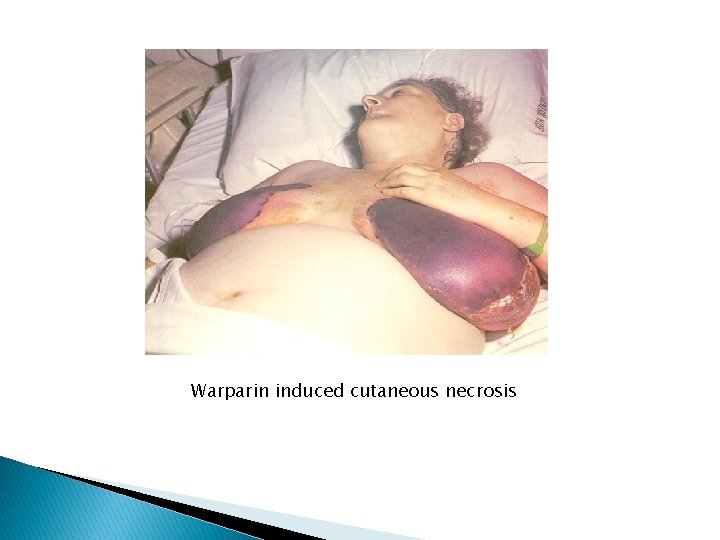
ACDR- related necrosis -After oral drug or at sites of injection Warfarin cutaneous necrosis: Idiosyncratic Onset: 3 -5 days of anticoagulation therapy. Due to a transient hypercoagulable state and thrombus formation. Risk factors: high initial dose, obesity, female, hereditary deficiency of protein C, protein S or antithrombin III.
Sharply demarcated, deep purple to black necrosis. Lesions vary with severity of reaction: petechaie to acchymoses to tender hemorrhagic infarcts to extensive necrosis ->deep tissue sloughing/ ulceration. Usually single. On areas of abundant fat. Acral areas spared. Coagulation studies: within normal limits
Differential Diagnosis - Purpura fulminans (DIC), - Hematoma in overly anticoagulated patient, - Necrotizing soft tissue infection, - Vasculitis, - Recluse spider bite. Course/ Prognosis May subside/heal by granulation or require surgical intervention. Life threatening if extensive in an elderly debilitated patient.
Warparin induced cutaneous necrosis
Warparin induced cutaneous necrosis
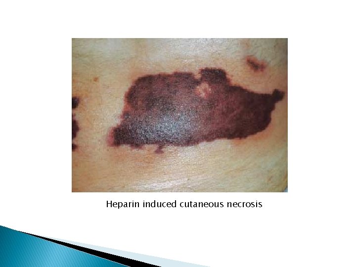
Heparin induced cutaneous necrosis
Other causes of ACDR-related necrosis -Heparin/ Interferon-α/ embolia cutis medicamentosa at site of injection - Ergotism: acral gangrene (suppositories perianal) - At pressure sites in deeply sedated patients.