
AMOEBAE 1 Entamoeba histolytica Morphologic forms 1 Active
AMOEBAE 1

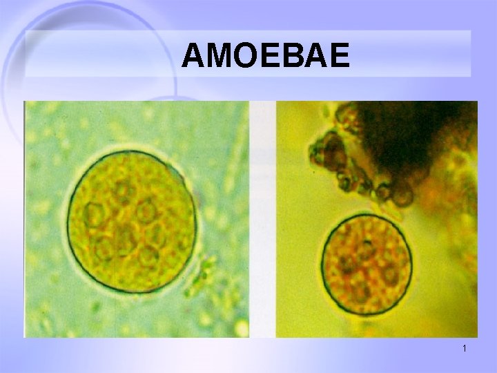
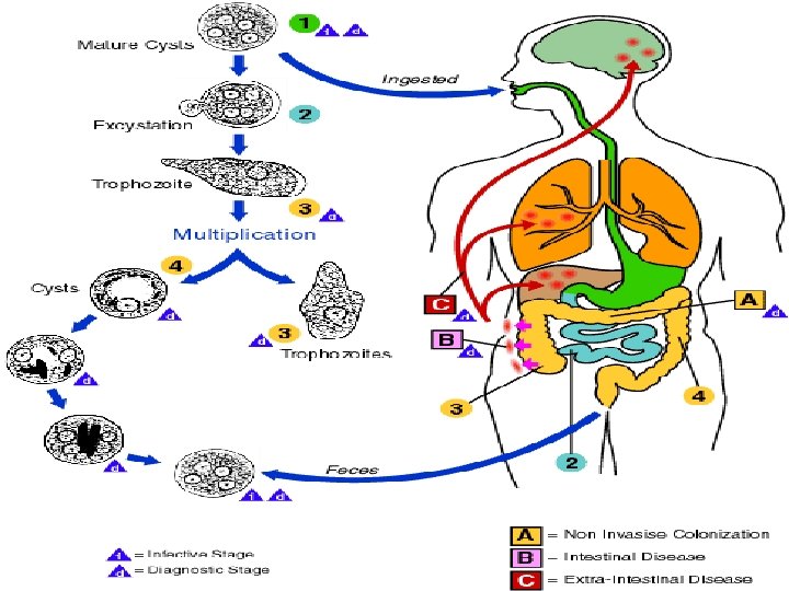
Entamoeba histolytica Morphologic forms: 1 - Active Trophozoite form 2 - Cyst form 2

3
4
Pathogenesis of Extra-intestinal Amoebiasis 5
Lungs Diaphragm Subphrenic abscess Skin Liver Pericardium Peritoneum Stomach Intestines Inferior vena cava Portal circulation Peritoneum General circulation Primary infection in colon Brain Spleen Suprarenal Kidney Perianal skin Genitals 6
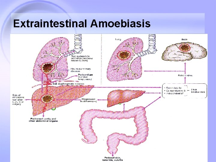
Extraintestinal Amoebiasis 7

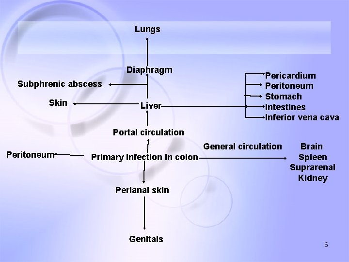
Hepatic Amoebiasis • Pathogenesis and pathology E. histolytica trophozoites portal vein pseudopodia amebic liver abscess liver tissue cytolytic enzymes rupture peritonitis 8
Hepatic Amoebiasis • Single abscess, different size , usually at the right lobe of the liver. • Microscopically three zones : ü Inner: necrotic material and no amoebae ü Intermediate: Degenerated liver cells, RBCs, WBCs ü Outer : normal layer of liver cells with trophozoites replicating • Typically pus is thick, chocolate brown, resembling anchovy sauce 9

Pulmonary Amoebiasis • Direct primary infection (emboli in blood circulation) • Secondary infection: direct extension from liver abscess through adhesions in diaphragm. • Single or multiple abscess • Expectoration of reddish-brown sputum is characteristic 10
Cerebral Amoebiasis • Occur from complication of liver and pulmonary amoebiasis. • Causes single or several abscess • Very uncommon 11
Spleen and Cutaneous Amoebiasis • Spleen abscess always seen with liver amoebiasis • Cutaneous amoebiasis seen in perianal site 12
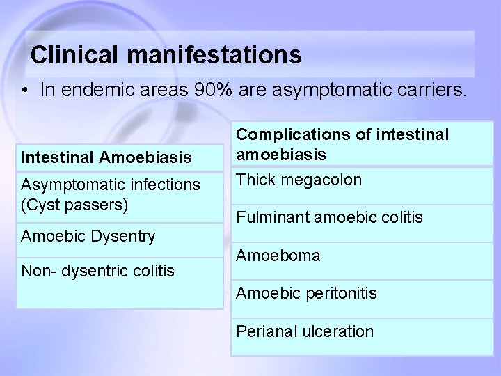
Clinical manifestations • In endemic areas 90% are asymptomatic carriers. Intestinal Amoebiasis Asymptomatic infections (Cyst passers) Amoebic Dysentry Non- dysentric colitis Complications of intestinal amoebiasis Thick megacolon Fulminant amoebic colitis Amoeboma Amoebic peritonitis Perianal ulceration 13

Clinical manifestations Extra-Intestinal Amoebiasis Non-suppurative amoebic hepatitis Pulmonary amoebiasis Amoebic liver abscess Cerebral amoebiasis Complications of Liver abscess Genitourinary amoebiasis ü Empyema ü Pericarditis ü Peritonitis 14
Amoebic dysentry • Incubation period: long and insidious onset • Symptoms; Local abdominal tenderness over sigmoid colon, caecum or appendix. • Stools frequency 6 -8 times a day, with blood and mucus and stool is copius in amount. 15

Amoebic liver abscess • Insidious onset • Associated with fever, sweating abdominal pain. • Liver enlarged, and tender. Gross pathology of amebic abscess of liver. Tube of "chocolate" pus from abscess 16
Diagnosis • Parasitic diagnosis • Culture and Isoenzymes diagnosis • Serodiagnosis • Radio-imaging diagnosis 17
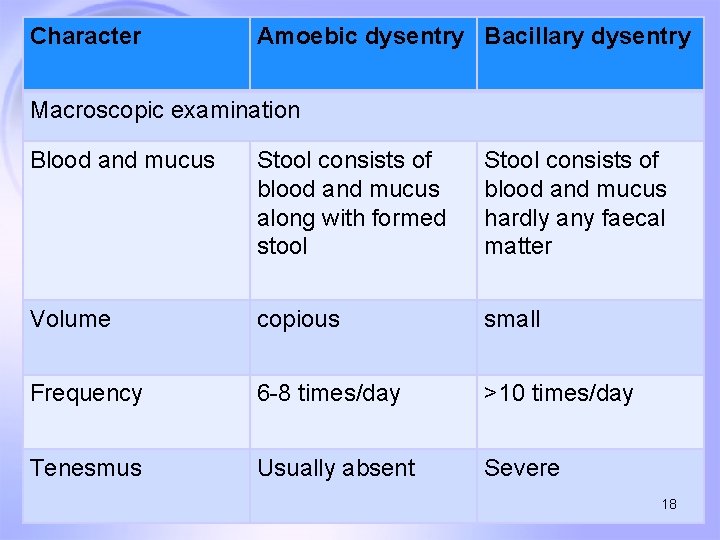
Character Amoebic dysentry Bacillary dysentry Macroscopic examination Blood and mucus Stool consists of blood and mucus along with formed stool Stool consists of blood and mucus hardly any faecal matter Volume copious small Frequency 6 -8 times/day >10 times/day Tenesmus Usually absent Severe 18
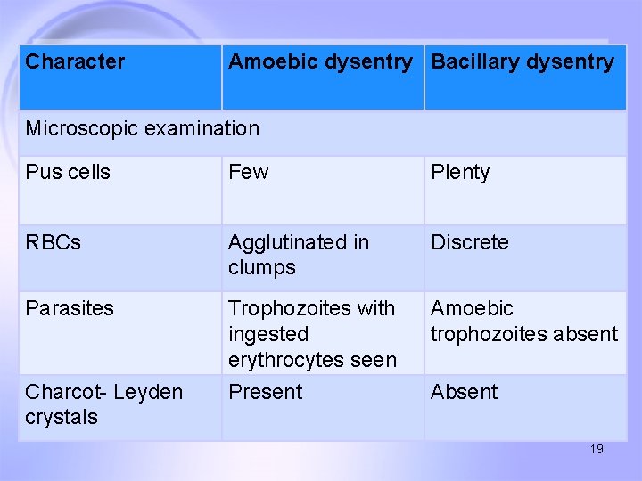
Character Amoebic dysentry Bacillary dysentry Microscopic examination Pus cells Few Plenty RBCs Agglutinated in clumps Discrete Parasites Trophozoites with ingested erythrocytes seen Amoebic trophozoites absent Charcot- Leyden crystals Present Absent 19
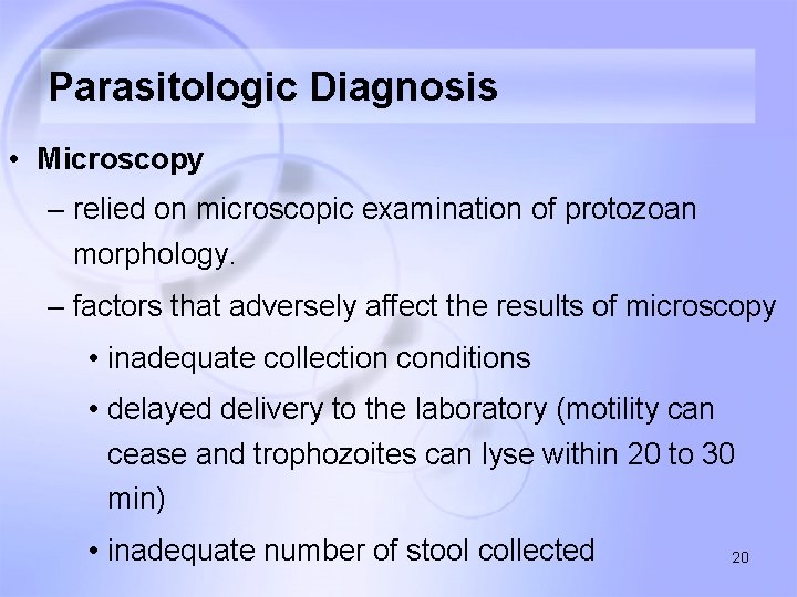
Parasitologic Diagnosis • Microscopy – relied on microscopic examination of protozoan morphology. – factors that adversely affect the results of microscopy • inadequate collection conditions • delayed delivery to the laboratory (motility can cease and trophozoites can lyse within 20 to 30 min) • inadequate number of stool collected 20
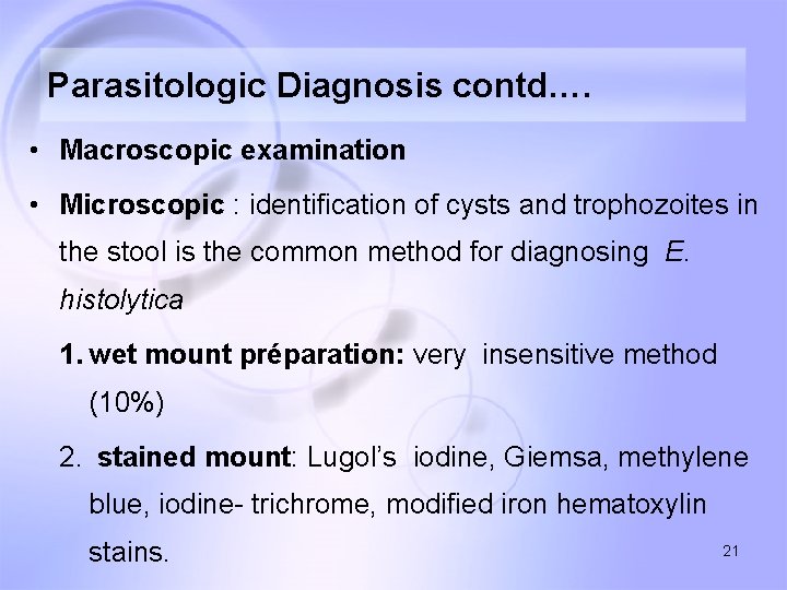
Parasitologic Diagnosis contd…. • Macroscopic examination • Microscopic : identification of cysts and trophozoites in the stool is the common method for diagnosing E. histolytica 1. wet mount préparation: very insensitive method (10%) 2. stained mount: Lugol’s iodine, Giemsa, methylene blue, iodine- trichrome, modified iron hematoxylin stains. 21
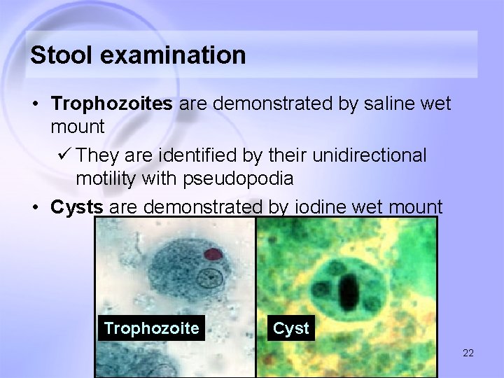
Stool examination • Trophozoites are demonstrated by saline wet mount ü They are identified by their unidirectional motility with pseudopodia • Cysts are demonstrated by iodine wet mount Trophozoite Cyst 22
Culture and Isoenzymes • Culture Medium: üXenic medium: Robinson medium, Diamond TYSGM-9 medium üaxenic medium: Diamond TYI-S-33 medium • isoenzyme patterns: ü obtained from amebic culture lysates by electrophoresis ü Differentiate E. histolytica from E. dispar and E moshkovskii, 23
Antibody Detection • Inability to distinguish past from current infection in endemic areas • Combination with detection of the parasite offers the best approach to diagnosis • Serum antibodies to E. histolytica can be detected in 75 to 85% of patients with symptomatic E. histolytica infection. • Assays used: IHA, ELISA, LA, CIE, CF, IFA 24
Antigen Detection • Antigen-based ELISA kits specific for E. histolytica use monoclonal antibodies • Advantages of Antigen-based ELISA: some of the assays differentiate E. histolytica from E. dispar; 25
Diagnosis: Extraintestinal amoebiasis • Specimens: Stool, Aspirates from liver abscess, sputum, peritoneal aspiration Cutaneous swabs. • Parasitic diagnosis: Demonstration of amoebic trophozoites in aspirated pus or sputum by microscopy or culture is specific diagnosis. • Only 15% of cases of amoebic liver abscess are positive to for trophozoites 26
Serodiagnosis • Immense value in diagnosis of extraintestinal amoebiasis • Antibody detection tests like ELISA, and Indirect haemagglutination tests are commonly done. • Amoebic antibody titre of 1: 128 or more is diagnostic of amoebic liver abscess. 27
Antigen detection tests • These are of more value in differentiating recent and past infection. • They are also of prognostic value in chemotherapeutic and surgical treatment. 28
Other tests • Leukocytosis, mild anemia, high ESR • Raised liver enzymes (ALT, AST), serum cholesterol levels. • Radio-diagnosis: ü USG abdomen: rapid and sensitive ü CT-scan, MRI 29
Symbiotic Amoebae in Human Intestines • • • Entamoeba coli Entamoeba hartmanni Endolimax nana Iodamoeba bütschlii Dientamoeba fragilis Blastocystis hominis 30

Entamoeba coli Trophozoites • broad blunt pseudopodia • small eccentric karyosome • irregular peripheral chromatin Cysts • 8 nuclei • Chromatid bodies less prominent 31
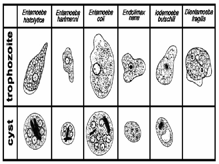
Intestinal Amebae 32
- Slides: 32