Cutaneous circulation Dr Pushpa Lata Sachan Associate professor


- Slides: 19
Cutaneous circulation Dr Pushpa Lata Sachan Associate professor C I M S & H , Lucknow
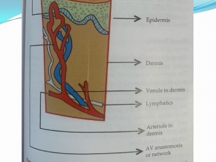
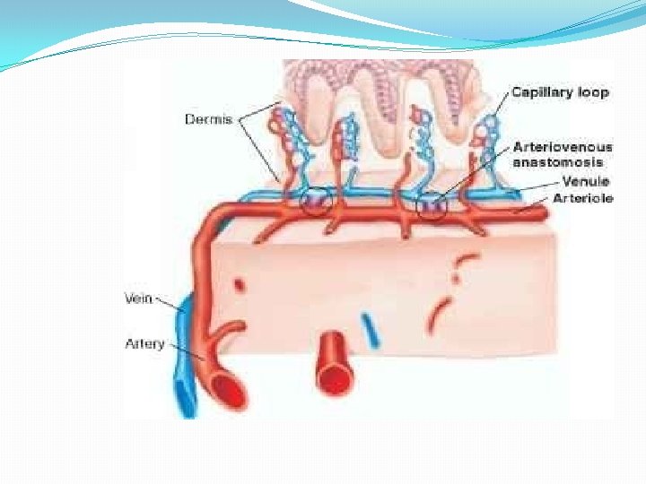
Cutaneous circulation • Functional anatomy –Blood supply of the skin of apical regions ( Fingers, feet , toes , palm, nose lip , ear lobes etc. ) is different from non apical regions (the body tarso) of the body. • Apical areas an arteriolar network exist at the boundary of dermis and subcutaneous tissue.
�From this network arterioles ascend from deep dermis to superficial layer of dermis. �Capillary loops originate from superficial dermal network and perfuse the dermal papilla and epidermis. �Non apical areas – here vascular pattern is modified. �Arteriovenous anastomoses mainly occur in superficial dermal tissue. �It Is very few or absent in non apical areas
�Normal blood flow to the skin varies from 1 to 150 ml per 100 g of tissue per min. �The skin blood vessel are supplied by sympathetic fibers. �No parasympathetic innervation is seen in the skin. �Activation of sympathetic fibers results in vasoconstriction. �Vasodilation occurs by decreasing the sympathetic activity.
Regulation of cutaneous blood flow �Is regulated by decreasing the sympathetic activity. �Cutaneous blood flow is regulated by neural , thermal and metabolic factors. �Neural regulation – cutaneous blood vessels are supplied by sympathetic vasoconstrictor fibers. �There is no vasodilator system supplying the skin blood vessels.
Thermal regulation �Thermal regulation – cutaneous blood flow is mainly regulated by body temperature. �Increased body temperature causes vasodilation and decreased body temperature causes vasoconstriction. �
HYPOTHALAMIC CONTROL MECHANISM Temperature Regulation centre of Hypothalamus
EXPOSURE
Metabolic regulation �Is not important for cutaneous circulaton. �Local production of bradykinin in the sweat causes cutaneous vasodilation.
Applied physiology �Vascular response to injury – �White response �Triple response � White response – Skin is stroked lightly with pointed object , stroke line become pale this is called as white reaction. �This occur due to decreased blood flow in the capillaries due to contraction of precapillary sphincter in response to injury. �The response is observed in about 15 second.
Triple response �When the skin is stroked with pointed object -the response to injury manifest as triple response. �This is called as triple response as it three component red , wheal and flare. �Red reaction – the skin becomes red in about 10 seconds. Redness occur due to capillary dilation that increases capillary blood flow. Capillary dilation occur due to direct response of capillaries to pressure.
�Wheal –swelling is called wheal. This occur within few minutes following red reaction. �It occur due to increased permeability of capillaries and post capillary venules. �Histamine released from local mast cells causes vasodilation and increases capillary permeability that result in extravasation of fluid.
�Flare - Spreading out of redness from the site of injury to surrounding area is called as flare. �It occur due to arteriolar dilation. � arteriolar dilation occur by activation of axon reflex. �From the site of injury impulse is conducted in the afferent fiber. �Sensory neuron give branches to blood vessel.
�The impulse in addition to its conduction to the spinal cord orthodomically, it also relayed antidromically to blood vessels �Axon reflex is an example of antidromic conduction of impulse. �The ending of sensory fibers on the blood vessels release substance P and CGRP that produce arteriolar dilation. �Redness spreads out from injury to surrounding skin in the form of flare.
Reactive hyperemia �This is defined as increased blood flow in an area when blood supply to the area is reestablished following a brief period of occlusion. �The blood flow to the skin increase when the circulation is reestablish after the short period of occlusion.
�Reactive hyperemia also occur in visceral organs. �It occur due to vasodilation produced by hypoxia during occlusion. �When circulation reestablished blood flow increases through dilated blood vessels and skin becomes red