
FACE AND SCALP Objectives 1 Name the branches
FACE AND SCALP
Objectives 1. Name the branches of the trigeminal nerve that provide sensory innervation to the skin of the face and describe the territories they supply 2. Name the arteries that supply the face and describe the origin, course, anatomical relations and branches of the facial and superficial temporal arteries 3. Name the veins that drain the face and describe the origin, course, anatomical relations and termination of the facial and retromandibular veins 4. Name the muscles of facial expression and describe their location, action and nerve supply 5. Describe the course and anatomical relations of the facial nerve 6. Name the extrapetrosal branches of the facial nerve and the muscles they supply
Objectives 7. Describe the location and anatomical relations of the parotid gland 8. Describe the course and termination of the parotid duct 9. Name the layers of the scalp 10. Name the blood vessels and nerves that supply the scalp
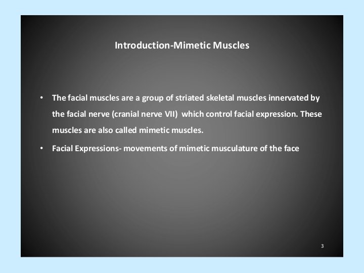
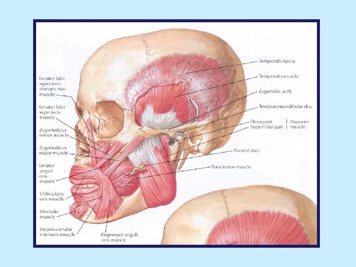
Muscles of the Face (of Facial Expression) • embedded in superficial fascia of face • originate from bones of skull and insert into skin • serve as sphincters or dilators of facial openings and move overlying skin to modify expression of face

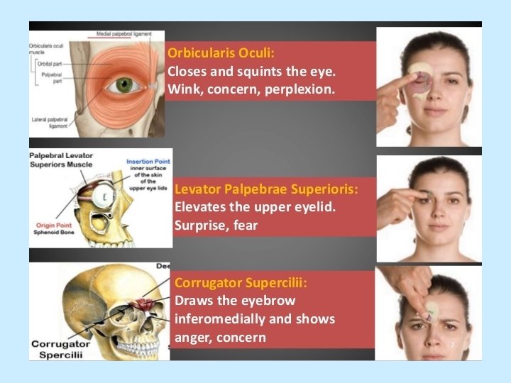


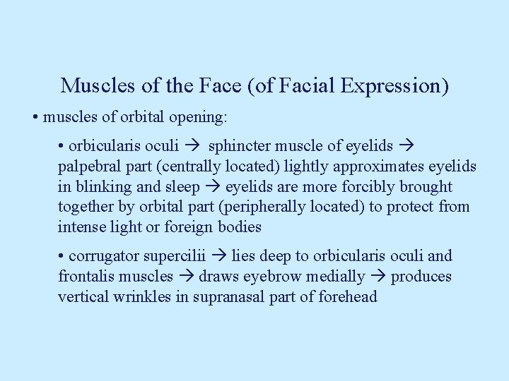
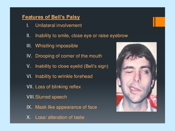
Muscles of the Face (of Facial Expression) • muscles of orbital opening: • orbicularis oculi sphincter muscle of eyelids palpebral part (centrally located) lightly approximates eyelids in blinking and sleep eyelids are more forcibly brought together by orbital part (peripherally located) to protect from intense light or foreign bodies • corrugator supercilii lies deep to orbicularis oculi and frontalis muscles draws eyebrow medially produces vertical wrinkles in supranasal part of forehead
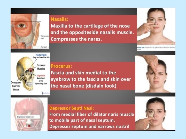
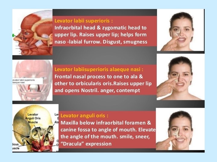
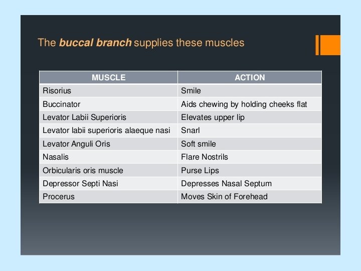
Muscles of the Face (of Facial Expression) • muscles of nose: • procerus located over root of nose produces transverse wrinkles across root of nose • nasalis muscle (transverse and alar parts) dilates nasal aperture (nose is flattened and widened) • muscles of mouth: • orbicularis oris fibers encircle oral orifice within substance of lips most fibers are derived from buccinator and other muscles around mouth sphincter muscle of lips compresses lips together and protrudes them • levator labii superioris alaeque nasi
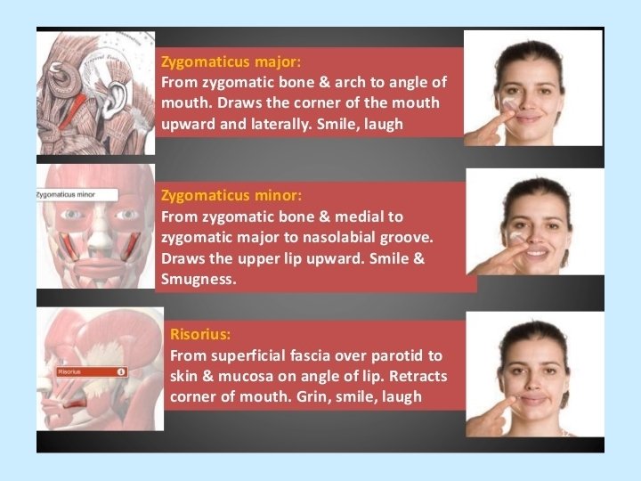
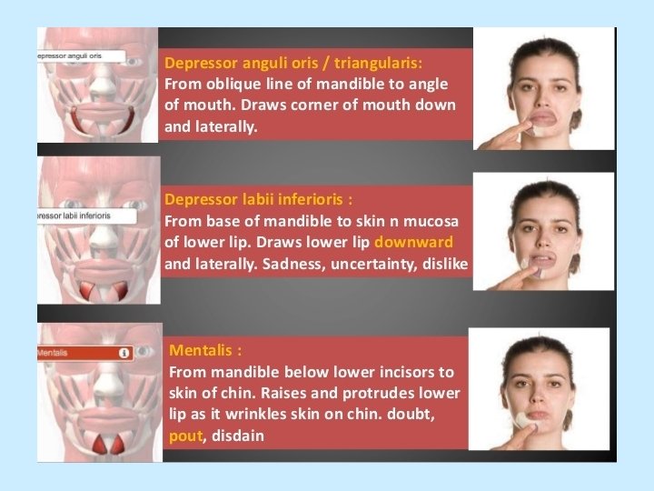
Muscles of the Face (of Facial Expression) • muscles of mouth (cont. ): • levator labii superioris • levator anguli oris • zygomaticus minor elevates upper lip • zygomaticus major draws angle of mouth upward and backward (smiling) • risorius retracts angle of mouth (grinning) • depressor anguli oris • depressor labii inferioris • mentalis draws up and puckers skin of chin (assists in protrusion and eversion of lower lip)
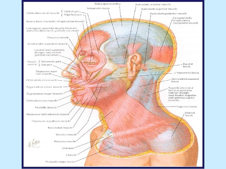
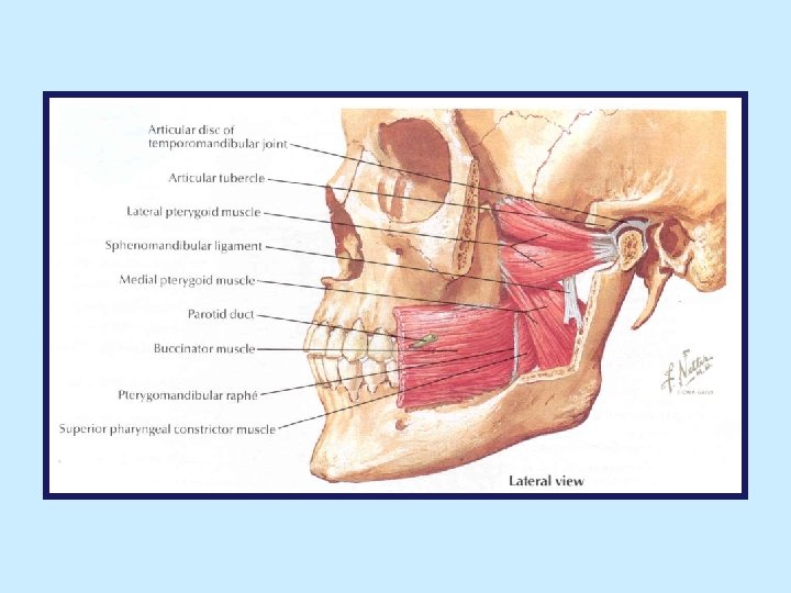
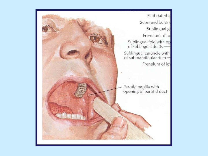
Muscles of the Face (of Facial Expression) • buccinator: • originates from outer surfaces of alveolar processes of maxilla and mandible, opposite molar teeth, and from pterygomandibular raphe (thin fibrous band that extends from pterygoid hamulus to posterior end of mylohyoid line of mandible) • muscle fibers pass forward and form muscle layer of cheek at angle of mouth fibers enter upper and lower lips and become continuous with orbicularis oris • pierced by parotid duct • compresses cheek against molar teeth (aids in mastication, together with tongue, by pushing food between teeth), assists in expelling air from oral cavity (whistling, blowing, playing wind instruments)
Vasculature of the Face

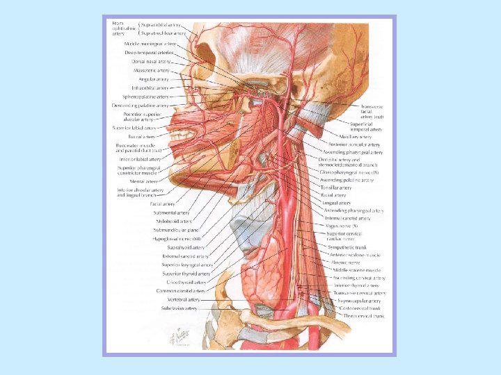
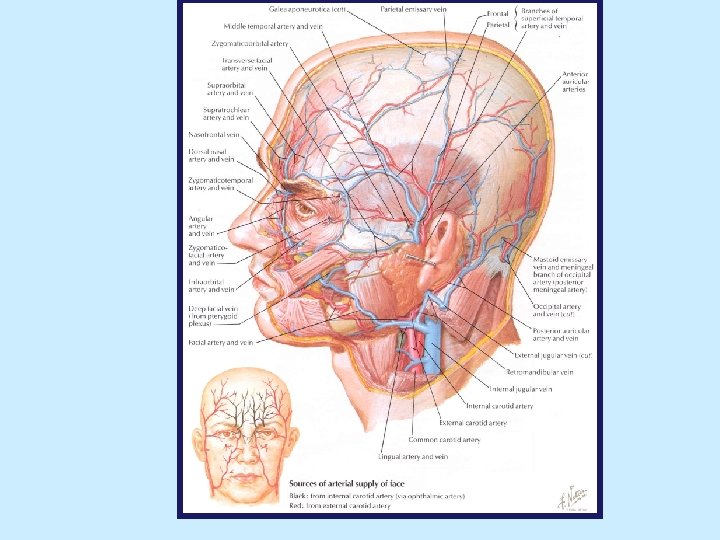
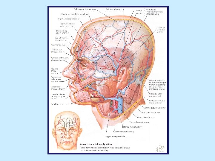
Arteries of the Face • Derived from branches of external and internal carotid arteries • terminal branches of arteries supplying face anastomose freely • many arteries accompany cutaneous nerves and have same name as nerves • facial branches of ophthalmic artery (branch of internal carotid artery): • lacrimal artery • supraorbital artery • supratrochlear artery • dorsal nasal artery (accompanies infratrochlear nerve) • external nasal artery
Arteries of the Face (Internal Carotid A con’t) • facial branches of ophthalmic artery (cont. ): • zygomaticofacial artery • zygomaticotemporal artery • facial branches of maxillary artery (larger terminal branch of external carotid artery): • infraorbital artery • buccal artery • mental artery (originates from inferior alveolar artery) • facial branches of superficial temporal artery (smaller terminal branch of external carotid artery): • transverse facial artery • zygomaticoorbital artery
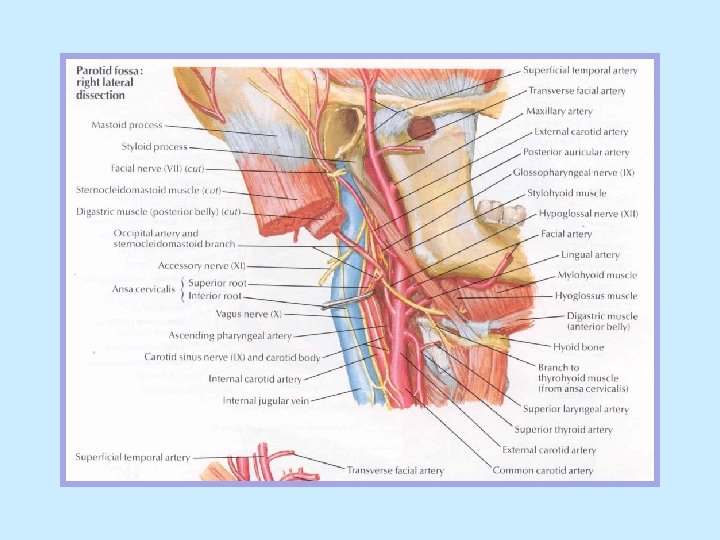
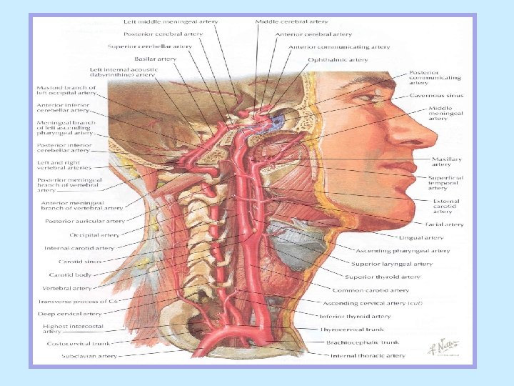
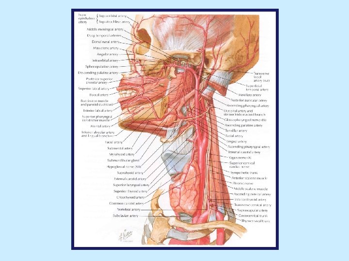
External Carotid Artery • begins at upper border of thyroid cartilage ascends in carotid triangle leaves carotid triangle as it passes deep (medial) to posterior belly of digastric and stylohyoid terminates within substance of parotid gland, posterior to neck of mandible, dividing into superficial temporal and maxillary arteries • branches: • anterior branches: superior thyroid, lingual and facial arteries • posterior branches: occipital and posterior auricular arteries • medial branch: ascending pharyngeal artery • terminal branches: superficial temporal and maxillary arteries
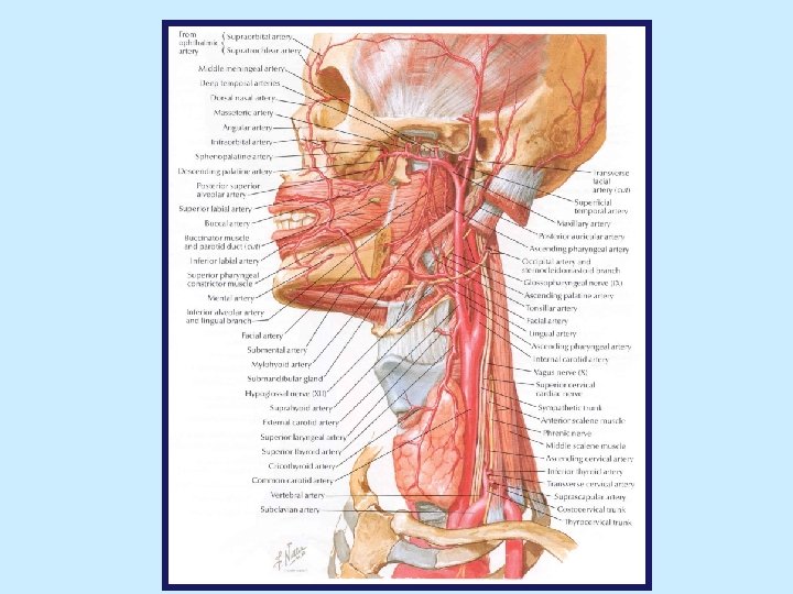
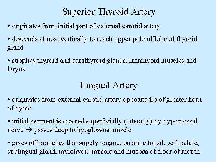
Superior Thyroid Artery • originates from initial part of external carotid artery • descends almost vertically to reach upper pole of lobe of thyroid gland • supplies thyroid and parathyroid glands, infrahyoid muscles and larynx Lingual Artery • originates from external carotid artery opposite tip of greater horn of hyoid • initial segment is crossed superficially (laterally) by hypoglossal nerve passes deep to hyoglossus muscle • gives off branches that supply tongue, palatine tonsil, soft palate, sublingual gland, mylohyoid muscle and mucosa of floor of mouth
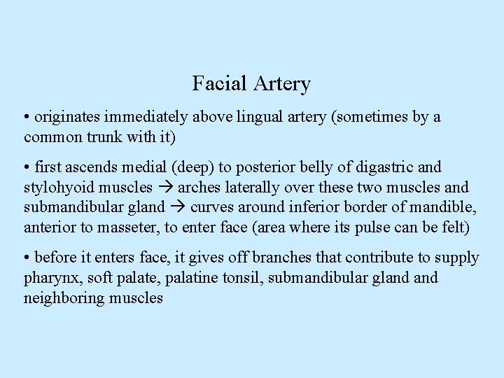
Facial Artery • originates immediately above lingual artery (sometimes by a common trunk with it) • first ascends medial (deep) to posterior belly of digastric and stylohyoid muscles arches laterally over these two muscles and submandibular gland curves around inferior border of mandible, anterior to masseter, to enter face (area where its pulse can be felt) • before it enters face, it gives off branches that contribute to supply pharynx, soft palate, palatine tonsil, submandibular gland neighboring muscles
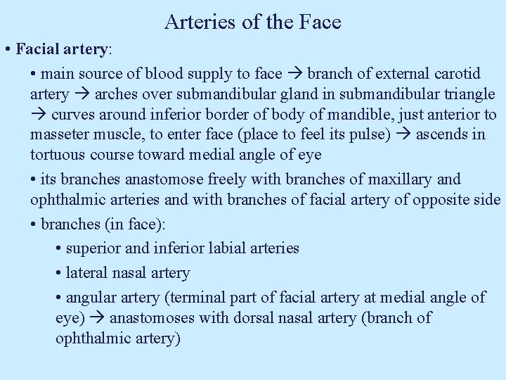
Arteries of the Face • Facial artery: • main source of blood supply to face branch of external carotid artery arches over submandibular gland in submandibular triangle curves around inferior border of body of mandible, just anterior to masseter muscle, to enter face (place to feel its pulse) ascends in tortuous course toward medial angle of eye • its branches anastomose freely with branches of maxillary and ophthalmic arteries and with branches of facial artery of opposite side • branches (in face): • superior and inferior labial arteries • lateral nasal artery • angular artery (terminal part of facial artery at medial angle of eye) anastomoses with dorsal nasal artery (branch of ophthalmic artery)
Occipital Artery • originates from posterior aspect of external carotid artery, close to inferior border of posterior belly of digastric • passes posteriorly and superiorly, deep (medial) to posterior belly of digastric and mastoid process reaches back of scalp its terminal branches accompany branches of greater occipital nerve and supply posterior part of scalp • along its curse, it gives off branches that supply auricle, middle ear, mastoid air cells, cranial dura mater and neighboring muscles
Posterior Auricular Artery • arises from posterior aspect of external carotid artery at upper border of posterior belly of digastric • passes posteriorly and superiorly to reach notch between cartilage of external acoustic meatus and mastoid process it supplies auricle, middle ear, scalp above and behind ear, parotid gland neighboring muscles Ascending Pharyngeal Artery • smallest branch of external carotid artery arises near its origin • ascends along pharyngeal wall toward cranial base, medial to internal carotid artery • supplies pharynx, soft palate, palatine tonsil, middle ear and cranial dura mater
Superficial Temporal Artery • upward continuation of external carotid artery • ascends anterior to ear with auriculotemporal nerve and superficial temporal vein passes superficial (lateral) to root of zygomatic process of temporal bone (area where pulse can be felt) • superior to zygomatic process of temporal bone, it divides into anterior (frontal) and posterior (parietal) branches supply frontal, temporal and parietal regions of scalp • also gives branches to parotid gland, external ear, face and temporomandibular joint
Maxillary Artery • largest of 2 terminal branches of external carotid artery • passes anteriorly, deep to neck of mandible runs through infratemporal fossa, and terminates in pterygopalatine fossa • gives off branches that supply external and middle ear, cranial dura mater, upper and lower teeth and gingiva, muscles of mastication, face, hard and soft palate, palatine tonsil and nasal cavity
Motor Innervation of the Face
Motor Innervation Muscles of the Face
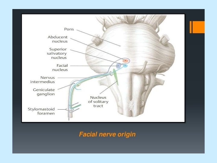
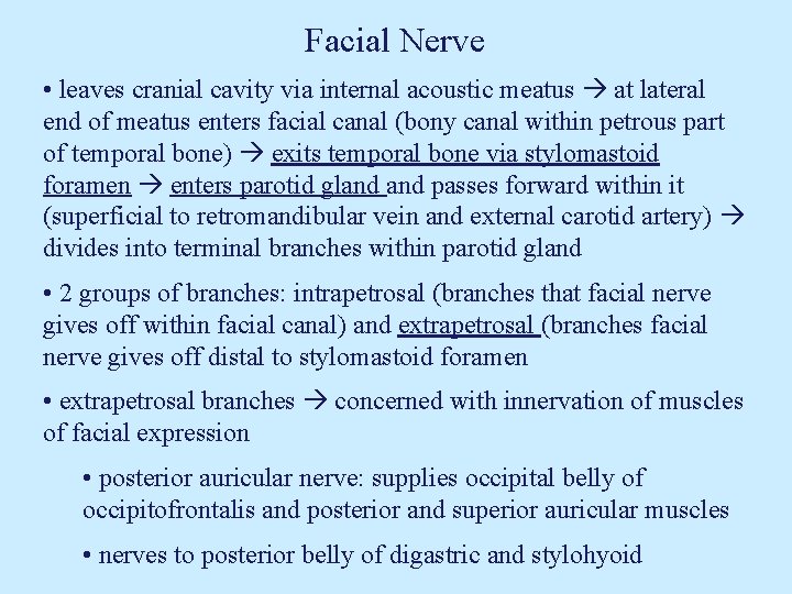
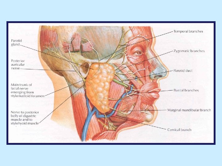
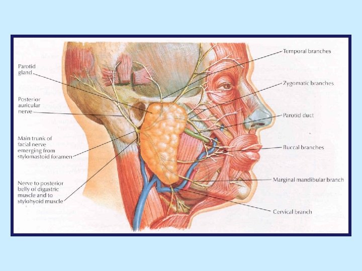
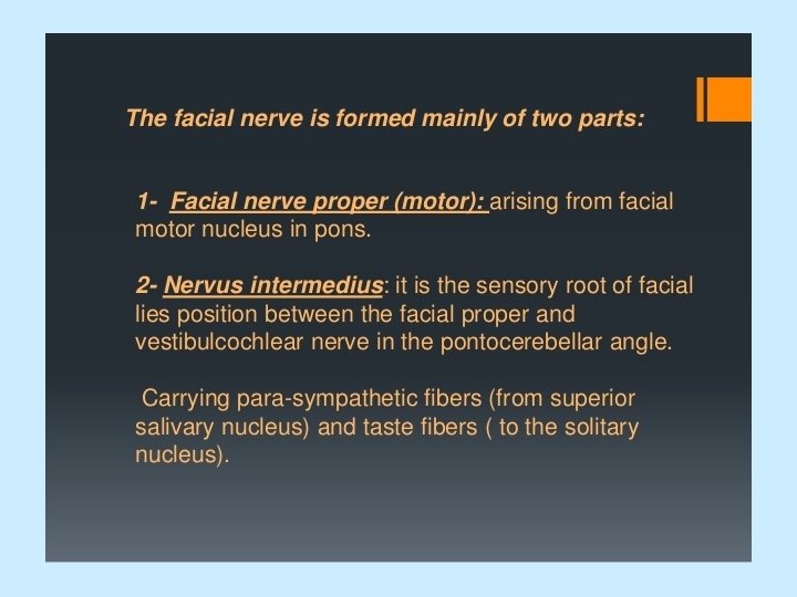
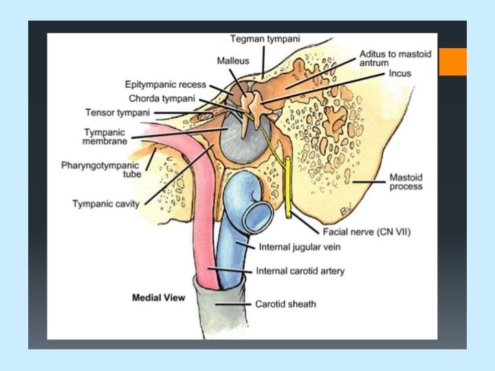
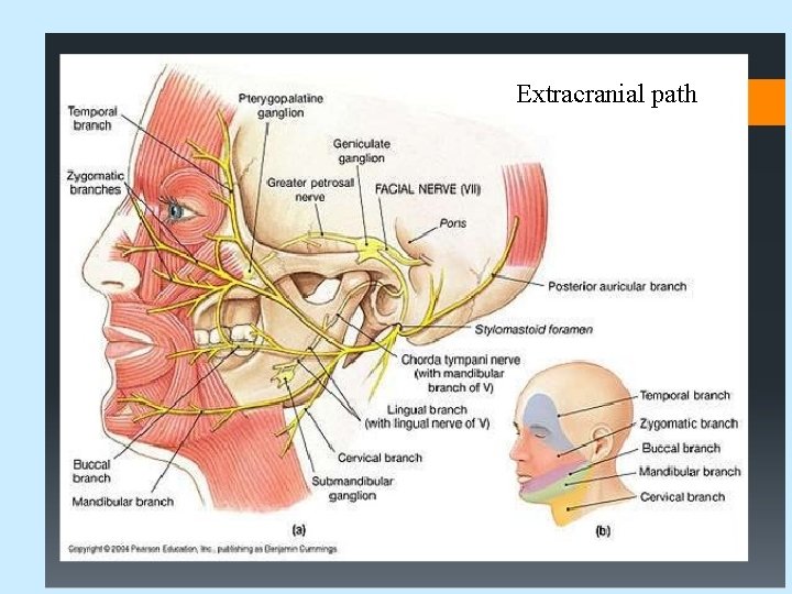
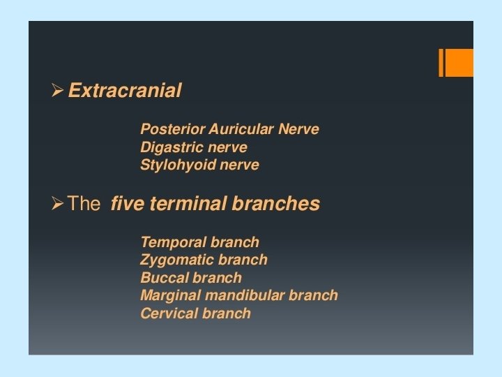
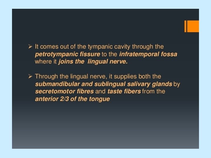
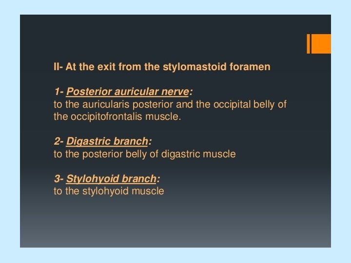
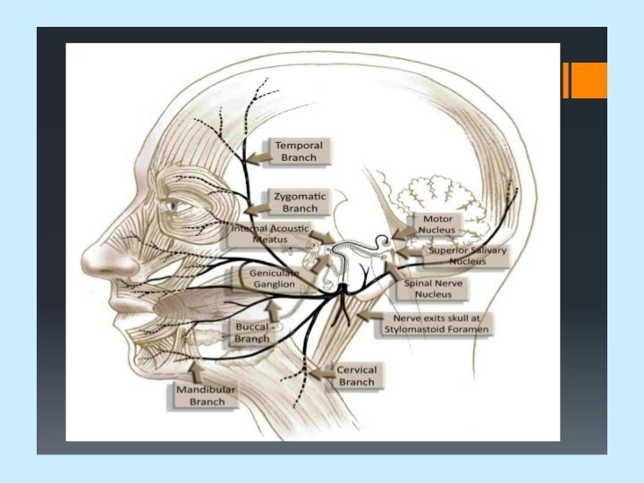
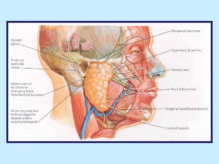
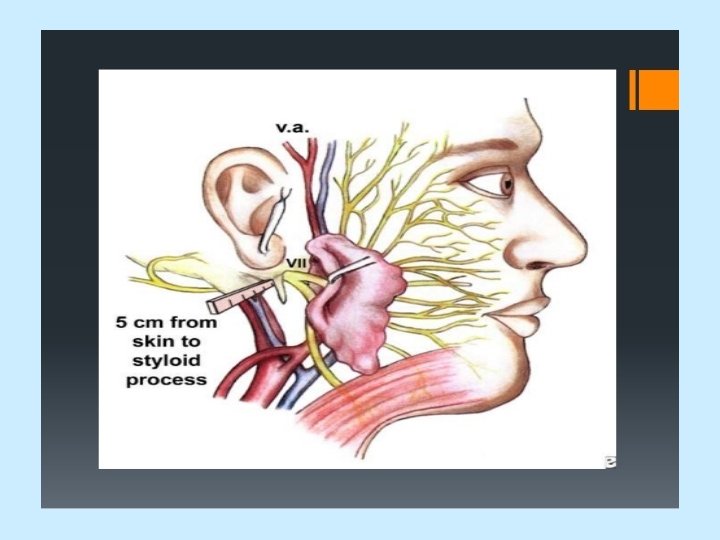
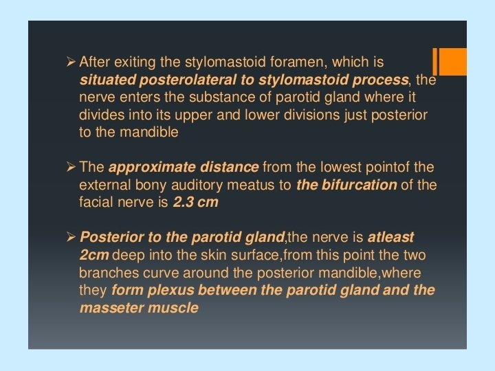
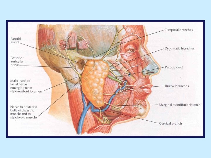
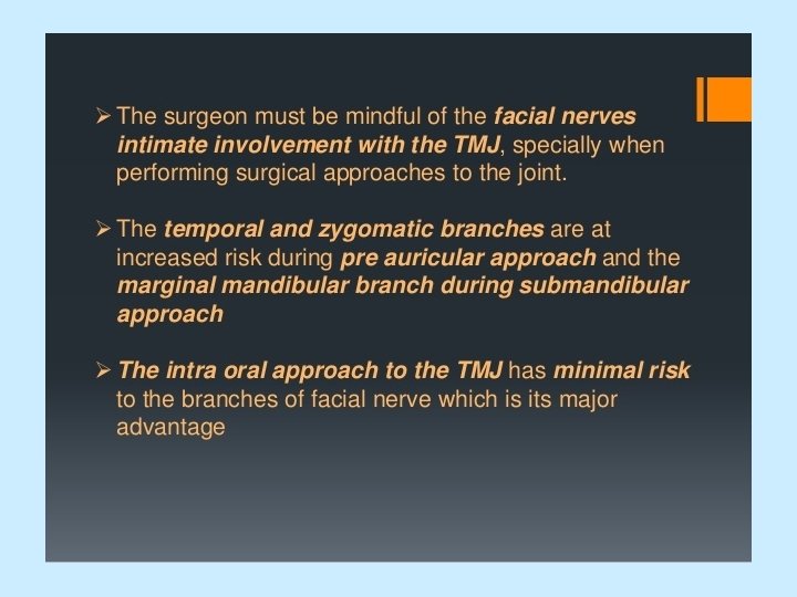
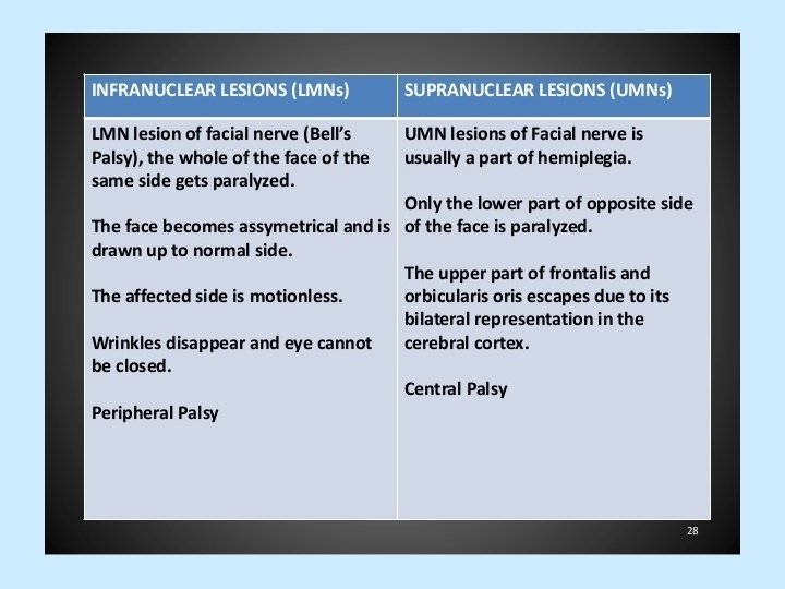
Facial Nerve • leaves cranial cavity via internal acoustic meatus at lateral end of meatus enters facial canal (bony canal within petrous part of temporal bone) exits temporal bone via stylomastoid foramen enters parotid gland passes forward within it (superficial to retromandibular vein and external carotid artery) divides into terminal branches within parotid gland • 2 groups of branches: intrapetrosal (branches that facial nerve gives off within facial canal) and extrapetrosal (branches facial nerve gives off distal to stylomastoid foramen • extrapetrosal branches concerned with innervation of muscles of facial expression • posterior auricular nerve: supplies occipital belly of occipitofrontalis and posterior and superior auricular muscles • nerves to posterior belly of digastric and stylohyoid
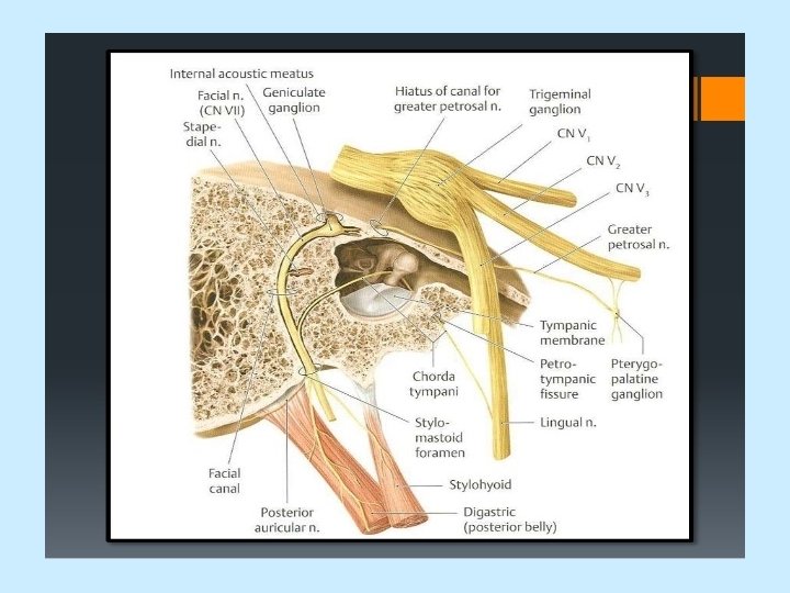
Extracranial path
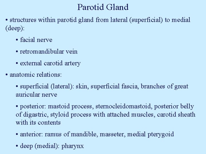
Parotid Gland • structures within parotid gland from lateral (superficial) to medial (deep): • facial nerve • retromandibular vein • external carotid artery • anatomic relations: • superficial (lateral): skin, superficial fascia, branches of great auricular nerve • posterior: mastoid process, sternocleidomastoid, posterior belly of digastric, styloid process with attached muscles, carotid sheath with its contents • anterior: ramus of mandible, masseter, medial pterygoid • deep (medial): pharynx
Scalp
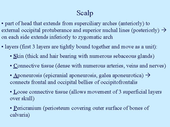
Scalp • part of head that extends from superciliary arches (anteriorly) to external occipital protuberance and superior nuchal lines (posteriorly) on each side extends inferiorly to zygomatic arch • layers (first 3 layers are tightly bound together and move as a unit): • Skin (thick and hair bearing with numerous sebaceous glands) • Connective tissue (dense with numerous arteries, veins and nerves) • Aponeurosis (epicranial aponeurosis, galea aponeurotica) connects frontal and occipital bellies of occipitofrontalis • Loose connective tissue (allows movement of 3 superficial layers over skull) • Pericranium (periosteum covering outer surface of bones of calvaria)
Vasculature and Sensory Innervation of the scalp
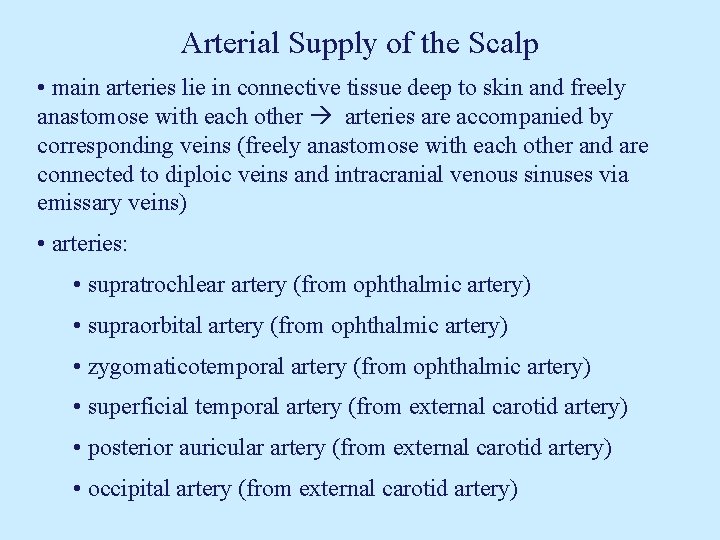
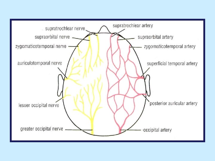
Arterial Supply of the Scalp • main arteries lie in connective tissue deep to skin and freely anastomose with each other arteries are accompanied by corresponding veins (freely anastomose with each other and are connected to diploic veins and intracranial venous sinuses via emissary veins) • arteries: • supratrochlear artery (from ophthalmic artery) • supraorbital artery (from ophthalmic artery) • zygomaticotemporal artery (from ophthalmic artery) • superficial temporal artery (from external carotid artery) • posterior auricular artery (from external carotid artery) • occipital artery (from external carotid artery)
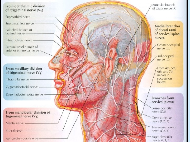
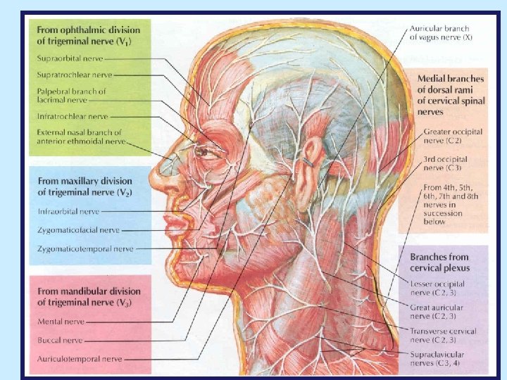
Cutaneous (Sensory) Innervation of the Head & Neck • skin of back of scalp and back of neck is supplied by dorsal rami of spinal nerves C 2 -C 5 (dorsal rami of C 1 and C 6 -C 8 normally have no cutaneous distribution) • skin of anterior and lateral aspects of neck is supplied by ventral rami of C 2 -C 4 spinal nerves via cutaneous branches of cervical plexus emerge from underneath posterior border of sternocleidomastoid muscle • cutaneous branches of cervical plexus: • lesser occipital nerve (C 2) follows posterior border of sternocleidomastoid supplies skin over lateral part of occipital region and upper part of medial surface of auricle
Cutaneous Innervation of the Head & Neck • cutaneous branches of cervical plexus (cont. ): • great auricular nerve (C 2, C 3) ascends superficial to sternocleidomastoid, parallel to external jugular vein supplies skin over angle of mandible and parotid gland, and both surfaces of lower part of auricle • transverse cervical nerve (C 2, C 3) runs anteriorly, superficial to sternocleidomastoid supplies skin of anterior and lateral aspects of neck • supraclavicular nerves (C 3, C 4) arise by a common trunk, which divides into medial, intermediate, and lateral supraclavicular nerves descend across side of lower neck supply skin at base of neck and skin that covers upper parts of pectoralis major and deltoid muscles

Putting It All Together
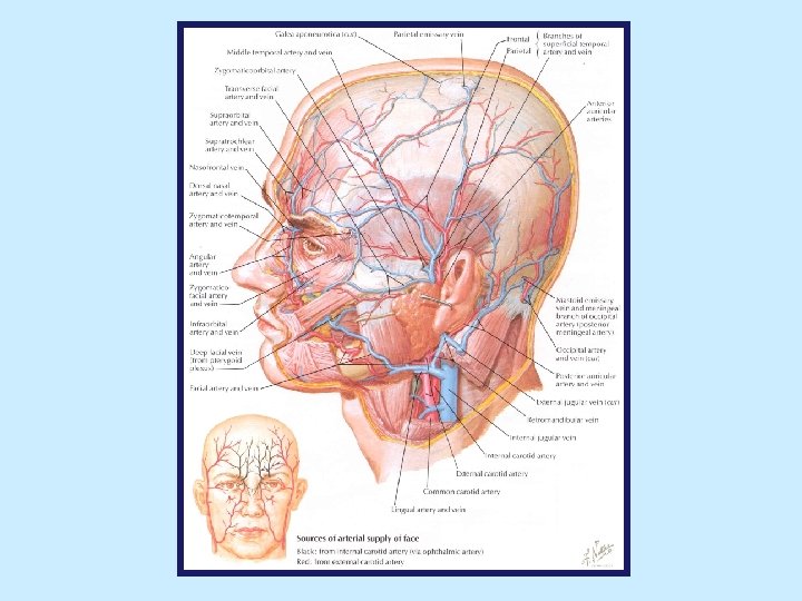
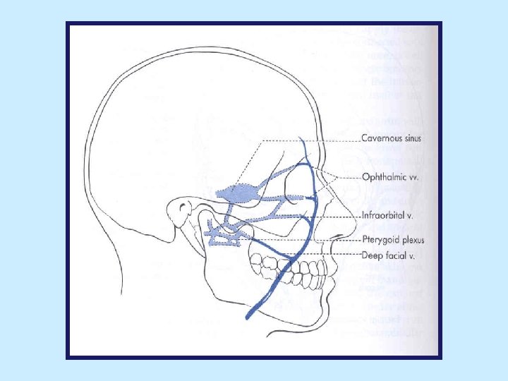
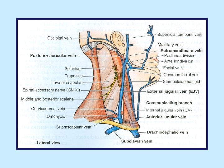
Veins of the Face • veins of face follow similar pattern as arteries facial branches of ophthalmic, maxillary and superficial temporal arteries are accompanied by corresponding veins • facial vein: • begins as angular vein at medial angle of eye by union of supraorbital and supratrochlear veins • descends posterior to facial artery to inferior border of mandible crosses superficial to submandibular gland joins anterior branch of retromandibular vein drains into internal jugular vein (after joining anterior branch of retromandibular vein it is known as common facial vein) • communicates with cavernous sinus via ophthalmic veins, deep facial vein, and infraorbital vein these connections represent pathways for spread of infections from face to cavernous sinus
Veins of the Face • retromandibular vein: • formed within parotid gland by union of superficial temporal and maxillary veins • descends posterior to ramus of mandible, inside parotid gland at level of angle of mandible divides into anterior and posterior branches • anterior branch joins facial vein to form common facial vein • posterior branch joins posterior auricular vein to form external jugular vein
Arterial Supply of the Scalp • main arteries lie in connective tissue deep to skin and freely anastomose with each other arteries are accompanied by corresponding veins (freely anastomose with each other and are connected to diploic veins and intracranial venous sinuses via emissary veins) • arteries: • supratrochlear artery (from ophthalmic artery) • supraorbital artery (from ophthalmic artery) • zygomaticotemporal artery (from ophthalmic artery) • superficial temporal artery (from external carotid artery) • posterior auricular artery (from external carotid artery) • occipital artery (from external carotid artery)
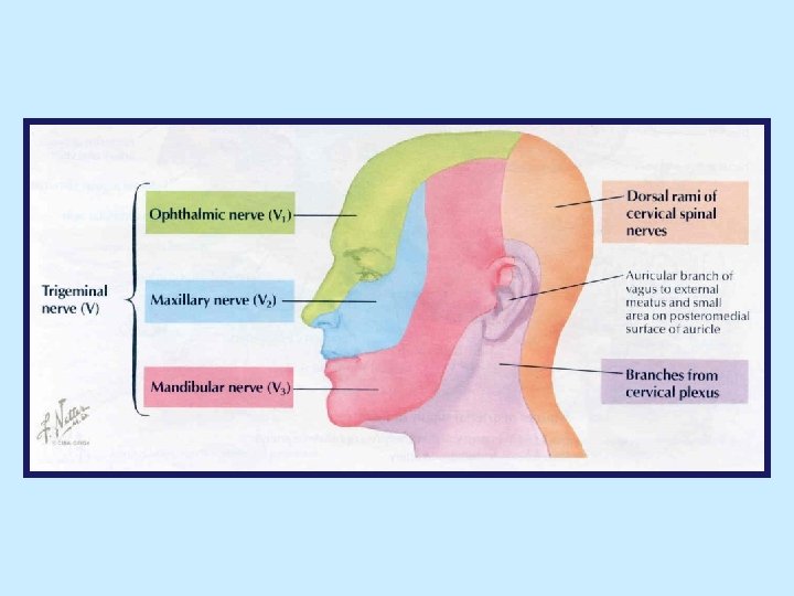
Cutaneous Innervation of the Face • supplied by branches of 3 divisions of trigeminal nerve, except small area over angle of mandible and parotid gland (supplied by great auricular nerve branch of cervical plexus) • ophthalmic nerve (V 1): • supplies forehead, anterior scalp, upper eyelid and most of the nose • branches: • lacrimal nerve: supplies lateral part of upper eyelid • supraorbital nerve: emerges from orbit through supraorbital notch (foramen) supplies central part of upper eyelid, forehead anterior scalp
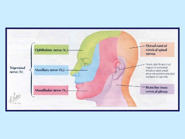
Cutaneous Innervation of the Face • ophthalmic nerve (V 1): • branches (cont. ): • supratrochlear nerve: curves around superior orbital margin, medial to supraorbital nerve supplies medial part of upper eyelid and forehead, close to midline • infratrochlear nerve: supplies medial part of upper eyelid and adjacent part of side of nose • external nasal nerve: emerges immediately inferior to nasal bone supplies side of nose as far as tip (apex)
Cutaneous Innervation of the Face • maxillary nerve (V 2): • supplies small part of nose (ala), lower eyelid, prominence of cheek, upper lip and anterior part of temporal region • branches: • infraorbital nerve: direct continuation of maxillary nerve emerges from infraorbital foramen and gives off branches that supply lower eyelid, upper lip and side of nose • zygomaticofacial nerve: supplies skin over prominence of cheek • zygomaticotemporal nerve: supplies skin over anterior part of temporal region
Cutaneous Innervation of the Face • mandibular nerve (V 3): • supplies skin of lower lip and chin, cheek, temporal region and part of external ear • branches: • mental nerve: branch of inferior alveolar nerve emerges from mental foramen and supplies lower lip and chin • buccal nerve: supplies cheek • auriculotemporal nerve: ascends posterior to superficial temporal vessels and anterior to auricle supplies parts of auricle, external acoustic meatus and tympanic membrane, most of temporal region of scalp, parotid gland temporomandibular joint
Scalp • part of head that extends from superciliary arches (anteriorly) to external occipital protuberance and superior nuchal lines (posteriorly) on each side extends inferiorly to zygomatic arch • layers (first 3 layers are tightly bound together and move as a unit): • Skin (thick and hair bearing with numerous sebaceous glands) • Connective tissue (dense with numerous arteries, veins and nerves) • Aponeurosis (epicranial aponeurosis, galea aponeurotica) connects frontal and occipital bellies of occipitofrontalis • Loose connective tissue (allows movement of 3 superficial layers over skull) • Pericranium (periosteum covering outer surface of bones of calvaria)
Sensory Nerve Supply of the Scalp • anterior to ears is supplied by branches of trigeminal nerve • posterior to ears is supplied by branches of upper cervical spinal nerves • nerves: • supratrochlear nerve (from V 1) • supraorbital nerve (from V 1) • zygomaticotemporal nerve (from V 2) • auriculotemporal nerve (from V 3) • lesser occipital nerve (from ventral ramus of C 2) • greater occipital nerve (dorsal ramus of C 2)
Motor Innervation to Facial Muscles
Facial Nerve • leaves cranial cavity via internal acoustic meatus at lateral end of meatus enters facial canal (bony canal within petrous part of temporal bone) exits temporal bone via stylomastoid foramen enters parotid gland passes forward within it (superficial to retromandibular vein and external carotid artery) divides into terminal branches within parotid gland • 2 groups of branches: intrapetrosal (branches that facial nerve gives off within facial canal) and extrapetrosal (branches facial nerve gives off distal to stylomastoid foramen • extrapetrosal branches concerned with innervation of muscles of facial expression • posterior auricular nerve: supplies occipital belly of occipitofrontalis and posterior and superior auricular muscles • nerves to posterior belly of digastric and stylohyoid
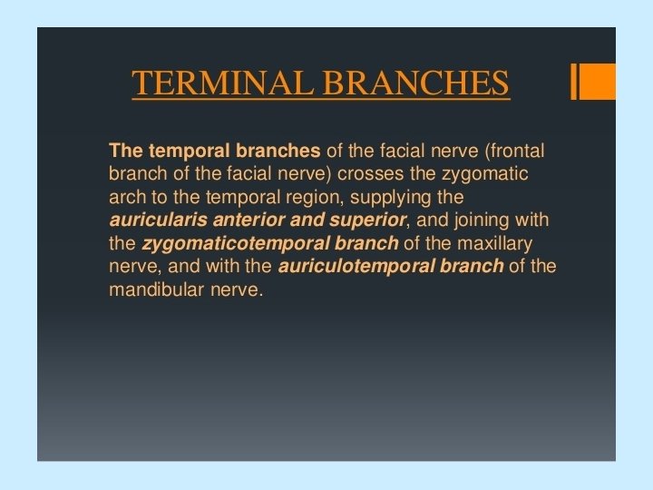
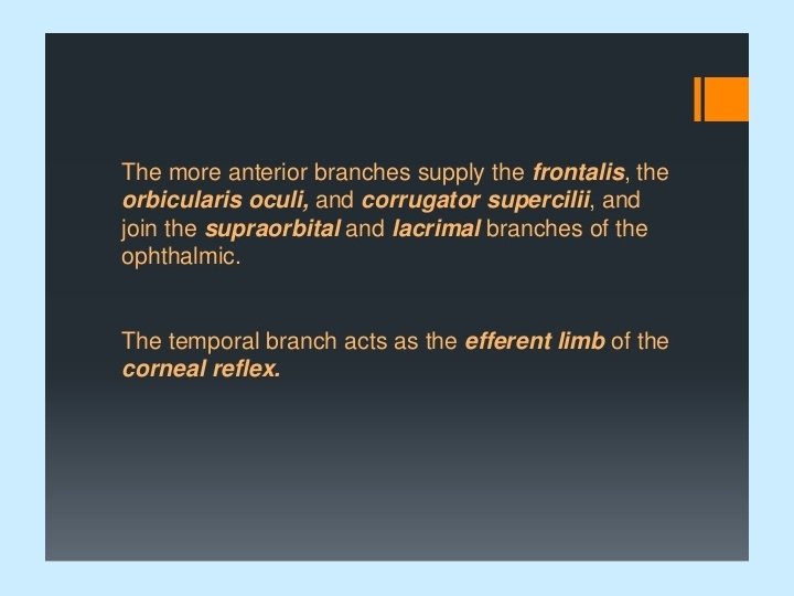
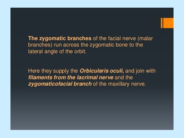
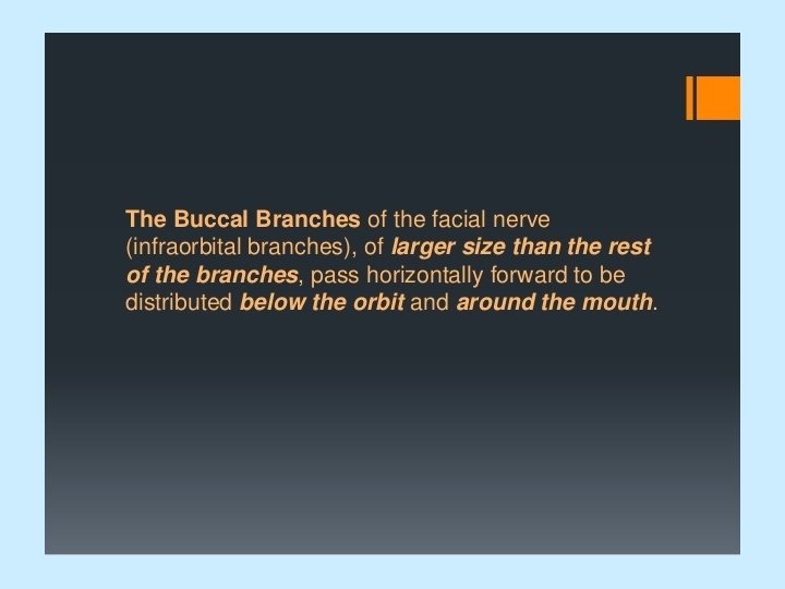
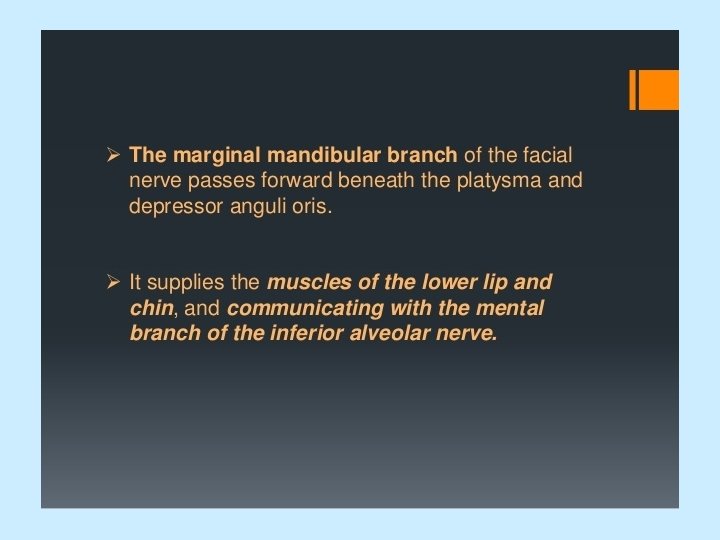
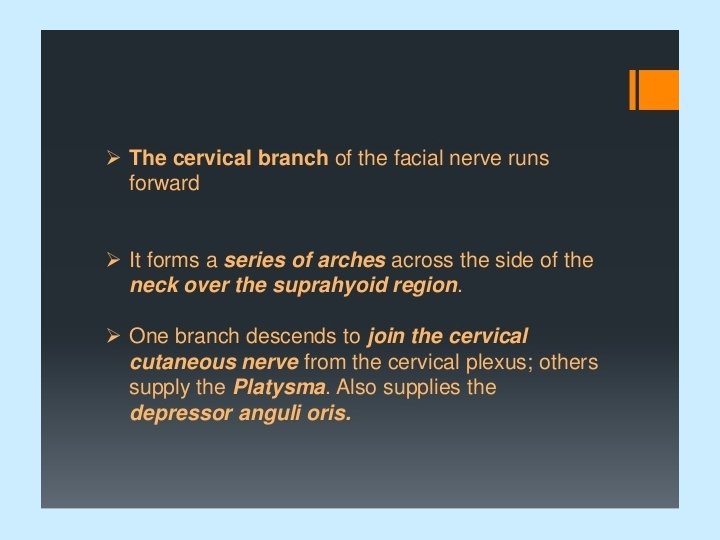
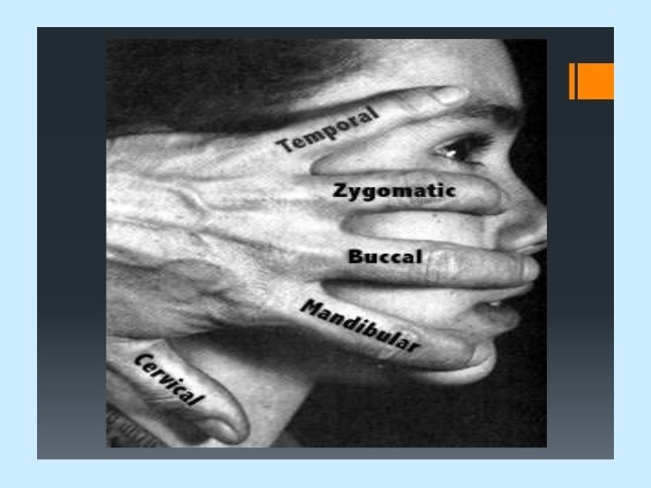
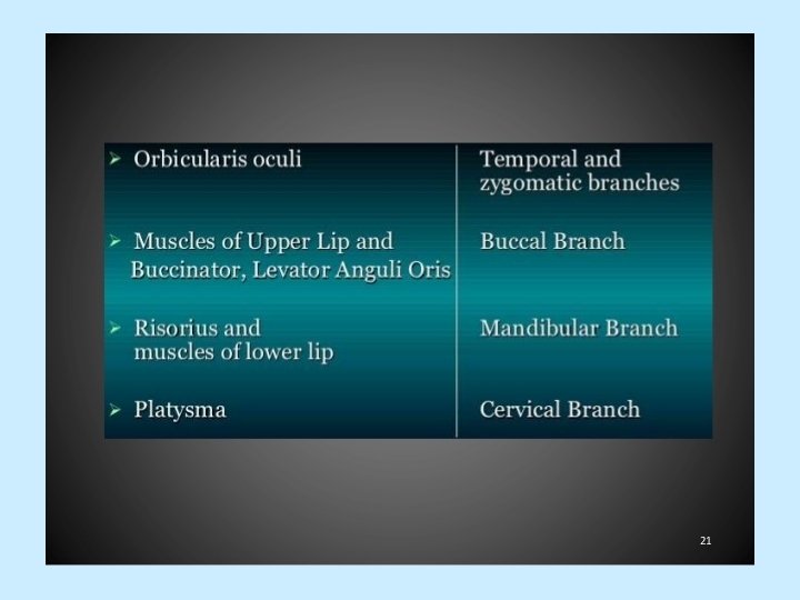
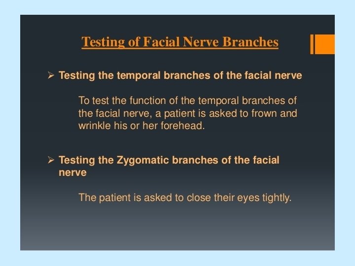
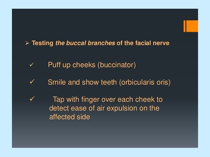
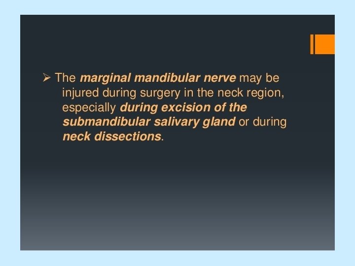
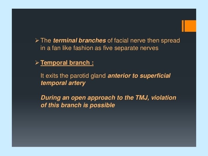
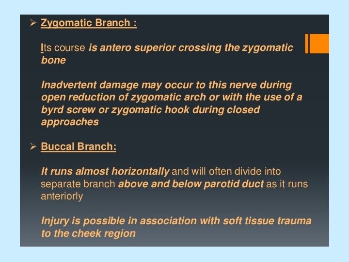
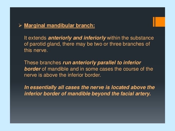
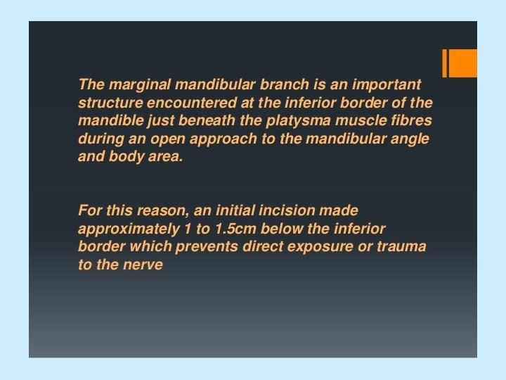
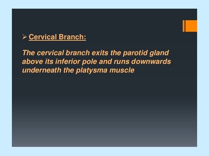
Facial Nerve • terminal branches: • temporal branches: supply anterior and superior auricular muscles, frontal belly of occipitofrontalis, orbicularis oculi and corrugator supercilii • zygomatic branches: supply orbicularis oculi and zygomaticus major and minor • buccal branches: supply buccinator and muscles of upper lip and nose • marginal mandibular branch: supplies muscles of lower lip and chin • cervical branch: supplies platysma
- Slides: 114