ACHALASIA CARDIA Dr Ramdas Rai Prof Unit Chief


- Slides: 57
ACHALASIA CARDIA Dr. Ramdas Rai Prof. & Unit Chief YMCH
• It is a primary esophageal motility disorder. • There is failure of relaxation of lower esophageal sphincter and absent peristalsis in the body of the esophagus. • It is mainly due to failure of integration of parasympathetic impulses causing functional obstruction (Achalasia means failure to relax).
Aetiology • Idiopathic (There is degeneration of Auerbach’s myentric plexus along the entire length of esophagus more so in LES) • Stress. • Vit B 1 deficiency. • Acquired : Chagas disease- caused by Trypanosoma Cruzi, which is common in South America called as sleeping sickness. • Diffuse esophageal spasm (Corckscrew esophagus).
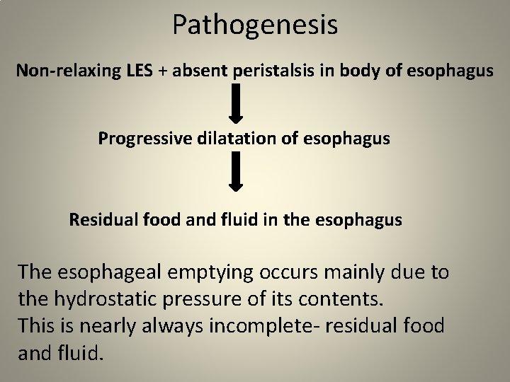
Pathogenesis Non-relaxing LES + absent peristalsis in body of esophagus Progressive dilatation of esophagus Residual food and fluid in the esophagus The esophageal emptying occurs mainly due to the hydrostatic pressure of its contents. This is nearly always incomplete- residual food and fluid.
• Eventually, retention esophagitis develops due to fermentation of food residues. • Achalasia cardia is a precancerous conditionseven times chances of getting squamous cell carcinoma
Pathology • Thickening of circular muscle fibres in distal esophagus. • Myenteric inflammation, depletion of ganglion cells, neural fibrosis, reduced nitric oxide & VIP (mediators of LES relaxation). • Dilatation of proximal esophagus with atony.
Clinical Features • More common in females. F: M= 3: 2 • 20 to 40 years of age group. • Dysphagia - presenting feature - slow and progressive - liquids>>> solids • Regurgitation – foul smelling esophageal content.
• • • Retrosternal discomfort. Recurrent respiratory tract infections. Anemia – glossitis, stomatitis, pallor. Weight loss. Malutrition. Walking while eating , chin thrusting , neck & shoulder extension , valsalva manoeuvre facilitates emptying of food from the oesophagus.
Triad of Achalasia cardia • Dysphagia • Regurgitation • Weight loss
Staging I Proximal dilatation < 4 cms II Dilatation between 4 -6 cms III Dilatation > 6 cms IV Sigmoid dilatation
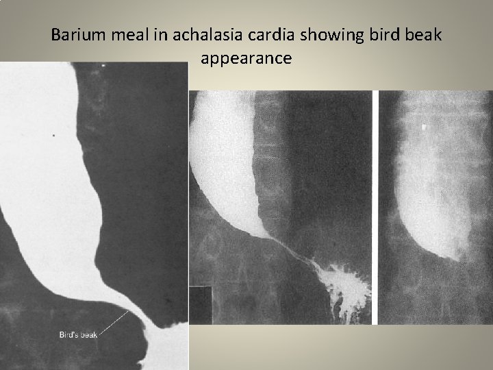
Investigations Barium swallow is diagnostic-shows Pencil like smooth narrowing of lower esophagus- bird beak appearance Dilatation of proximal esophagus Absence of fundic gas bubble Sigmoid esophagus or megaesophagus
Barium meal in achalasia cardia showing bird beak appearance
• Chest X-ray : patches of pneumonia , double mediastinal strip of dilated esophagus with air fluid level in posterior mediastinum on lateral view, pseudotumour, absence of fundal gas shadow. • Esophageal manometry shows unrelaxed LES with high resting pressure – very useful & gold standard. It shows failure of LES to relax completely during swallowing & complete absence of peristalsis. LES pressure is > 35 mm of mm. Hg
• Esophagoscopy is done to confirm the diagnosis & to rule out carcinoma esophagus. It shows totally closed LES with atonic, dilated proximal esophagus. Biopsy of mucosa at LES should be done.
Treatment Non surgical: • Endoscopic injection of botulinum toxin to sphincter- high recurrence rate • Calcium channel blocker like nifedipine – short term relief • Forcible dilatation-
• Forcible dilatation- stretches spasmodic segment - gradual repeated dilatations are required. Dilatation upto 54 French bougie can be done. - 2 types of dilatations are used: Ø Plummer’s pneumatic dilatation Ø Negus hydrostatic balloon dilatation
• Plummer’s pneumatic dilatations done using balloons of 30 -40 mm diameters. It is inserted over a guide wire. Risk of perforation , need for repeated dilatations are the problems. • Negus hydrostatic dilatation is done to dilate O -G junction. It is not very well accepted method as chances of perforation is high. Success rate is 65 percent. 30 mm diameter balloon is inflated for 3 minutes.
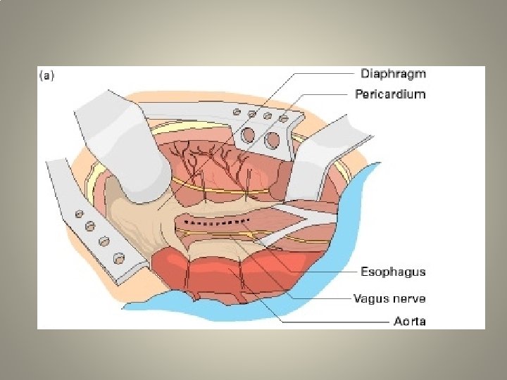
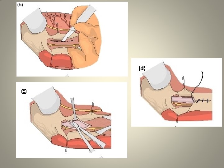
Surgery : v. Modified Heller’s operation: • Oesophagocardiomyotomy. • Success rate is 85 %. • Either through thoracic or abdominal approach. • Thickened muscle fibres are cut logitudinally for about 8 -10 cm, 2 cm proximal to thickened muscle to 1 cm distal to O-G junction. • Care taken not to open the mucosa. (Anterior myotomy is done now. Original Heller’s is both anterior & posterior myotomies.
• Nissen’s or Toupet’s fundoplication is done along with the above procedure to prevent reflux. • Laparoscopic/Thoracoscopy , cardiomyotomy is ideal. • Resection is done when failure of myotomy occurs or when metaplasia is present. v. Transhiatal total oesophgectony with gastriric pull up & oesophago-gastric anastomosis in the neck.
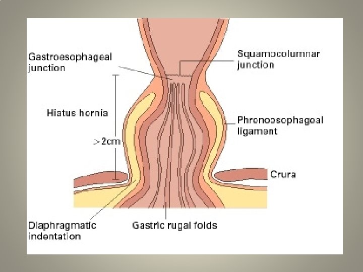
Hiatus Hernia
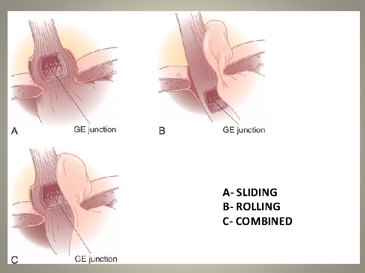
TYPES • Sliding hernia (85%) • Rolling hernia (10 -12%) • Combined
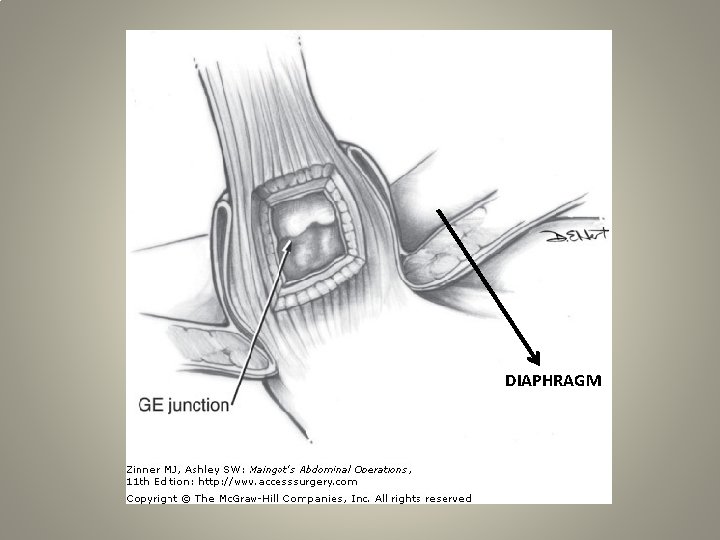
Sliding Hernia • Sliding hernia is commonly assosciated with GERD • Here the cardia migrates back & forth between the posterior mediastinum & peritoneal cavity.
DIAPHRAGM
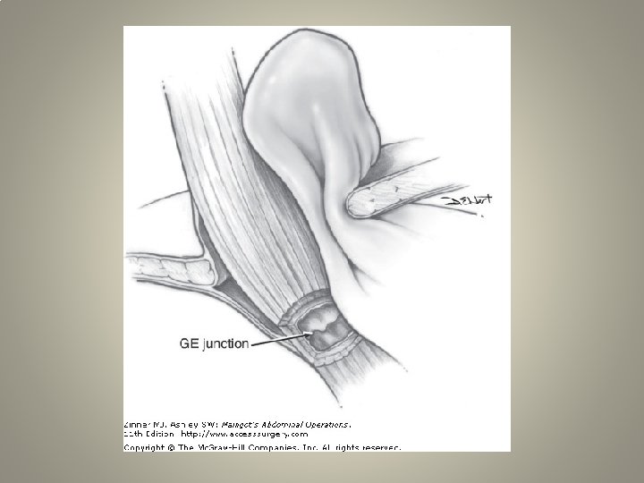
Rolling Hernia (Paraoesophageal Hernia) It is herniation of stomach fundus or rarely other abdominal contents colon/spleen through a hiatus , usually towards left side.
A- SLIDING B- ROLLING C- COMBINED
Clinical Features • • Common in elderly Abdominal pain & chest pain Hiccough, early satiety Regurgitation , post prandial bloating Cardiac abnormality (arrhythmia) Dysphagia Dyspnoea 40% presents with acute features with perforation/gangrene/bleeding.
Complications • • • Gangrene of stomach Perforation into the mediastinum Perforation into the peritoneum Gastric volvulus Ischaemic logitudinal ulcer in the herniated stomach in rolling hernia is called as Cameron ulcer.
Investigations • Plain X-ray- lateral & PA erect view showing retrocardiac air-fluid level. • Barium meal study. • ECG. • 3 D CT scan.
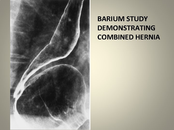
BARIUM STUDY DEMONSTRATING COMBINED HERNIA
Treatment is always surgical. Excision of sac & repair of the defect. If gangrenous , gastrectomy is required. Either abdominal or thoracic or laparosopic approach can be used. • Mesh reinforcement to hiatus to close the defect may be needed. • •
GASTRO-ESOPHAGEAL REFLUX DISEASE
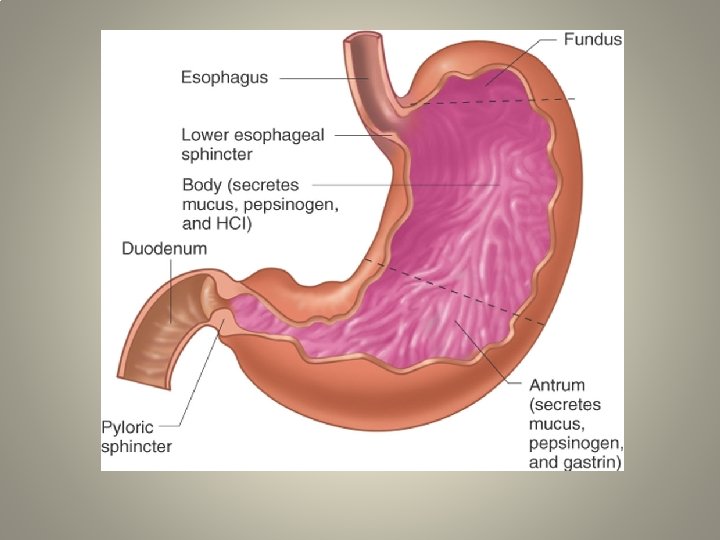
• Reflux of stomach contents into the lower esophagus. • Normal competence of the gastro-esophageal junction is maintained by the lower esophageal sphincter(LES).
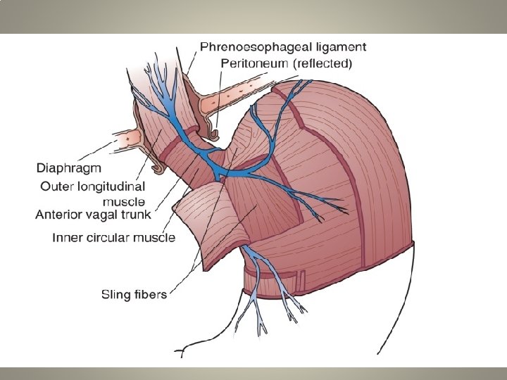
Etiology Anatomical factors: • Altered obliquity of the gastro-esophageal junction(Angle of HIS). • Altered length of the intra-abdominal esophagus. • Reduced pinching action of the right crus of the diaphragm(Pinch-Cock effect). • Alteration of the phreno-esophageal ligament. • Alteration in sling mechanism of the gastric musculature.
Physiological factors: • Altered transient relaxation period in LES. • Reduced LES pressure – hypothyroidism, secretin, cholecystokinin, Ca+2 channel blockers, deriphylline, caffeine, fatty meal etc. • Delayed gastric emptying – diabetes, neuromuscular blockade, gastroparesis, medications. • Increased gastric distension and gastric acid hypersecretion.
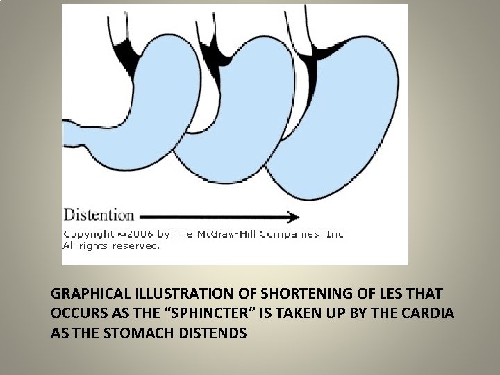
GRAPHICAL ILLUSTRATION OF SHORTENING OF LES THAT OCCURS AS THE “SPHINCTER” IS TAKEN UP BY THE CARDIA AS THE STOMACH DISTENDS
Miscellaneous factors: • • • Alcohol consumption. Smoking. Stress. Obesity. Pregnancy.
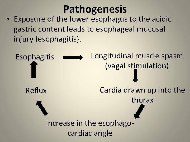
Pathogenesis • Exposure of the lower esophagus to the acidic gastric content leads to esophageal mucosal injury (esophagitis). Esophagitis Reflux Longitudinal muscle spasm (vagal stimulation) Cardia drawn up into the thorax Increase in the esophagocardiac angle
Clinical Features • Triad of – retrosternal burning pain(heart burn), epigastric pain and regurgitation. • Symptoms typically occur within seconds of ingestion of food. • Heart burn- most common symptom - provoked by consumption of fats, spicy food, alcohol. - aggravates on exercise, stooping forward, supine position.
• Nocturnal reflux. • Post prandial fullness. • Odynophagia. • Laryngeal symptoms - hoarseness of voice, cough, sore throat. • Hematemesis.
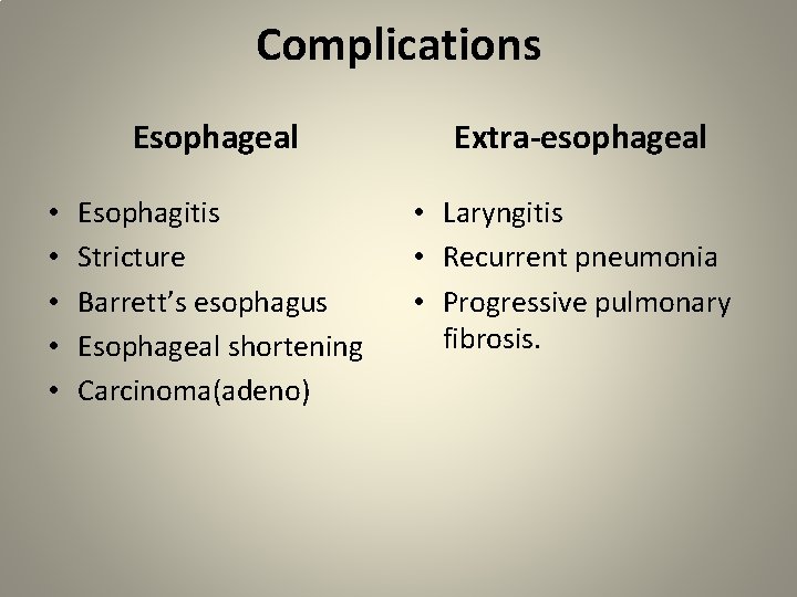
Complications • • • Esophageal Extra-esophageal Esophagitis Stricture Barrett’s esophagus Esophageal shortening Carcinoma(adeno) • Laryngitis • Recurrent pneumonia • Progressive pulmonary fibrosis.
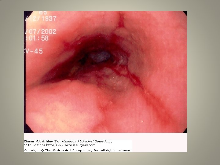
Investigations • Barium swallow. • Endoscopy- assess mucosal injury, exclude other diseases, sliding hernia, biopsy. • Esophageal manometry – to asses the transient relaxation period. • 24 hours esophageal p. H monitoring.
Treatment General measures: life-style modifications • Control of obesity • Avoid smoking and alcohol consumption. • Avoid consumption of tea, coffee, chocolate, spice. • Small frequent meals.
Medical management: • H 2 antagonists; antacids. • Prokinetic drugs: metoclopramide, domperidone, cisapride, itopride. • Mucosa protection : sucralfate, colloid bismuth. • Proton-pump inhibitors: very effective - omeprazole, rabeprazole, pantoprazole, esomeprazole.
Endoscopic therapies : • • Endoluminal suturing. Plexiglass microspheres Stretta catheter. Endoscopic full thickness plication.
Surgical management: Indication: • Failure of drug treatment • Sliding hernia • Barrett’s ulcer • Severe pain • Presence of complications like bleeding, stricture etc.
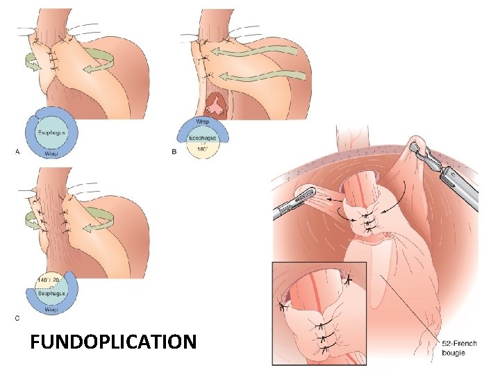
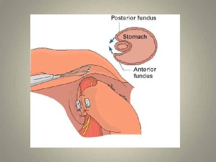
1. Fundoplication : laparotomy/laparoscopy • Nissen’s – total 360 degree posterior fundoplication • Toupet’s – patial 180 degree posterior fundal/posterolateral. • Lind- posterior and anterior. 2. Collis-Nissen vertical gastroplasty and fundoplication. 3. Belsey Mark 4 operation.
FUNDOPLICATION
Differential diagnosis • • Achalasia cardia Carcinoma esophagus Peptic ulcer Gallstones Pancreatic disease Gastritis Cardiac angina
THANK YOU