
EVALUTE THE RESULT OF THE ENDOSCOPE AND IMPRINT

- Slides: 38
EVALUTE THE RESULT OF THE ENDOSCOPE AND IMPRINT CYTOLOGY TESTING IN GASTRIC CANCER IN NATIONAL CANCER HOSPITAL Ph. D. Le Quang Hai Ph. D. Pham Cam Phuong M. S. Bui Tien Dung
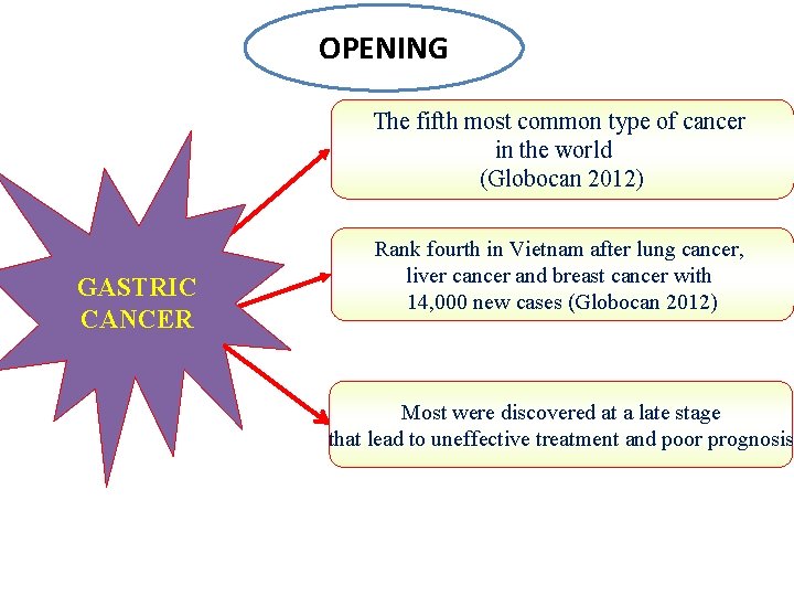
OPENING The fifth most common type of cancer in the world (Globocan 2012) GASTRIC CANCER Rank fourth in Vietnam after lung cancer, liver cancer and breast cancer with 14, 000 new cases (Globocan 2012) Most were discovered at a late stage that lead to uneffective treatment and poor prognosis
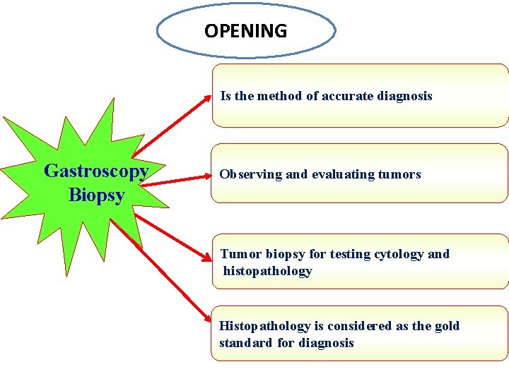
OPENING Is the method of accurate diagnosis Gastroscopy Biopsy Observing and evaluating tumors Tumor biopsy for testing cytology and histopathology Histopathology is considered as the gold standard for diagnosis
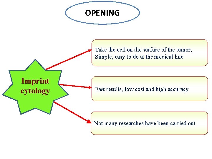
OPENING Take the cell on the surface of the tumor, Simple, easy to do at the medical line Imprint cytology Fast results, low cost and high accuracy Not many researches have been carried out
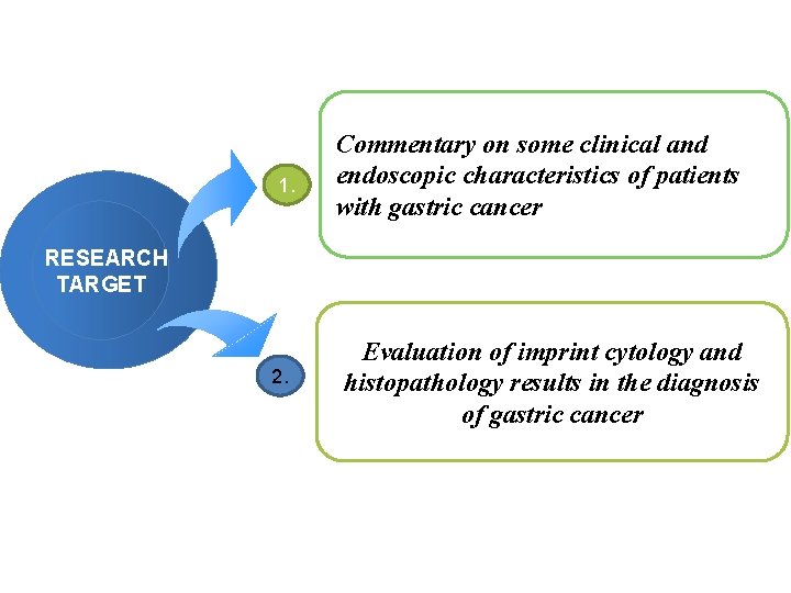
1. Commentary on some clinical and endoscopic characteristics of patients with gastric cancer RESEARCH TARGET 2. Evaluation of imprint cytology and histopathology results in the diagnosis of gastric cancer
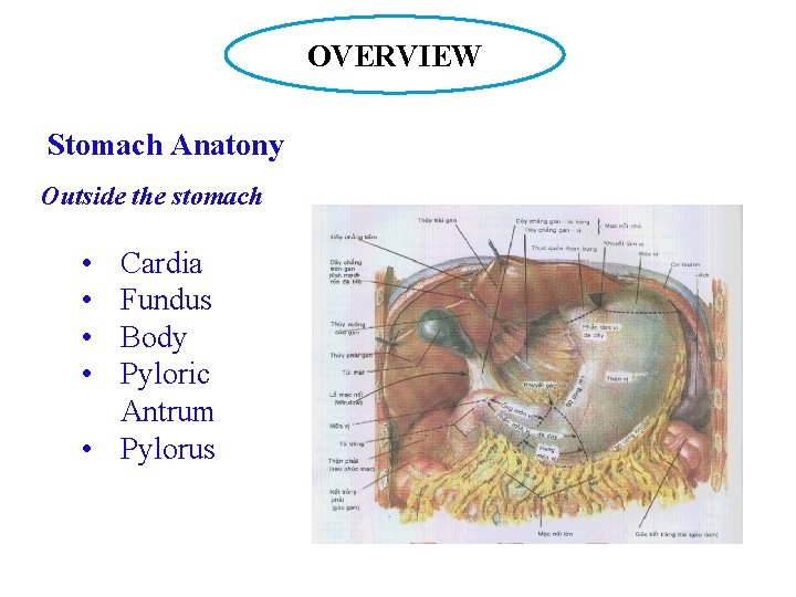
OVERVIEW Stomach Anatony Outside the stomach • • Cardia Fundus Body Pyloric Antrum • Pylorus
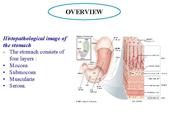
OVERVIEW Histopathological image of the stomach - The stomach consists of four layers : • Mocosa • Submocosa • Muscularis • Serosa.
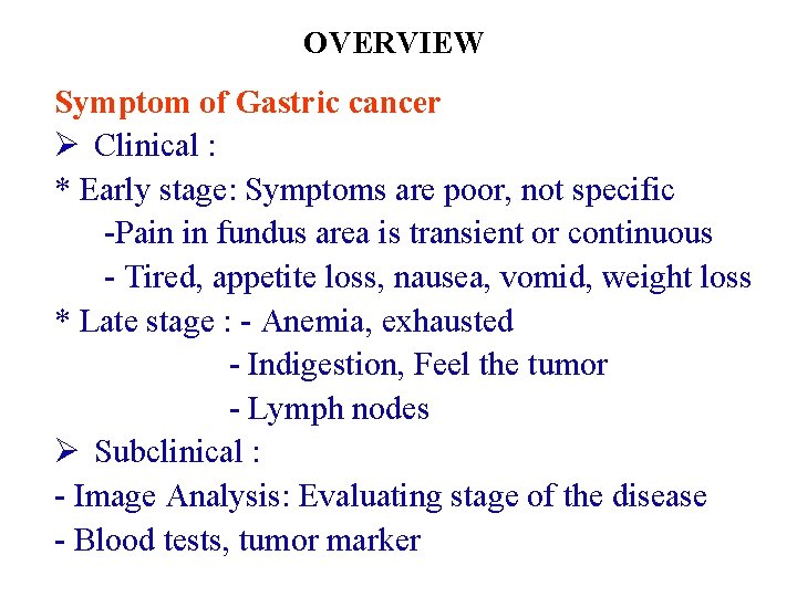
OVERVIEW Symptom of Gastric cancer Ø Clinical : * Early stage: Symptoms are poor, not specific -Pain in fundus area is transient or continuous - Tired, appetite loss, nausea, vomid, weight loss * Late stage : - Anemia, exhausted - Indigestion, Feel the tumor - Lymph nodes Ø Subclinical : - Image Analysis: Evaluating stage of the disease - Blood tests, tumor marker
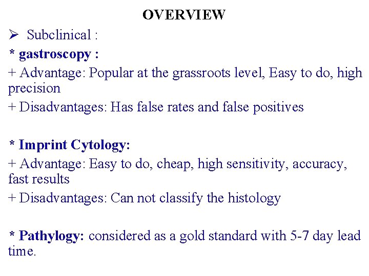
OVERVIEW Ø Subclinical : * gastroscopy : + Advantage: Popular at the grassroots level, Easy to do, high precision + Disadvantages: Has false rates and false positives * Imprint Cytology: + Advantage: Easy to do, cheap, high sensitivity, accuracy, fast results + Disadvantages: Can not classify the histology * Pathylogy: considered as a gold standard with 5 -7 day lead time.
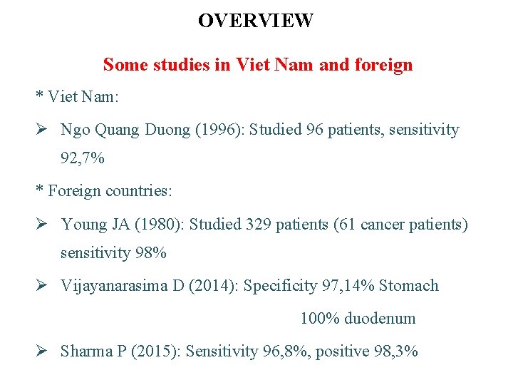
OVERVIEW Some studies in Viet Nam and foreign * Viet Nam: Ø Ngo Quang Duong (1996): Studied 96 patients, sensitivity 92, 7% * Foreign countries: Ø Young JA (1980): Studied 329 patients (61 cancer patients) sensitivity 98% Ø Vijayanarasima D (2014): Specificity 97, 14% Stomach 100% duodenum Ø Sharma P (2015): Sensitivity 96, 8%, positive 98, 3%
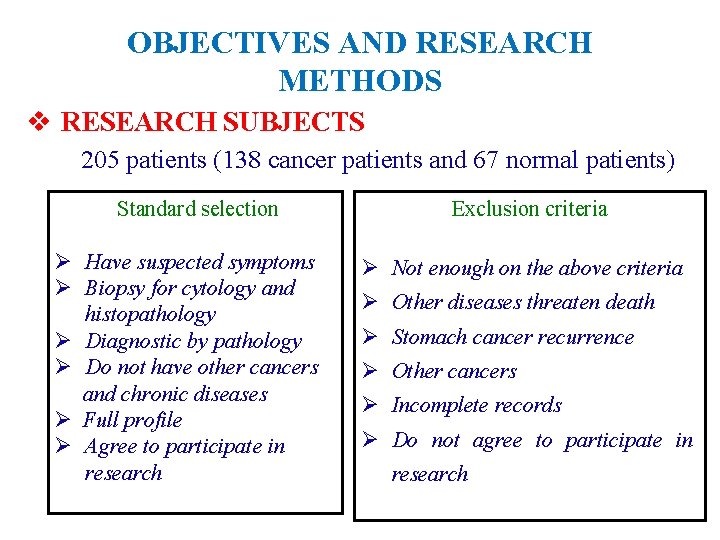
OBJECTIVES AND RESEARCH METHODS v RESEARCH SUBJECTS 205 patients (138 cancer patients and 67 normal patients) Standard selection Ø Have suspected symptoms Ø Biopsy for cytology and histopathology Ø Diagnostic by pathology Ø Do not have other cancers and chronic diseases Ø Full profile Ø Agree to participate in research Exclusion criteria Ø Not enough on the above criteria Ø Other diseases threaten death Ø Stomach cancer recurrence Ø Other cancers Ø Incomplete records Ø Do not agree to participate in research
OBJECTIVES AND RESEARCH METHODS Ø Research location: Nation Cancer Hospital Ø Study time : From 01/08/2015 to 31/8/2016 Ø Research design: Descriptive research Ø Database: According to research sample Ø Research ethics: Has been agreed by the authorities The information obtained is only for research purposes
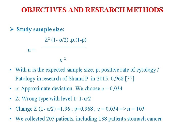
OBJECTIVES AND RESEARCH METHODS Ø Study sample size: Z 2 (1 - α/2). p. (1 -p) n= 2 • With n is the expected sample size; p: positive rate of cytology / Patology in research of Shama P in 2015: 0, 968 [77] • : Approximate deviation. We choose = 0, 034 • Z: Wrong type with level 1: 1 -α/2 • Change Z (1 - α/2) =1, 96 ; p=0, 968 ; = 0, 034 => n = 103 • We collected 205 patients, including 138 patients stomach cancer
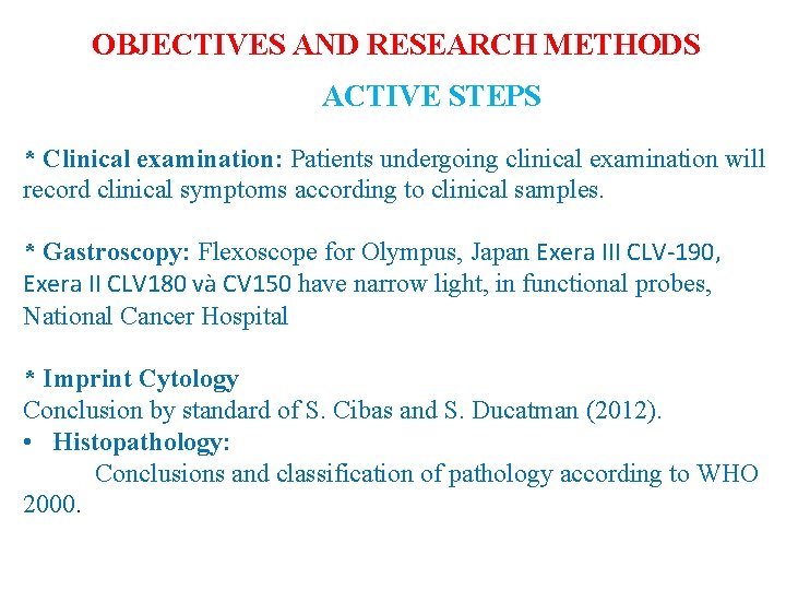
OBJECTIVES AND RESEARCH METHODS ACTIVE STEPS * Clinical examination: Patients undergoing clinical examination will record clinical symptoms according to clinical samples. * Gastroscopy: Flexoscope for Olympus, Japan Exera III CLV-190, Exera II CLV 180 và CV 150 have narrow light, in functional probes, National Cancer Hospital * Imprint Cytology Conclusion by standard of S. Cibas and S. Ducatman (2012). • Histopathology: Conclusions and classification of pathology according to WHO 2000.
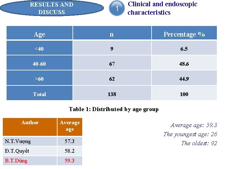
RESULTS AND DISCUSS 1 Clinical and endoscopic characteristics Age n Percentage % <40 9 6. 5 40 -60 67 48. 6 >60 62 44. 9 Total 138 100 Table 1: Distributed by age group Author Average N. T. Vượng 57. 3 Đ. T. Quyết 58. 2 B. T. Dũng 59. 3 Average age: 59. 3 The youngest age: 26 The oldest: 92
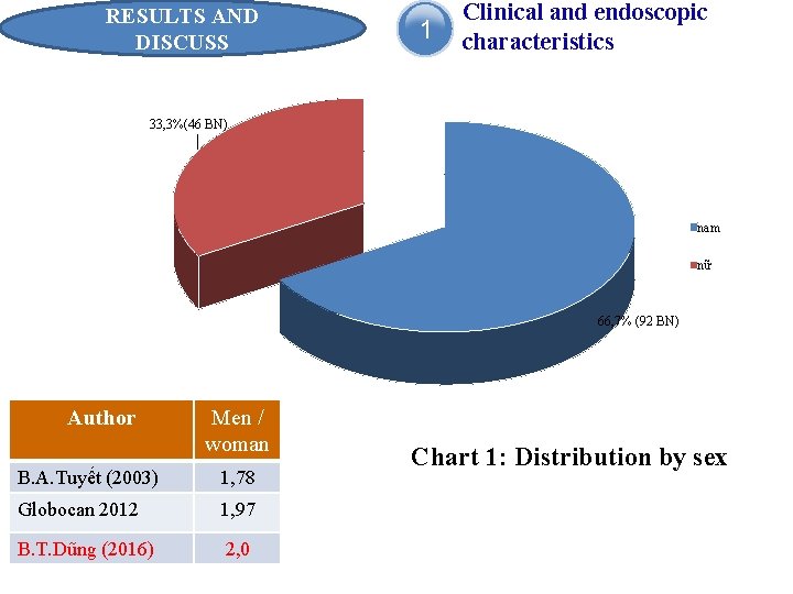
RESULTS AND DISCUSS 1 Clinical and endoscopic characteristics 33, 3%(46 BN) nam nữ 66, 7% (92 BN) Author Men / woman B. A. Tuyết (2003) 1, 78 Globocan 2012 1, 97 B. T. Dũng (2016) 2, 0 Chart 1: Distribution by sex
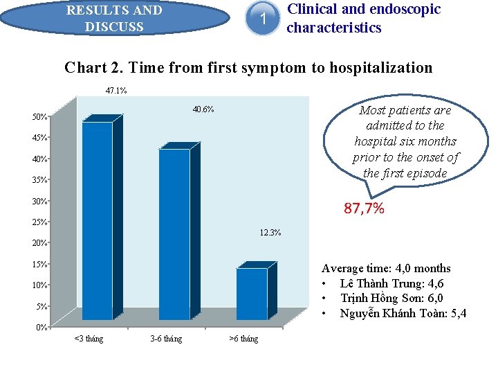
RESULTS AND DISCUSS 1 Clinical and endoscopic characteristics Chart 2. Time from first symptom to hospitalization 47. 1% Most patients are admitted to the hospital six months prior to the onset of the first episode 40. 6% 50% 45% 40% 35% 30% 87, 7% 25% 12. 3% 20% 15% Average time: 4, 0 months • Lê Thành Trung: 4, 6 • Trịnh Hồng Sơn: 6, 0 • Nguyễn Khánh Toàn: 5, 4 10% 5% 0% <3 tháng 3 -6 tháng >6 tháng
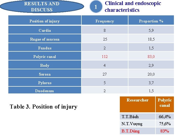
RESULTS AND DISCUSS 1 Clinical and endoscopic characteristics Position of injury Frequency Proportion % Cardia 8 5, 9 Rugae of mucosa 25 18, 5 Fundus 2 1, 5 Polyric canal 112 83, 0 Body 4 2, 9 Serosa 27 20, 0 Pylorus 5 3, 7 Duodenum 2 1, 5 Researcher Table 3. Position of injury Polyric canal T. T. Bách 66, 4% N. T. Vượng 75, 6% B. T. Dũng 83%
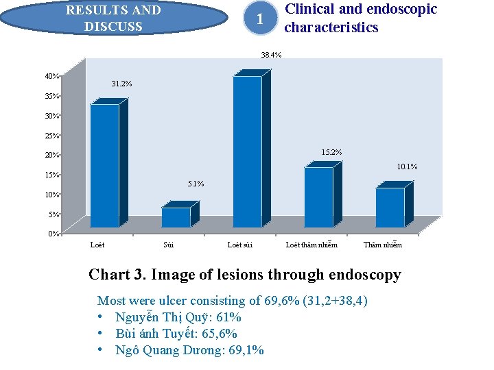
RESULTS AND DISCUSS 1 Clinical and endoscopic characteristics 38. 4% 40% 31. 2% 35% 30% 25% 15. 2% 20% 10. 1% 15% 5. 1% 10% 5% 0% Loét Sùi Loét sùi Loét thâm nhiễm Thâm nhiễm Chart 3. Image of lesions through endoscopy Most were ulcer consisting of 69, 6% (31, 2+38, 4) • Nguyễn Thị Quỹ: 61% • Bùi ánh Tuyết: 65, 6% • Ngô Quang Dương: 69, 1%
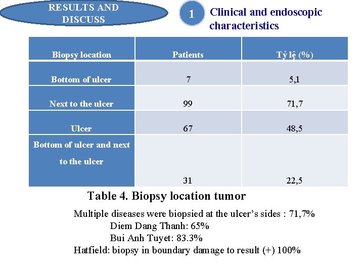
RESULTS AND DISCUSS 1 Clinical and endoscopic characteristics Biopsy location Patients Tỷ lệ (%) Bottom of ulcer 7 5, 1 Next to the ulcer 99 71, 7 Ulcer 67 48, 5 31 22, 5 Bottom of ulcer and next to the ulcer Table 4. Biopsy location tumor Multiple diseases were biopsied at the ulcer’s sides : 71, 7% Diem Dang Thanh: 65% Bui Anh Tuyet: 83. 3% Hatfield: biopsy in boundary damage to result (+) 100%
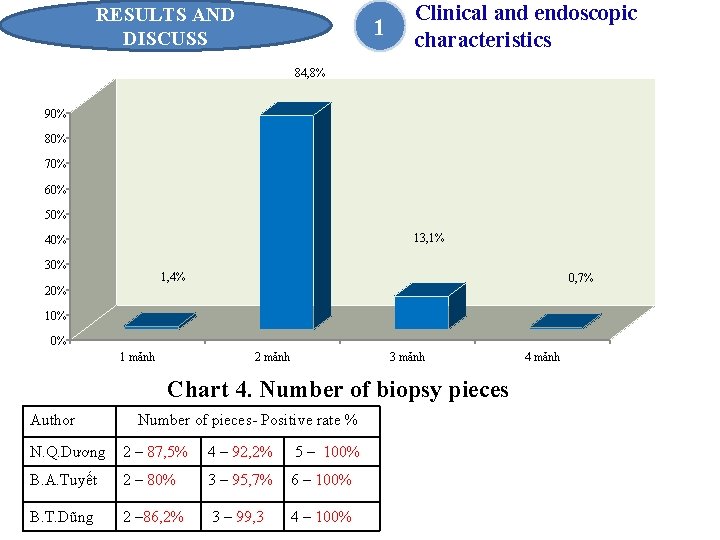
RESULTS AND DISCUSS 1 Clinical and endoscopic characteristics 84, 8% 90% 80% 70% 60% 50% 13, 1% 40% 30% 1, 4% 0, 7% 20% 10% 0% 1 mảnh 2 mảnh 3 mảnh Chart 4. Number of biopsy pieces Author Number of pieces- Positive rate % N. Q. Dương 2 – 87, 5% 4 – 92, 2% 5 – 100% B. A. Tuyết 2 – 80% 3 – 95, 7% 6 – 100% B. T. Dũng 2 – 86, 2% 3 – 99, 3 4 – 100% 4 mảnh
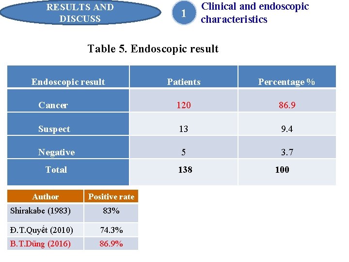
RESULTS AND DISCUSS 1 Clinical and endoscopic characteristics Table 5. Endoscopic result Patients Percentage % Cancer 120 86. 9 Suspect 13 9. 4 Negative 5 3. 7 Total Author 138 Positive rate Shirakabe (1983) 83% Đ. T. Quyết (2010) 74. 3% B. T. Dũng (2016) 86. 9% 100
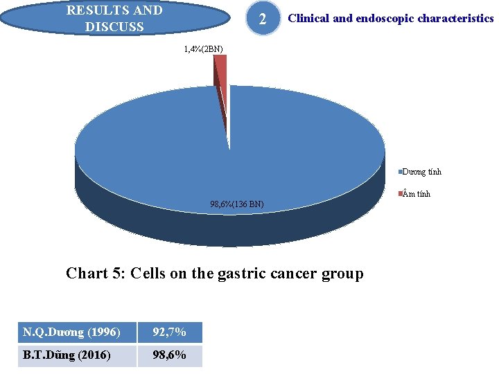
RESULTS AND DISCUSS 2 Clinical and endoscopic characteristics 1, 4%(2 BN) Dương tính m tính 98, 6%(136 BN) Chart 5: Cells on the gastric cancer group N. Q. Dương (1996) 92, 7% B. T. Dũng (2016) 98, 6%
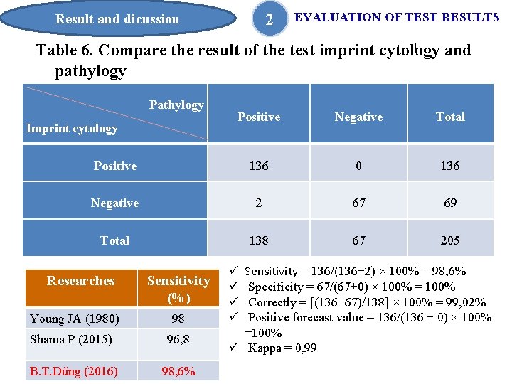
2 Result and dicussion EVALUATION OF TEST RESULTS l Table 6. Compare the result of the test imprint cytology and pathylogy Pathylogy Positive Negative Total Positive 136 0 136 Negative 2 67 69 Total 138 67 205 Imprint cytology Researches Sensitivity (%) Young JA (1980) 98 Shama P (2015) 96, 8 B. T. Dũng (2016) 98, 6% Sensitivity = 136/(136+2) × 100% = 98, 6% Specificity = 67/(67+0) × 100% = 100% Correctly = [(136+67)/138] × 100% = 99, 02% Positive forecast value = 136/(136 + 0) × 100% =100% ü Kappa = 0, 99 ü ü
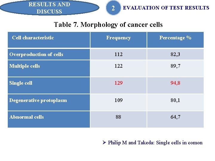
RESULTS AND DISCUSS 2 EVALUATION OF TEST RESULTS Table 7. Morphology of cancer cells Cell characteristic Frequency Percentage % Overproduction of cells 112 82, 3 Multiple cells 122 89, 7 Single cell 129 94, 8 Degenerative protoplasm 109 80, 1 Abnormal cells 88 64, 7 Ø Philip M and Takeda: Single cells in comon
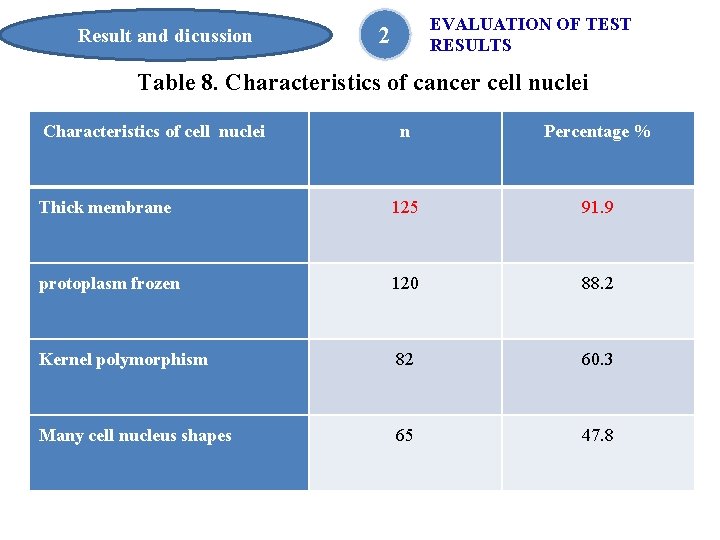
Result and dicussion EVALUATION OF TEST RESULTS 2 Table 8. Characteristics of cancer cell nuclei Characteristics of cell nuclei n Percentage % Thick membrane 125 91. 9 protoplasm frozen 120 88. 2 Kernel polymorphism 82 60. 3 Many cell nucleus shapes 65 47. 8
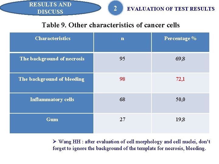
RESULTS AND DISCUSS 2 EVALUATION OF TEST RESULTS Table 9. Other characteristics of cancer cells Characteristics n Percentage % The background of necrosis 95 69, 8 The background of bleeding 98 72, 1 Inflammatory cells 68 50, 0 Gum 27 19, 8 Ø Wang HH : after evaluation of cell morphology and cell nuclei, don’t forget to ignore the background of the template for necrosis, bleeding.
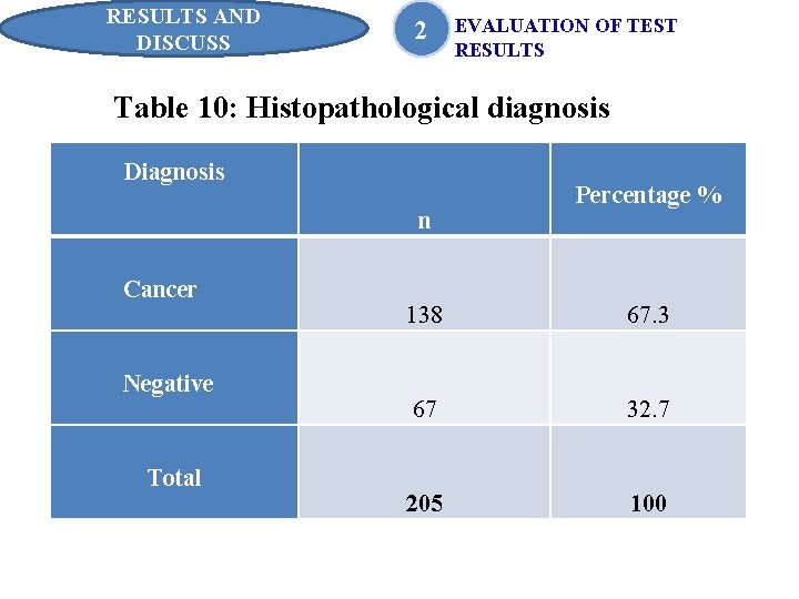
RESULTS AND DISCUSS 2 EVALUATION OF TEST RESULTS Table 10: Histopathological diagnosis Diagnosis n Cancer Negative Total Percentage % 138 67. 3 67 32. 7 205 100
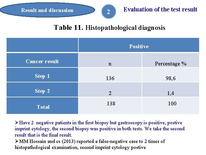
Result and discussion 2 Evaluation of the test result Table 11. Histopathological diagnosis Positive Cancer result n Percentage % Step 1 136 98, 6 Step 2 2 1, 4 138 100 Total ØHave 2 negative patients in the first biopsy but gastroscopy is positive, postive imprint cytology, the second biopsy was positive in both tests. We take the second result that is the final result. ØMM Hossain and cs (2013) reported a false-negative case to 2 times of histopathological examination, second imprint cytology postive
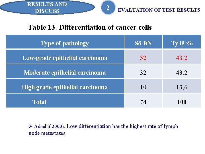
RESULTS AND DISCUSS 2 EVALUATION OF TEST RESULTS Table 13. Differentiation of cancer cells Type of pathology Số BN Tỷ lệ % Low-grade epithelial carcinoma 32 43, 2 Moderate epithelial carcinoma 32 43, 2 High grade epithelial carcinoma 10 13, 6 74 100 Total Ø Adachi( 2000): Low differentiation has the highest rate of lymph node metastases
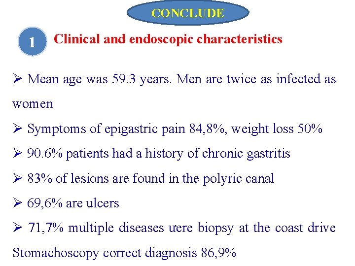
CONCLUDE 1 Clinical and endoscopic characteristics Ø Mean age was 59. 3 years. Men are twice as infected as women Ø Symptoms of epigastric pain 84, 8%, weight loss 50% Ø 90. 6% patients had a history of chronic gastritis Ø 83% of lesions are found in the polyric canal Ø 69, 6% are ulcers Ø 71, 7% multiple diseases ưere biopsy at the coast drive Stomachoscopy correct diagnosis 86, 9%
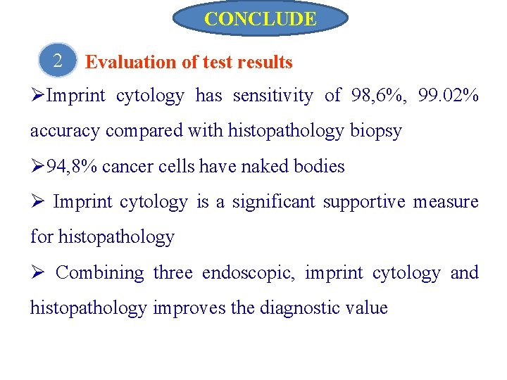
CONCLUDE 2 Evaluation of test results ØImprint cytology has sensitivity of 98, 6%, 99. 02% accuracy compared with histopathology biopsy Ø 94, 8% cancer cells have naked bodies Ø Imprint cytology is a significant supportive measure for histopathology Ø Combining three endoscopic, imprint cytology and histopathology improves the diagnostic value
REQUEST 1. Biopsy for high value in diagnosis for gastric cancer, should be more commonly used for medical routes for the early diagnosis of gastric cancer. 2. Imprint cytology should be performed along with endoscopic and histopathologic biopsies. 3. At baseline medical facilities have not yet pathology, endoscopic biopsies and imprint cytology are valuable in the diagnosis of early gastric cancer screening.
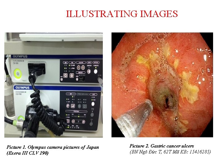
ILLUSTRATING IMAGES Picture 1. Olympus camera pictures of Japan (Exera III CLV 190) Picture 2. Gastric cancer ulcers (BN Ngô Đức T, 62 T Mã KB: 15416285)
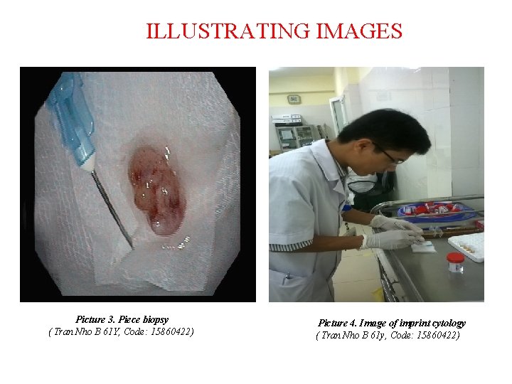
ILLUSTRATING IMAGES Picture 3. Piece biopsy ( Tran Nho B 61 Y, Code: 15860422) Picture 4. Image of imprint cytology ( Tran Nho B 61 y, Code: 15860422)
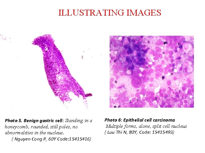
ILLUSTRATING IMAGES Photo 5. Benign gastric cell: Standing in a honeycomb, rounded, still poles, no abnormalities in the nucleus. ( Nguyen Cong P, 60 Y Code: 15415416) Photo 6: Epithelial cell carcinoma Multiple forms, alone, split cell nucleus ( Luu Thi N, 80 Y, Code: 15415493)
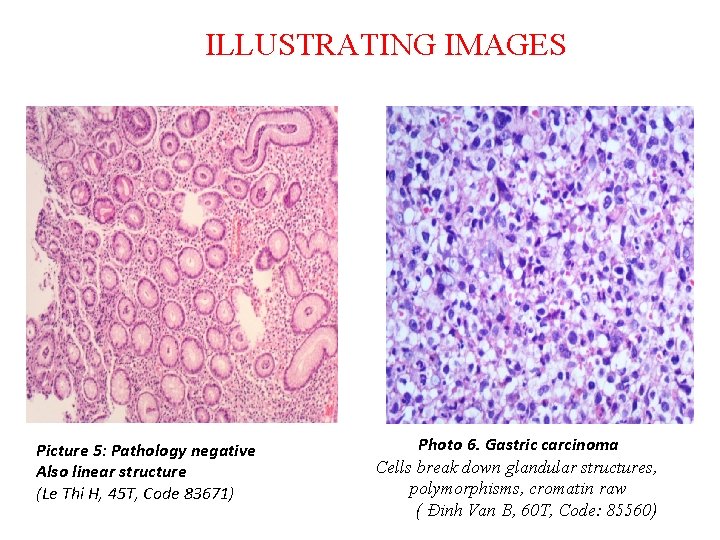
ILLUSTRATING IMAGES Picture 5: Pathology negative Also linear structure (Le Thi H, 45 T, Code 83671) Photo 6. Gastric carcinoma Cells break down glandular structures, polymorphisms, cromatin raw ( Đinh Van B, 60 T, Code: 85560)
Thank you very much ! 38